PPT-Knee Anatomy-Posterior view
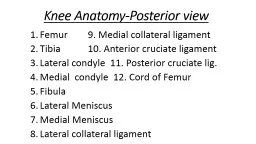
Femur 9 Medial collateral ligament Tibia 10 Anterior cruciate ligament Lateral condyle 11 Posterior cruciate lig Medial condyle 12 Cord of Femur Fibula Lateral Meniscus
Download Presentation
"Knee Anatomy-Posterior view" is the property of its rightful owner. Permission is granted to download and print materials on this website for personal, non-commercial use only, provided you retain all copyright notices. By downloading content from our website, you accept the terms of this agreement.
Presentation Transcript
Transcript not available.