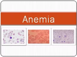
/
General Approach to Anemia AND Iron Deficiency General Approach to Anemia AND Iron Deficiency

General Approach to Anemia AND Iron Deficiency - PowerPoint Presentation

faustina-dinatale
faustina-dinatale . @faustina-dinatale
Follow
496 views
Uploaded On 2015-10-18

General Approach to Anemia AND Iron Deficiency - PPT Presentation

  Seventh International Symposium in Continuing Nursing EducationMarch 2014 32014 Donald W McLaren MD Objectives To discuss how to evaluate and determine cause of anemia To discuss Iron deficiency ID and Iron Deficiency Anemia IDA causes evaluation signs symptoms treatment an ID: 164299

deficiency anemia b12 iron anemia deficiency iron b12 amp http uptodate search rbc retic www 2014 count hgb blood

Share:

Link:

Embed:

Download Presentation from below link

Download Presentation The PPT/PDF document "General Approach to Anemia AND Iron Defi..." is the property of its rightful owner. Permission is granted to download and print the materials on this web site for personal, non-commercial use only, and to display it on your personal computer provided you do not modify the materials and that you retain all copyright notices contained in the materials. By downloading content from our website, you accept the terms of this agreement.


Presentation Transcript

Slide1

General Approach to Anemia AND Iron Deficiency


Seventh International Symposium in Continuing Nursing Education/March, 2014

3/2014

Donald W. McLaren, MDSlide2

Objectives

To discuss how to evaluate and determine cause of anemia

To discuss Iron deficiency (ID) and Iron Deficiency Anemia (IDA) - causes, evaluation, signs, symptoms, treatment and prevention at different ages

To discuss briefly few other common causes of anemia Slide3
Slide4

Burden of anemia globally

24% of world population; 1.62 billion anemic

Greatest burden pregnant women and children < 5

Highest prevalence

Africa

and SE Asia

In these locations > 65% of children < 5 years are anemicSlide5

http://www.k4health.org/toolkits/anemia-prevention/anemia-causes-prevalence-impactSlide6

Definitions

Anemia

– reduced # circulating RBCs or decreased circulating hemoglobin as measured by decreased Hgb, Hct or RBC count

Hgb – concentration of hemoglobin (gm/dl)

Hct – % of sample of whole blood (WB) occupied by intact RBCs.

RBC count: # RBCs in specified volume of blood – usually millions of RBC / μL of whole blood (WB)Slide7

Indices

MCV (mean corpuscular volume)

– volume of average RBC measured or calculated as MCV (femtoliters; 10

-15

) = 10 X Hct ÷ RBC; indication of cell size

MCH (Mean Corpuscular Hemoglobin) - mass of Hgb in a RBC

MCHC (mean corpuscular hemoglobin concentration) – measure of concentration of hemoglobin in a given volume of packed RBCsSlide8

Microcytosis

:

MCV; RBC size small

Macrocytosis

:

MCV; RBC size large

Normocytosis

: MCV WNL; RBC size normal

Hypochromic

: paler than normal – ↓Hgb as measured by

MCH, MCHC

Hyperchromic

:

Hgb –

MCH, MCHCSlide9

RDW (RBC distribution width)

– measure of variability of size of RBCs - normal about 11.5-14.5 – increased in nutritional deficiencies (like Fe, B12)

Anisocytosis – when there is a lot of variation of sizes of RBC as seen on microscope -

RDW

Peripheral smear

– blood smear stained to be looked at to see cell size, shape, abnormal cells, etc.Slide10

Reticulocyte

– immature RBC usually released into blood 1 day prior to becoming mature RBC

Reticulocyte count

– number of reticulocytes in peripheral blood expressed as % of total RBCsSlide11

What is included in a CBC

Hgb, Hct

RBC count

Indices – MCV, MCH, MCHC

RDW

WBC and differential or diff indicating how many of each type of WBC is there

Platelet count

Normal values vary with age and sexSlide12

http://upload.wikimedia.org/wikipedia/en/4/49/CBC_With_Differential.jpgSlide13

http://www.childrensmn.org/manuals/lab/hematology/018981.aspSlide14
Slide15
Slide16

RBC and appearance in peripheral smear

http://en.wikipedia.org/wiki/Reticulocyte_indexSlide17

Reticulocyte unstained and stained

http://en.wikipedia.org/wiki/Reticulocyte_indexSlide18
Slide19
Slide20

http://www.med-ed.virginia.edu/courses/path/innes/nh/morphology.cfmSlide21

Define anemia

Hgb, Hct (H/H) > 2 SD below the mean

H/H < 12g/dl or 36% for adult women

H/H < 13.5(14.0) and 41(42)% for men

Higher if live at altitude, smokers, athletes

Consider volume shifts: bleed, pregnancy

African-Americans Hgb 0.5-1.0 g/dl < white

Can drop 15%, be significant drop, still WNL Slide22

Other basics of erythropoiesis (making of red cells)

EPO (

erythropoietin

) from kidney

↑ if

anemia.

If marrow functioning, raw materials present (iron, B12) EPO stimulates marrow to produce more RBCs – leads to increased reticulocytes.

Reticulocytes released from bone marrow and circulate normally 1 day before becoming mature RBC

Normal RBCs circulate for about 110-120 days before being removed from circulationSlide23

Some more basics

Ordinarily rate of production = rate of removal

Retics replace about 1% of cells daily so normal retic count around 1% (0.5-1.5%)

If increased RBC destruction (i.e. SCD) must replace more. If cell lifespan 20 days must replace 5% / day

Will now discuss W/U of anemiaSlide24

Causes of Anemia – only 3

Decreased RBC production

– bone marrow not keeping up with normal RBC loses (hypoproliferative)

Increased RBC destruction

or

decreased survival time

(hemolysis)

Blood loss

(hemorrhage)

Combinations not uncommon Slide25

Essential Lab for Evaluation of Anemia

CBC

Peripheral smear

read by someone who knows what they are looking for

Reticulocyte count

Can classify anemia based on these 3

Other testing depending on resultsSlide26

2 General Approaches to Evaluate Anemia

Kinetic (physiological) approach

addresses the

mechanism

or

cause

of the anemia. Retic count used to divide into hemolysis or blood loss (

retic) vs. hypoproliferative (

retic count)

Morphologic approach

– categorizing based on

RBC size

as determined by

MCV

and peripheral smear

Will discuss a combination of twoSlide27

Kinetic approach

Low retic = decreased RBC production

Lack of nutrients (Fe, B12, folate)

Bone marrow disorders or suppression

Low EPO level

High retic = increased destruction of RBC or blood loss Slide28

Morphological Approach to Anemia

Determine RBCs size: small, normal or large based on MCV, peripheral smear

For adults MCV range about 80-96

DDx if microcytic anemia fairly short

Retic count helpful to further sort out normocytic and macrocytic anemias.

Sometimes not straightforward – classic Fe deficiency microcytic, but if mild and early is normocyticSlide29

Always begin with Hx and Px

Are they stable? Are there S/S of anemia?

Known or obvious bleeding?

Is anemia acute, chronic or subacute

Any chronic illness?

Ethnicity

Alcohol, medications, history transfusions

Good dietary history

Big spleen, liver disease, jaundice, tender bone, H-S megaly, lymphadenopathySlide30

Look at rest of CBC/peripheral smear – not just RBCs

Abnormal cells

may indicate SCD, spherocytosis, leukemia, and on occasion make the diagnosis for you

Pancytopenia

changes the differential diagnosis – likely need hematologist

Destruction in peripheral blood

Aplastic anemia, hematologic malignancy, B12 deficiency, anorexia, radiation, chemotherapy, myeloproliferative diseaseSlide31
Slide32
Slide33
Slide34

Microcytic anemia

Relatively few causes

*Fe deficiency

anemia

(IDA) (early can be normocytic)

↓RBC, ↓Indices, ↑ RDW

*Thalassemias

(

globin synthesis)

*Lead toxicity

(

heme synthesis)

*Anemia of chronic disease (or normocytic)

Sideroblastic anemiasSlide35
Slide36

Macrocytic (↑MCV) DDx

*Folate, B12 deficiency (

Usual cause of MCV > 115)

Extreme reticulocytosis

Alcoholism, liver disease,

hypothyroidism

*HIV antiviral therapy

*Most chemotherapeutic agents

Myelodysplastic disorders

Adults > 65 may have higher MCV than younger – use 100 rather than 96 for ULNSlide37

Can narrow diagnosis based on peripheral smear and retic count

*Megaloblastic

(macro-ovalocytes and hypersegmented neutrophils) or

Low retic

B12 or folate deficiency

HIV meds,

Anticonvulsants

(folate depletion) (dilantin, valproate), Cancer drugs (

+

anemia)

Nonmegaloblastic

with round macrocytes and macroreticulocytes –

(

low retic

) Alcohol, thyroid, liver disease

(

high retic

) some hemolytic

Myeloproliferative disorders - bone marrowSlide38
Slide39

Normocytic Anemia DDx with reticulocytosis

Huge category – Narrow ddx with retic count

*Blood loss, hypersplenism

*Increased destruction, loss of RBCs (hemolysis)

Congenital or intrinsic

(

Hemoglobinopathy

: SS, SC;

RBC membrane disorders

: spherocytosis;

RBC enzyme deficiency

(G-6-PD, pyruvate kinase)

Acquired (extrinsic):

Mechanical

,

macrovascular

,

Micro- angiopathic

(DIC, HUS, TTP)

autoimmune hemolytic anemias, drug related

(Aldomet, penicillins, cephalosporins)] PNH

Mixture of micro/macrocytic anemiasSlide40

Normocytic Anemia With Low Retic

Decreased production normal sized cells (*

anemia of chronic illness, aplastic anemia, bone marrow infiltration)

*Uncompensated increase in plasma volume (pregnancy)

Low EPO states (CRF, liver or kidney disease, Endocrine deficiency)

Myeloproliferative disorders, dysplasia, fibrosis, marrow replacement

Bone marrow

can get dx 90-92% of timeSlide41

Summary (read RPI as retic for now)

Microcytic Normocytic Macrocytic

↑ RPI or ARC

↑ RPI or ARC

↓ RPI

or ARC

↓ RPI

Or ARC

Then it is easy to look up differential for each combination of MCV and Retic. *RPI = Reticulocyte production index; ARC = Absolute retic countSlide42

Many sites for DDx once anemia classified MCV and RPI

Just Google causes of ____ anemia.

Microcytic anemia or

microcytosis

http://www.aafp.org/afp/2010/1101/p1117.html

Normocytic anemia or

normocytosis

http://www.aafp.org/afp/2000/1115/p2255.html

Macrocytic anemia or

macrocytosis

http://www.aafp.org/afp/2009/0201/p203.htmlSlide43

Use of retic count

Normal 0.5-1.5% in adult

Normally keeps up with RBC loss so that Hgb/Hct stable

Can increase significantly to replace lost RBCs in hemolysis, blood loss if a) marrow working, b) sufficient EPO, c) nutrients

How do we know in the face of anemia if elevated retic count is appropriately high vs. insufficient for degree of anemia. If Hct is 25, what should retic be? 3? 7? 15?Slide44

Most common anemias in U.S.

Copyright © 2000-2013 The Cleveland Clinic Foundation. http://www.clevelandclinicmeded.com/medicalpubs/diseasemanagement/hematology-oncology/anemia/ Slide45

Corrected retic count or RPI (reticulocyte production index)

Can use either absolute retic count (RBC X % retics) or RPI to clarify situation

Reticulocyte count is a %. So, if you decrease RBCs (anemia) with the same number of retics gives falsely elevated %

How many retics being released from bone marrow vs. normal - measure of how many RBCs being made in the bone marrow

1

st

of 2 corrections is for low RBC count – to give a “corrected retic count” or “retic index” = Retic count X Hct/45 (normal Hct) Slide46

10% reticsSlide47

Retic Count X Hct/normal Hct = 20%

X 5/10 = 10%Slide48

Reticulocyte production index (RPI)

Second correction needed in anemia, retics are released early in a less mature state

Retics normally stay in the blood for 1 day

If released early stay in circulation longer before reaching maturity

Divide previously corrected retic count by maturation factor corrects for early release

Hct 36-45: 1.0; Hct 26-35: 1.5 Hct 16-25: 2.0

Hct

<

15: 2.5Slide49

http://faculty.washington.edu/rrichard/anemia.pptSlide50

RPI

RPI =

retic count X Hct/45

Maturation correction

1-2% is normal in non-anemic individual

In anemia,

RPI< 2 means there is an inadequate response

to correct anemia and indicates hypoproliferative anemia

In anemia,

RPI> 3 appropriate/adequate compensatory response to anemia

and represents destruction or decreased survival or loss of red cellsSlide51

http://allaboutblood.com/tag/corrected-reticulocyte-index/Slide52

Iron (Fe) Deficiency anemia (IDA) in Infants and Young Children

IDA most common cause anemia in world

1-3 years old: 9% in U.S. have ID, 3% IDA

Risk factors: poverty, Black or Hispanic race, obesity, prematurity, immigrants

Fe balance

75% bound in heme proteins Hgb & myoglobin

Most rest is storage iron in ferritin, Hemosiderin

Tiny amount bound to transferrin for transport

3% in critical enzyme systemsSlide53

Adults: 5% of daily Fe needs to come from dietary sources: absorption = loss

Children: 30% of daily needs come must from diet due to growth spurt and body muscle mass increasesSlide54

Fe Absorption

Very little normally leaves, enters body daily – little lost in feces, urine. Most recycled by macrophages

Mechanisms affecting intestinal absorption, transport a) body Fe stores, b) rate of erythropoiesis, c) Bioavailability of dietary Fe, d) hepcidin

Absorption increases with

erythropoiesis.Slide55

Intestinal Fe absorption and need for iron

Bioavailability: Heme sources (meat, poultry, fish) (30%) > than non-heme sources (vegetable) (10%)

Hepcidin: made by liver – inhibits GI Fe absorption and storage Fe release

Healthy term infants have enough Fe stores for 5-6 months if mother has enough Fe. Premies much less.Slide56

Some Fe sources and infant needs

Breast milk: low Fe; 50% bioavailability

Formulas: 12-36X more Fe; 4-6% bioavailability.

Full term breastfed babies need 1 mg/kg up to 15 mg beginning at age 4 months

Breastfed premies: Need 2-4 mg/kg up to 15 mg starting at age 1 month of age

Age 1-3: 7; 4-8: 10; 9-13: 8 mg/daySlide57

Causes of IDA

Insufficient Fe intake

Poor Fe absorption (poor dietary sources or other reasons)

Introduction of unmodified cow’s milk at < age 12 months

FOB loss 2

o

to

cow’s milk protein-induced colitis

(6 moa: 30% : 5% FOB + if on Cow’s milk vs. formula in infants)

Breast milk too long

– without supplementation Slide58

Causes of IDA Continued

Giving > 600 ml/day or > 6 breast feeds / day at 8-12 months of age.

Preschool - > 720 cc milk daily

(low concentration and bioavailability of Fe + possible

intestinal blood loss increased

Absorbed most from duodenum so decreased absorption with Celiac, Crohn’s, giardia, resection proximal SB, H. pylori.

Blood loss - IBD, cow’s milk protein-induced colitisSlide59

Prevention

Exclusive breast feeding till age 4-6 mo.

Supplement with Fe at 4 months

With iron fortified cereal after 6 mo

Only Fe fortified formulas (avoid cow’s milk) if not breastfed till age 1 year

After 6 mo

>

one feeding of foods rich in Vitamin C daily (increase Fe absorption)

After 6 months consider pureed meats

Age 1-5 limit to 24 oz cows milk/daySlide60

Development of IDA

Use up storage iron first

Iron limited erythropoiesis

IDA (last stage and first to recover)

Initially normocytic / normochromic

Eventually classic microcytic hypochromic hypoproductive anemiaSlide61

Signs/symptoms

Most asymptomatic as develops slowly

Lethargy, irritability, tachypnea, poor feeding

Pallor (not reliable sign till severe),

↑HR

exercise capacity (even pre-anemic)

Pica (appetite for substances not fit for food - paper, clay, dirt), pagophagia (pica for ice is common and specific for ID) – often precede anemia and resolve early upon treatment

Beeturia (red urine with beets)

Fe deficiency can cause RLS - 9X

↑incidenceSlide62

Complications of IDA and treatment

Neurodevelopmental issues

Psychomotor, mental development impairment, cognitive issues even in adolescents

Fe supplementation can prevent but may not correct once established.

Improvement of attention, concentration and cognitive function with Fe supplementation

Auditory and visual dysfunction

Decreased work capacity in adults

Immunity – Fe may increase bacterial, malaria infection risk. Transferrin has bacteriostatic effects lost if saturated by FeSlide63

Screening

2/3 with ID don’t have IDA (age 1-3 9% ID, 2-3% IDA)

2/3 those anemic have another diagnosis

Anemia risk assessment at ages 4,

15

18, 24,

30

mo then yearly. All premies at risk.

Use focused dietary history

Standards already listed

< 2 servings of Fe rich foods daily after 6 months or < 3 servings age 1-5 years

Intake of sweets, lots of fatty snacks, soft drinksSlide64

Diagnosis/Lab

Universal lab screening of all 9-12 mo.

Hgb, Hct or

CBC

If at risk re-screen at 15-18 months

If high risk of Fe deficiency also check ferritin as one can be ID without IDA

Fe deficiency anemia typically has low MCV, MCH, MCHC and high RDWSlide65
Slide66

If IDA suspected

Can treat in this age empirically and test to confirm only if not responding well

If need to prove or if severe check

Ferritin best single test

– measure of Fe stores. (Acute phase reactant so if another illness can be falsely elevated -

in IDA)

Fe

↓ in IDA

TIBC

(total iron binding capacity)

in IDA

Fe/TIBC= Fe, transferrin saturation

↓ in IDASlide67

Treatment

If anemic dietary history, correct diet

Lead exposure questionnaire or level

If age > 2 some recommend

stool for FOB X 3

, retic count, peripheral smear

If in doubt or severe (

<

7 Hgb) check Fe, TIBC, ferritin (< 12), transferrin sat along with FOB X 3.Slide68

Treatment continued

Most cost effective means to tx if consistent diet history and no indication of Pb toxicity is a presumptive trial of iron ($5.00)

3 -6 mg/kg elemental Fe/day up to 150 mg

FeSO

4

daily or BID (FeSO

4

20% elemental Fe)

Continue Fe several (3-4) months after anemia resolved to replace stores

Maximize absorption - give between meals, with juiceSlide69

Iron therapy

Repeat CBC 1 month (sooner if severe) for

>

1 gm/dl increase in Hgb – if not re-evaluate

Can use other forms of Fe – i.e. Fe Gluconate

Avoid enteric coated if possible

Rarely causes GI upset at this age (10%)

MUST at same time institute dietary changes

Repeat periodically till normal for age

Once normal continue Fe 3-4 months to replace Fe storesSlide70

Nonresponders

Check adherence including diet

Intercurrent illness that can lower Hgb?

Check Ferritin, Hgb electrophoresis, Vit B12, RBC folate, Fe, TIBC, ferritin

Fe/TIBC = transferrin saturation

Several stool specimens for occult bloodSlide71

Non-responders continued

Eliminate all cow’s milk protein from diet

R/O Celiac disease

ESR and albumin – to screen for IBD

Consider rare causes, incorrect dx (Thalassemia, Chronic disease)

Rare to need parenteral Fe

Rare to need transfusion even if Hgb 4-5 g/dl unless in distressSlide72

Adolescents

risk due to expansion of blood volume, increased muscle mass with growth

Eating patterns (vegetarian, anorexia)

Incidence rises with age in females

ID 11% IDA 3% 16-19 in girl vs. < 1% boys

Obesity, training athletes, periods

↑ risk

Screen ALL at risk, girls q 5 yr beginning at 13, boys once during growth spurtSlide73

Adolescents

Cutoff Hgb < 11 and cutoff ferritin < 12

For tx least expensive is Fe sulfate 325 Fe Sulfate = 65 mg elemental Fe.

Ca inhibits, ascorbic acid

↑ absorption

If not tolerating iron consider

Taking with food + ascorbic acid

Feosol 45 mg elemental Fe/5cc better tolerated

More likely not to tolerate, need transfusion (< 7 Hgb + distress, end organ issues)Slide74

Adults

Main difference in adult – Must look for cause: cause blood loss till proven otherwise. Overt sources of blood loss

Severe traumatic hemorrhage

Hematemesis, melana, or hematochezia

Hemoptysis

Severe menorrhagia, pregnancies, lactation

Gross hematuria

Frequent blood donating

Marathon runningSlide75

Occult blood loss – not obvious

Usually GI tract in men and post-menopausal women

Heavy periods in pre-menopausal women

While reduced Fe absorption and deficient diet may be cause, must do GI work-up to avoid missing malignancy

Overall risk of

malignancy

not high (

12%

), but much higher than without IDASlide76

Evaluation of occult blood loss

First study to do based on history – Colonoscopy vs. EGD

In one study of 100 persons, source found in 62%: 25 with colonoscopy, 36 with EGD, 1 with both; 11 had cancer

For most do

colonoscopy first

because almost all need even if EGD finds lesion

If cause not found EGDSlide77

Evaluation of occult blood loss

If no cause found

consider SB capsule endoscopy

R/O

Celiac disease, H. Pylori

, atrophic gastritis esp. if refractory to Fe therapy (100% Celiac, 70% H. Pylori refractory)

Hookworms

cause increase losses

Can consume 0.3-0.5 ml blood/day

Often cause

eosinophilia

Consider Foods/meds that interfere with Fe absorptionSlide78

Other causes

Gastric bypass

Pulmonary hemosiderosis

Intravascular hemolysis leading to urinary loss of Fe

Congenital Fe deficiency not responsive to Fe or defects in Fe absorption or utilizationSlide79

Transient erythroblastopenia of childhood

Temporary (always) RBC aplasia occurring at ages 6 mo – 4 yrs

Fairly common but don’t know true incidence as many cases not detected

Normocytic anemia

Cause not known. Viral? Toxic?

Mild neutropenia possible; normal or slightly high plateletsSlide80

TEC

Hgb 6-8 with reticulocytopenia

Normal MCV except during recovery due to retics (distinguishes from congenital pure red cell aplasia – macrocytic anemia)

Lasts 1-2 months then recovery

80% recover within 1 month

Transfusion rarely necessarySlide81

Thalassemia

Major forms deadly or very sick

Minor and trait look a bit like IDA

Beta thal minor – has elevated Hgb A2, F on Hgb electrophoresis, target cells, low MCV and – W/U for IDA

Alpha more complex

If failed Fe tx important to diagnose to prevent eventual iron overload from increases absorption and repeated tx.Slide82

B12 (and folate) deficiency

Macrocytic anemia with low retic

Check B12, folate in all with high MCV

B12 variable so if low need repeat

Folate: leafy green veges, fruit, enriched sources

B12 animal products: eggs, meat, fish, milk

(homocysteine, MMA) Both high in B12 and only HC in folate deficiency. Slide83

Can effect all 3 hematopoietic cell lines

B12 low from poor intake, gastrectomy, bariatric surgery, H. Pylori, breastfed, fish tapeworm, drugs (PPI), hereditary causes, pernicious anemia, Celiac

Folate – Poor intake, drugs (MTX), increased needs, Celiac

Folate needed in purine synthesis; B12 needed as cofactor in activation of folate

Neurological damage from B12 deficiency which can be permanentSlide84

S/S/lab in B12 deficiency

B12 – anemia but not always

Macrocytosis and oval cells

Neuro symptoms

Hypersegmented WBC (> 5 nuclei)

B12 signs/symptoms: Glossitis,

+

anemia, memory loss, irritability, ataxia, dementia, peripheral neuropathy

Danger that neurological symptoms of B12 can be permanent once establishedSlide85
Slide86

Cobalamin levels

 — ”Commercial labs use different methods for measuring Cbl. …there are different normal ranges and no "gold standard

Accordingly, therapeutic trials of Cbl are warranted when testing results are in conflict with the clinical diagnosis.”

Interpretation of B12 (Cbl) levels :

“●>300 pg/mL (>221 pmol/L) — normal result; Cbl deficiency unlikely (ie,

probability of 1 to 5 percent

)

●200 to 300 pg/mL (148 to 221 pmol/L) — borderline result; Cbl deficiency possible

●<200 pg/mL (<148 pmol/L) — low; consistent with Cbl deficiency (

specificity of 95 to 100 percent

)”

Schrier SL. “Diagnosis and treatment of vitamin B12 and folate deficiency.”

UpToDate

accessed on 1/3/2014

http://www.uptodate.com/contents/diagnosis-and-treatment-of-vitamin-b12-and-folate-deficiency?source=search_result&search=vitamin+B12+deficiency&selectedTitle=1~150Slide87

Anemia of chronic disease

Occurs with inflammation (lupus), malignancy, infection (Tb). But even DM, anemia of elderly.

Mechanism

Cytokines decrease EPO production (relative decrease)

Decreased response to EPO

Interference with Fe absorption and trapping by macrophages due to increased hepcidin formation

Perhaps some decreased RBC survival

Characteristics

Most normocytic normochromic hypoproliferative – (microcytic

Fe level, TIBC and transferrin saturation (20% low) decreased but ferritin normal or elevated as acute phase reactant

Decreased absolute retic count, RPI

Usually mild, non-progressive around 11 - (20% < 8%)Slide88

Anemia in Malaria

Can cause severe anemia especially in SSA and often on top of chronic anemia

Multiple mechanisms: acute hemolysis, G-6PD deficiency, extravascular clearance in spleen, intravascular destruction, suppression of erythropoiesis, others

Fe deficiency may be protective against malaria infection – reduced parasitemia, rate of severe malaria by 38%

Especially severe in those with SCDSlide89

Anemia in Sickle Cell Disease

Chronic hemolysis with Hct 20-30% and 3-15% reticulocytosis , elevated indirect bili

2 causes of acute severe anemia - present with pallor, weakness, lethargy-can be fatal

Splenic sequestration crisis

– vaso-occlusion in spleen with rapid increase in size and drop in Hgb of at least 2, low platelets, reticulocytosis. Tend to recur within year

Aplastic crisis

– Parvovirus B19 – decreased reticulocytes. Often need transfusion though retics return in 2-14 daysSlide90

G6PD glucose-6-phosphate dehydrogenase deficiency

Hemolysis precipitated by certain drugs, infection or occasionally a chronic hemolytic anemia

13 % of black men and 2% black women – different genotype in Mediterranean, Arabic and Asian descent

Primaquine – always check G-6-PD levels prior to givingSlide91

Summary

While anemia can be quite complex, we presented a fairly simple evaluation of anemia based on the cell size or MCV and mechanism of the anemia based on the retic count of RPI

Anemia is very common with considerable morbidity around the world

We discussed several specific anemias especially IDA, it’s causes, prevention,

complications, diagnosis

and treatmentSlide92

Abrams SA. “Iron requirements and iron deficiency in adolescents.”

UpToDate

accessed 1/3/2014

http://www.uptodate.com/contents/iron-requirements-and-iron-deficiency-in-adolescents?source=search_result&search=iron+deficiency&selectedTitle=5~150

Brill JR, Baumgardner DJ. “Normocytic Anemia.”

American Family Physician accessed 1/7/2014

http://

www.aafp.org/afp/2000/1115/p2255.html

Field JJ, Vichinsky EP, DeBaun MR. “Overview of the management and prognosis of sickle cell disease.

UpToDate

accessed 1/7/2014

http://www.uptodate.com/contents/overview-of-the-management-and-prognosis-of-sickle-cell-disease?source=search_result&search=sickle+cell+disease&selectedTitle=4~150Slide93

Harper JL. “Pediatric megaloblastic anemia.”

Medscape Reference

accessed 1/7/2014

http://emedicine.medscape.com/article/959918-overview

Inoue S. “Pediatric chronic anemia.”

Medscape reference

accessed 1/7/2014

http://emedicine.medscape.com/article/954598-overview

Kaferle J, Strzoda CE. “Evaluation of Macrocytosis.”

American Family Physician

accessed 1/7/2014

http://www.aafp.org/afp/2009/0201/p203.htmlSlide94

Mahoney DH. “Iron deficiency in infants and young children: screening, clinical manifestations, and diagnosis.

UpToDate

accessed 1/7/2014

http://www.uptodate.com/contents/iron-deficiency-in-infants-and-young-children-screening-prevention-clinical-manifestations-and-diagnosis?source=search_result&search=iron+deficiency&selectedTitle=3~150

Mahoney DH. “Iron deficiency in infants and young children: Treatment.”

UpToDate

accessed 1/7/2014

http://www.uptodate.com/contents/iron-deficiency-in-infants-and-young-children-treatment?source=search_result&search=iron+deficiency&selectedTitle=4~150

Patrick A. “Approach to anaemia Iron-B12-Folate. Accessed 1/7/2014

http://www.macmurray.co.nz/images/userfiles/file/anaemia%20and%20B12%20-%20Dr%20Alasdair%20Patrick.pdfSlide95

Recht M. “Overview of hemolytic anemia in children.

UpToDate

accessed 1/7/2014

http://www.uptodate.com/contents/overview-of-hemolytic-anemias-in-children?source=search_result&search=hemolytic&selectedTitle=4~150

Roberts DJ. “Anemia in malaria.”

UpToDate

accessed 1/7/2014

http://www.uptodate.com/contents/anemia-in-malaria?source=search_result&search=anemia+of+malaria&selectedTitle=1~150

Sandoval C. “Anemia in children due to decreased red blood cell production.

UpToDate

accessed 1/7/2014

http://www.uptodate.com/contents/anemia-in-children-due-to-decreased-red-blood-cell-production?source=search_result&search=anemia+due+to+reduced+red+cell&selectedTitle=2~150Slide96

Sandoval C. “Approach to the child with anemia.”

UpToDate

accessed 1/3/2014

http://www.uptodate.com/contents/approach-to-the-child-with-anemia?source=search_result&search=anemia&selectedTitle=2~150

Schrier SL. “Anemias due to decreased red cell production.

UpToDate

accessed 1/7/2014

http://www.uptodate.com/contents/anemias-due-to-decreased-red-cell-production?source=search_result&search=anemia+due+to+reduced+red+cell&selectedTitle=3~150

Schrier SL, Camaschella C. “Anemia of chronic disease (anemia of [chronic] inflammation)

UpToDate

accessed 1/7/2014

http://www.uptodate.com/contents/anemia-of-chronic-disease-anemia-of-chronic-inflammation?source=search_result&search=anemia+of+chronic&selectedTitle=1~100Slide97

Schrier SL. “Approach to the adult patient with anemia.”

UpToDate

accessed 1/3/2014

http://www.uptodate.com/contents/approach-to-the-adult-patient-with-anemia?source=search_result&search=adult+anemia&selectedTitle=1~150

Schrier SL. “Approach to the diagnosis of hemolytic anemia in the adult.”

UpToDate

accessed 1/7/2014

http://www.uptodate.com/contents/approach-to-the-diagnosis-of-hemolytic-anemia-in-the-adult?source=search_result&search=approach+anemia&selectedTitle=3~150

Schrier SL. “Causes and diagnosis of iron deficiency anemia in the adult.”

UpToDate

accessed 1/3/2014

http://www.uptodate.com/contents/causes-and-diagnosis-of-iron-deficiency-anemia-in-the-adult?source=search_result&search=approach+to+the+diagnosis+of+iron+deficiency+anemia&selectedTitle=1~150Slide98

Schrier SL. “Diagnosis and treatment of vitamin B12 and folate deficiency.”

UpToDate

accessed on 1/3/2014

http://www.uptodate.com/contents/diagnosis-and-treatment-of-vitamin-b12-and-folate-deficiency?source=search_result&search=vitamin+B12+deficiency&selectedTitle=1~150

Schrier SL. “Etiology and clinical manifestations and clinical manifestations of vitamin B12 and folate deficiency

UpToDate

accessed 1/3/2014

http://www.uptodate.com/contents/etiology-and-clinical-manifestations-of-vitamin-b12-and-folate-deficiency?source=search_result&search=vitamin+B12+deficiency&selectedTitle=2~150

Schrier SL. “Mean corpuscular volume.”

UpToDate

accessed 1/7/2014 http://www.uptodate.com/contents/mean-corpuscular-volume?source=search_result&search=mean+corpuscular&selectedTitle=1~150Slide99

Schrier SL, Auerbach M. “Treatment of the adult with iron deficiency anemia.”

UpToDate

accessed 1/7/2014

http://www.uptodate.com/contents/treatment-of-the-adult-with-iron-deficiency-anemia?source=search_result&search=iron+deficiency+anemia&selectedTitle=2~150

Vichinsky EP. “Overview of the clinical manifestations of sickle cell disease.”

UpToDate

accessed 1/7/2014

http://www.uptodate.com/contents/overview-of-the-clinical-manifestations-of-sickle-cell-disease?source=search_result&search=sickle+cell&selectedTitle=1~150

Vranken MV. “Evaluation of microcytosis.”

American Family Physician

accessed 1/7/2014

http://www.aafp.org/afp/2010/1101/p1117.htmlSlide100

Ware RE. “Autoimmune hemolytic anemia in children.”

UpToDate

accessed 1/7/2014

http://www.uptodate.com/contents/autoimmune-hemolytic-anemia-in-children?source=search_result&search=hemolytic+anemia&selectedTitle=4~150

“Evaluation of the anemic patient.” accessed 1/16/2014 www.medicine.wisc.edu/~williams/

anemia

1.ppt‎

“The Reticulocyte count.”

All about blood.

Accessed 1/7/2014

http://allaboutblood.com/tag/corrected-reticulocyte-index/

“Reticulocyte index.”

Wikipedia

accessed 1/7/2014

http://en.wikipedia.org/wiki/Reticulocyte_indexSlide101

Thalassemias

Check for + FH

Ferritin high or normal

RBC proportionately low in Fe deficiency, inappropriately high in thalassemia so > 3 times Hgb

Two types – Alpha and Beta Slide102

Beta thal (defect in forming beta chain of globin)

Beta Thal major serious illness with severe usually transfusion dependent anemia.

Minor or silent carrier (heterozygote)

Hct: usually > 30

Hgb electrophoresis: A2 and F high

MCV: < 75

RBC: usually increased

Target cells

RDW: usually normal

Risk: overtreating with Fe when not needed Slide103

Alpha thal

More complex as there are 4 genes, not 2 – 2 from each parent

4 gene deletion Hgb Barts and fatal early

3 gene deletion Hemoglobin H and severe

2 gene deletion

alpha thal minor

– hypochromic microcytic anemia

1 gene deletion silent carrier or minimus – usually normal; dx only by DNA analysis

Hgb electrophoresis normalSlide104

Anemia of Malaria

Major cause of severe malaria especially in sub-Saharan Africa.

Often on top of already existing chronic anemia of various etiologies.

Multiple mechanisms causing hemolysis and bone marrow suppression

Normocytic/normochromic without reticulocytosisSlide105

Multifactorial

Acute hemolysis in Blackwater fever

G-6PD deficiency and use of Quinine

Extravascular clearance (spleen)

Intravascular destruction of RBC

Clearance of uninfected RBC (10 uninfected cells removed/infected cell

Activation of monocyte/macrophage system

Suppression of erythropoiesis – inadequate EPO, direct inhibition, dyserythropoiesis, cytokine suppression, decreased responsiveness to EPO

Iron deficiency

B12 deficiency may contributeSlide106

Fe deficiency may be protective against malaria infection – reduced parasitemia, rate of severe malaria by 38%, all-cause mortality by 60%.

Fe supplementation actually increases malaria morbidity and mortality

Also higher hepcidin – wait till no malaria to treat with Fe

While SS trait is protective, if one has SCD and gets malaria, it tends to be much more severe, deadly than in general populationSlide107

Hemolytic anemias

Often rapid onset, jaundice, bilirubin pigment GB stones, splenomegaly, fragmented cells in peripheral smear

Increased LDH; Reduced haptoglobin 90% specific at diagnosing.

Normal LDH, serum haptoglobin > 25 mg/dl 92% sensitive in ruling out hemolysis in adults

Many kinds beyond scope of talkSlide108

CKD, endocrine disorders can present similarly

A few need bone marrow to diagnose

Treat underlying condition unless severe

Treat other complicating factors

Occasionally EPO if very low Hgb

If EPO give iron – may need to be parenteral due to hepcidin blocking absorptionSlide109

If rate hemolysis > rate of ability of bone marrow to replace destroyed cells develop anemia (AIHA)

CBC, retic, Coombs

, urinalysis

, blood smear.

AST and LDH up but not ALT

Some types AIHA respond to steroidsSlide110

B12 and folate deficiency

Macrocytic anemia with low retic

All with high MCV should have level B12, folate levels (RBC level better)

B12 variable so if low need two levels

Folate: leafy green veges, fruit and enriched sources

B12 in animal products, eggs, meat, fish, milk

If not sure true deficiency check homocysteine and MMA (methylmalanic acid) – both high if B12 low, homocysteine only if folate deficientSlide111

If borderline therapeutic trial reasonable

Can effect all 3 hematopoietic cell lines

B12 low from poor intake, gastrectomy or gastric bypass, bariatric surgery, H. Pylori, breastfed, fish tapeworm, drugs (PPI), Hereditary causes, pernicious anemia

Folate – mainly poor intake and some drugs (MTX), increased needs, Celiac

Danger is neurological damage from B12 deficiency which can be permanentSlide112

Folate needed in purine synthesis; B12 needed as cofactor in activation of folate

B12 – anemia but not always

Macrocytosis and oval cells

Neuro symptoms

Hypersegmented WBC (> 5 nuclei)Slide113

Folate – only anemia symptoms

B12 – neuro sx can be permanent

Glossitis

+

anemia – but more had MCV > 100

Memory loss

Irritability

Ataxia

Dementia

Peripheral neuropathy Slide114

Mechanisms of anemia in Sickle Cell Disease

All have chronic hemolysis with mild-moderate anemia (Hct 20-30%) and reticulocytosis (3-15%), elevated unconjugated bili, increased LDH and low haptoglobin.

Usually normocytic normochromic

High hemoglobin F

2 causes of acute severe anemia and present with pallor, weakness, lethargy can be fatal

Splenic sequestration crisis – vaso-occlusion in spleen so size increases rapidly – up to 30% get prior to splenic fibrosis due to multiple episodes of splenic infarction

(splenic enlargement, drop Hgb of at least 2, low platelets, reticulocytosis – can get hypovolemic shock. If get one tend to get recurrence within 12 monthsSlide115

Aplastic crisis – B19 – decreased reticulocytes < 10,000. Often need transfusion though retics return in 2-14 days

Some believe a third crisis and some do not called hyperhemolytic crisis

Teach parents to recognize enlarging spleenSlide116

Fe deficiency in adults

Fe stores in liver, spleen, bone marrow

Normal 3-4 grams

Hgb in circulating cells – 2 grams

Iron containing proteins – 400 mg

Plasma iron bound to transferrin – 3-7 mg

Remainder storage iron in form of ferritin or hemosiderin.Slide117

Stages of iron deficiency

Loss of stores (20% of menstruating women in U.S. have no stores)

Iron deficient RBC production

After stores gone enough in labile Fe pool to continue till further losses.

Some with very low ferritin without anemia have fatigue and decreased exercise tolerance

Further Fe loss- normocytic anemia with normal absolute retic count (low ferritin, Fe, high TIBC, low transferrin saturation,

Microcytic anemia with low retic countSlide118

For adults draw all at once – CBC, ferritin, Fe, TIBC

Test of absorption – 325 mg then repeat Fe 1-4 hours – increase 100 mcg/dL adequate

Nothing else causes low ferritin levelsSlide119

Best test is ferritin – if known inflammation divide by 3

Sensitivity 59%, specificity 99%

Some recommend using higher cutoff to make more sensitive

Transferrin saturation normal 25-45

High transferrin second in accuracy to ferritin but BCP and pregnancy raise itSlide120

Symptoms

Asymptomatic

HA, weakness, irritability, fatigue

Impaired exercise tolerance

Pica/Pagophagia

Beeturia – excretion of red urine if eats beets (absorption increased and ferric iron decolorizes

Fe deficiency can cause RLS – in one study of 24%, 9X higher than in general population – may even respond to iron if not iron deficientSlide121

Iron deficiency adult

The safety of routine iron in places with endemic malaria remains uncertain

Times that intravenous Fe might be considered: if not tolerating oral iron, IBD, chemotherapy induced anemia, unresponsive to oral Fe, if blood loss exceeds amount one can replace, after gastric bypass surgery or subtotal gastric resectionSlide122

Treatment

Not enteric coated

Not with food (inhibit absorption H2 receptor blockers, antacids, PPI, Ca, some antibiotics like tetracycline, fiber, tea, coffee, eggs, milk

Fe

++

best absorbed – give abscorbic acid

If not tolerated:

Try one with less iron like Fe gluconate (28-36 mg)

Try Ferrous sulfate elixir

Give with food

Fe fumarate 106 mg; sulfate 65; gluconate 28-36 mg elemental iron / tablet

Dose 150-200 / day elemental iron

Lower dose for elderlySlide123

If no response

Incorrect diagnosis

Non-adherence

Coexisting disease interfering

Not absorbed for physical reasons

Iron loss > replacement amount

Malabsorption of iron (Celiac)

Inherited conditionSlide124

Response to treatment

Pagophagia, disappear almost immediately

Better sense well being first few days

7-10 days maximal reticulocytosis

Retic count increases within 3-5 days

Hgb increases after 1-2 weeks by 2 - 3 gm/dl