/
Structure of Skeletal Muscles Structure of Skeletal Muscles

Structure of Skeletal Muscles - PowerPoint Presentation

felicity
felicity . @felicity
Follow
0 views
Uploaded On 2024-03-13

Structure of Skeletal Muscles - PPT Presentation

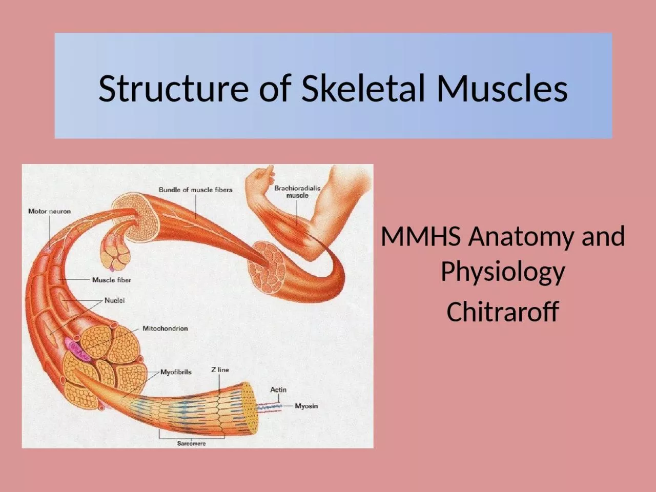
MMHS Anatomy and Physiology Chitraroff Fascia Fascia separates muscles from each other A type of Loose Connective Tissue Fascia may extend beyond the muscle to form tendons Fascia may form fibrous sheets called ID: 1047745

called muscle actin sarcomere muscle called sarcomere actin myofilaments form myosin skeletal line connective fascia fasciculus contraction surrounded tissue

Share:

Link:

Embed:

Download Presentation from below link

Download Presentation The PPT/PDF document "Structure of Skeletal Muscles" is the property of its rightful owner. Permission is granted to download and print the materials on this web site for personal, non-commercial use only, and to display it on your personal computer provided you do not modify the materials and that you retain all copyright notices contained in the materials. By downloading content from our website, you accept the terms of this agreement.


Presentation Transcript

1. Structure of Skeletal MusclesMMHS Anatomy and PhysiologyChitraroff

2. FasciaFascia separates muscles from each other.A type of Loose Connective TissueFascia may extend beyond the muscle to form tendons.Fascia may form fibrous sheets called Aponeuroses. (ex. Lumbodorsal Fascia)Fascia that closely surrounds one specific muscle is called epimysium. (epi = outside of)

3. Skeletal Muscle OrganizationEach muscle is made of smaller bundles called fascicles (sing. Fasciculus).Each fasciculus is surrounded by connective tissue called perimysium. (peri = surrounds)Fasciculi contain individual muscle cells called fibers surrounded by connective tissue called endomysium. (endo = inside)Each muscle fiber is further divided into myofibrils that may extend down the entire length of the muscle cell.

4. Skeletal Muscle Anatomy

5. Muscle MyofilamentsEach myofibril contains 2 types of protein myofilaments. (myo = muscle)Actin = thin myofilamentMyosin = thick myofilament Collectively, these make up a functional muscle unit called a Sarcomere.Myofilaments form the sarcomere = the basic unit of muscle that is responsible for muscle contraction.

6. The Sarcomere : The Functional Muscle UnitOne sarcomere extends from Z-Line to Z-Line.Each Z-Line provides a point of attachments for actin myofilaments.Myosin myofilaments are located between each actin myofilament.I-Band = Actin Only in this region.A-Band = where Actin and Myosin overlap.H-Zone = myosin only. See Sarcomere Diagram (Fig. 6.3 on p. 183)

7. The Sarcomere

8. The Sarcomere

9. The Muscle Contraction