/
Topical Interleukin-1 receptor antagonist therapy and corneal healing after photorefractive Topical Interleukin-1 receptor antagonist therapy and corneal healing after photorefractive

Topical Interleukin-1 receptor antagonist therapy and corneal healing after photorefractive - PowerPoint Presentation

genderadidas
genderadidas . @genderadidas
Follow
343 views
Uploaded On 2020-08-04

Topical Interleukin-1 receptor antagonist therapy and corneal healing after photorefractive - PPT Presentation

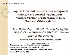
Brett Davies Capt USAF MC Vasudha Panday Maj USAF MC Charles Reilly Lt Col USAF MC Matthew Caldwell Maj USAF MC Wilford Hall Air Force Medical Center San Antonio TX The authors have no financial interest to disclose ID: 797095

corneal healing wound interleukin healing corneal interleukin wound haze keratectomy laser usaf results 1ra photorefractive antagonist exam ophthalmology refs

Share:

Link:

Embed:

Download Presentation from below link

Download The PPT/PDF document "Topical Interleukin-1 receptor antagonis..." is the property of its rightful owner. Permission is granted to download and print the materials on this web site for personal, non-commercial use only, and to display it on your personal computer provided you do not modify the materials and that you retain all copyright notices contained in the materials. By downloading content from our website, you accept the terms of this agreement.


Presentation Transcript

Slide1

Topical Interleukin-1 receptor antagonist therapy and corneal healing after photorefractive keratectomy in New Zealand White rabbits

Brett Davies, Capt, USAF, MC; Vasudha Panday, Maj, USAF, MC; Charles Reilly, Lt Col, USAF, MC ; Matthew Caldwell, Maj, USAF, MCWilford Hall Air Force Medical CenterSan Antonio, TX

The authors have no financial interest to discloseTopical IL-1ra provided by TheraKine, Inc.The conclusions do not necessarily reflect the views of the USAF or the DOD

ASCRS 2010, Boston, MA

Slide2

Purpose

This is a randomized, blinded study designed to compare topical Interleukin-1 antagonist (IL-1ra) against standard steroid treatment following photorefractive keratectomy (PRK). Vs

Slide3

Steroids

Glaucoma CataractsSecondary infectionDelayed wound healing

Slide4

Interleukin-1

Slide5

Methods

Slide6

Outcome Measures

Time to re-epithelizationWeekly slit lamp exam for haze

Pentacam evaluation prior to and at weeks 1,2,3, and 7

Histologic

exam at week 7

Slide7

Results

No difference in epithelial healing timeTime to re-

epithelization:

Slide8

Results- Slit lamp exam

p=0.392p<0.005

p<0.005

p=0.536

Subjective Haze

Slide9

Results-

Pentacam Haze

p<0.0001

Slide10

Results-Histology

Steroids

2.5mg

1.25mg

0.25mg

Central Foam Layer

p = 0.725

Thickest Foam Layer

p = 0.997

Slide11

Conclusions

Further studies needed to determine side effects of IL-1ra as well as response in human corneaIL-1ra does not increase epithelial healing timeNo difference in foam layer between treatment groupsSteroids significantly better than other groups on subjective haze analysis at weeks 4 and 5IL1-ra reduced objective haze (Pentacam

) similar to steroids at 1 month post opLower doses of IL1-ra reduced objective haze more than higher dose

Slide12

References

Dinarello CA, Wolff SM. The role of interleukin-1 in disease. N Engl J Med 1993; 328: 106Le J, Vilcek J. Tumor necrosis factor and interleukin 1: cytokines with multiple overlapping biological activities. Lab Invest 1987; 56: 234.Kijlstra A. The role of cytokines in ocular inflammation. Br J Ophthalmol 1994; 78: 885Girard MT, Matsubara

M, Fini ME. Transforming growth factor-beta and interleukin-1 modulate metalloproteinase expression by corneal stromal cells. Invest Ophthalmol Vis Sci

1991; 32: 2441.

Yamada, J, Dana MR,

Sotozono

C, Kinoshita S. Local suppression of IL-1 by receptor antagonist in the rat model of corneal alkali injury. Experimental Eye Research2003; 76(2): 161-7.

Biswas

PS,

Banerjee

K,

Zheng

M, Rouse BT. Counteracting corneal

immunoinflammatory

lesion with interleukin-1 receptor antagonist protein. J

Leukoc

Biol. 2004; 76(4): 868-75

Netto

MV. Wilson SE. Corneal wound healing relevance to

wavefront

guided laser treatments. [Review] [58 refs] Ophthalmology Clinics of North America 2004; 17(2): 225-31

Kaji

Y. Yamashita H.

Oshika

T. Corneal wound healing after

excimer

laser keratectomy. [Review] [28 refs] Seminars in Ophthalmology 2003; 18(1): 11-6

Alio

JL. Perez-

Santonja JJ. Tervo T. Tabbara KF. Vesaluoma M. Smith RJ. Maddox B. Maloney RK. Postoperative inflammation, microbial complications, and wound healing following laser in situ keratomileusis

. [Review] [82 refs] Journal of Refractive Surgery 2000; 16(5): 523-38Wachtlin J. Langenbeck K. Schrunder S. Zhang EP. Hoffmann F.

Immunohistology of corneal wound healing after photorefractive keratectomy and laser in situ keratomileusis. Journal of Refractive Surgery 1999; 15(4): 451-8Kato T. Nakayasu

K. Hosoda Y. Watanabe Y. Kanai A. Corneal wound healing following laser in situ keratomileusis (LASIK): a histopathological study in rabbits. British Journal of Ophthalmology 1999; 83(11): 1302-5

Gan

, L.,

Hamberg-Nystrom

, H,

Fagerholm

, P, Van

Setten

, G.

Cellular proliferation and leukocyte infiltration in the rabbit cornea after photorefractive keratectomy.

Acta

Ophthalmologica

Scandinavica

. 79(5):488-492, October 2001