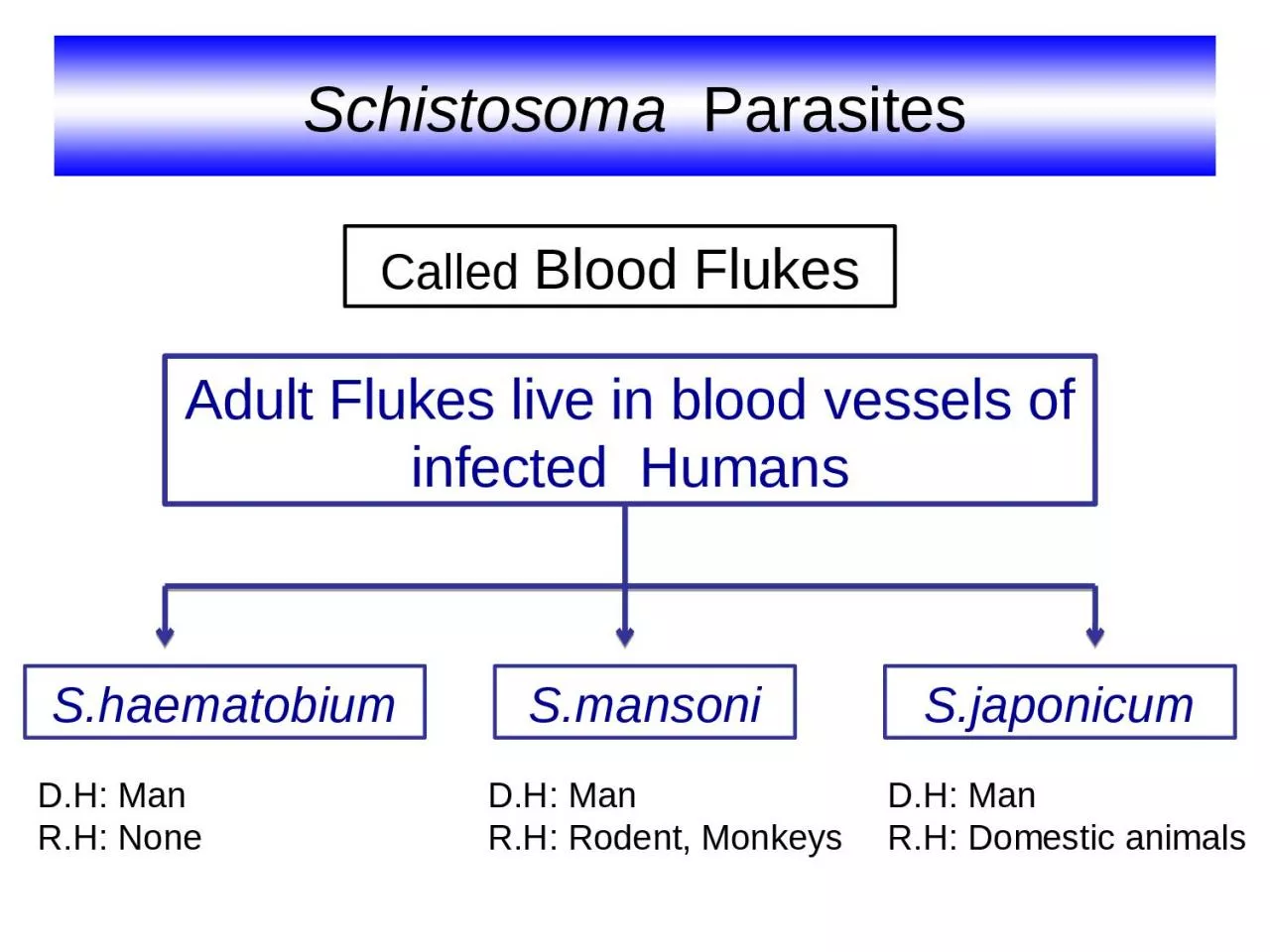
/
Schistosoma   Parasites Adult Flukes live in blood vessels of infected  Humans Schistosoma   Parasites Adult Flukes live in blood vessels of infected  Humans

Schistosoma Parasites Adult Flukes live in blood vessels of infected Humans - PowerPoint Presentation

genevieve
genevieve . @genevieve
Follow
66 views
Uploaded On 2023-08-30

Schistosoma Parasites Adult Flukes live in blood vessels of infected Humans - PPT Presentation

Shaematobium Smansoni Sjaponicum Called Blood Flukes DH Man RH Rodent Monkeys DH Man RH Domestic animals DH Man RH None Definitive Host Man Habitat Superior amp Inferior mesenteric plexus of veins ID: 1014626

blood stage eggs mansoni stage blood mansoni eggs schistosoma tissue egg amp lesions infection mesenteric embolic due mansonis laboratory

Share:

Link:

Embed:

Download Presentation from below link

Download Presentation The PPT/PDF document "Schistosoma Parasites Adult Flukes liv..." is the property of its rightful owner. Permission is granted to download and print the materials on this web site for personal, non-commercial use only, and to display it on your personal computer provided you do not modify the materials and that you retain all copyright notices contained in the materials. By downloading content from our website, you accept the terms of this agreement.


Presentation Transcript

1. Schistosoma ParasitesAdult Flukes live in blood vessels of infected HumansS.haematobiumS.mansoniS.japonicumCalled Blood FlukesD.H: ManR.H: Rodent, MonkeysD.H: ManR.H: Domestic animalsD.H: ManR.H: None

2. Definitive Host Man Habitat Superior & Inferior mesenteric plexus of veinsWhere adult S.mansoni liveBlood vesselsInferior mesenteric plexus of veinsWhere adult S.japonicum live

3. Has separate sexes.Lives in blood.Has non-operculated eggs.Has no Redia stage.Has no encysted metacercaria. Common species affecting man:S. mansoni, S. haematobium, S. japonicum, S. intercalatum and S. mekongi.Schistosoma differs from other trematodes in:

4. Sexes: separateFemale: cylindricalMale: flattened, folded (gynaecophoric canal).Suckers: oral & ventral (smaller in females).Digestive system: No muscular Pharynx Intestinal ceaca reunite in a single caecum.Adult Morphology

5. SpeciesS. mansoniS. japonicumSize8x1 mm15xo.5 mmTestes6-9 testes in cluster6-8 testes in a lineCuticlecourse tuberclesNo tuberclesMale

6. FemaleSpeciesS. mansoniS. japonicumSize14x0.15 mm26x0.3 mmOvaryovary ant. 1/3Ovary in the middleVitelline glandsvit. gl post 2/3Vit. gl. in post 1/2

7. EggS. mansoniS. japonicumIn stoolSize150x60 85x65 ShapeOvalLateral spineOvoidMinute knobShelltranslucent, non operculatedContentMature embryo (miracidium)

8. MiracidiumS. mansoniBiomphalaria alexandrinaSnail I. HS. japonicumOncomelania

9. Infective stage (Cercaria):Formed of body and forked tail (furcocercous)5-6 pairs of penetration glands.Skin penetration is aided by:1-Proteolytic enzymes.2-Surface tension of drying water.3-Movement of the tail

10. Worms in their habitatFemale lays eggsEggs in stoolsMiracidia hatchMiracidium infects snail i.h.Cercarial sheddingInfection in contaminated waterSchistosomulum reaches circulationWorms mate in the liver

11. Development of Schistosoma inside the body of infected humanCercariae penetrates human skinSchistosomulaVenous circulationSystemic circulationLiver Portal veinVesical plexusIIIIIVIIInf. mesenteric plexusAorta ♂♀In urine or stoolDirection of venous blood flowS.haematobiumS.mansoni

12. Pathogenesis and Clinical PictureThere are 4 progressive stages:I- Stage of invasionLocal dermatitis, irritation.Papular rash.(1-4 days)

13. II- Stage of migrationBy schistosomula(3-4 weeks)Metabolic products: toxic and allergic manifestations asurticaria, fever, headache, muscle pain. In the lung: verminous pneumonitis, minute haemorrhagecough, haemoptysis.In the liver: enlarged and tender.

14. III- Stage of egg deposition and extrusionThe patient may complain of:Generalized malaise, fever, rigors, urticaria, abdominal pain and liver tenderness.A- Eggs are deposited in the venous plexusUrticaria (acute stage: 1-2 month)

15. Katayama SyndromeBlood vesselSoluble egg antigens are released in blood streamOccurs mainly in S.japonicum infection1- ♀lays large number of eggs2- greater proximity to the liverimmune complexDeposited in the tissuesTissue damageThe patient suffers from:Fever, chills, diarrhoea, generalized lymphadenopathyDue to:Thus also called acute toxoemic schistosomiasisantibodiesCirculate

16. B- Stage of egg extrusion:Dysentery with blood and mucus in stoolIn mesenteric plexus of veinsIntestinal wallDamage & haemorrhageS.mansoni & S.japonicumEggs escape from the veins to the perivascular tissue due to:1-Pressure within the venule2-Effect of the spine3-oozing lytic secretion of the miracidium within the eggEgg extruded

17. IV- Stage of tissue reaction (chronic stage)Occurs around Schistosoma eggs deposited in various tissues.Attract inflammatory cellsDeposition of fibrous tissue Damage of affected organ and its fibrosisLoss of its functionEgg shellmiracidiumInflammatory cellsShell & miracidial antigensDelayed-type hypersensitivityGranuloma formation in tissue(months – years)Granuloma develops

18. IV- Stage of tissue reaction (chronic stage)In S.mansoni infection Eggs trapped in Intestinal wallEggs extruded in stool12Eggs fail to fix to venule wall are swept by blood3Cause Embolic lesions Polyps in the colon in S.mansoni infectionNormal mucosa of colon

19. Embolic Lesions in the LiverPortal hypertensionHepatosplenomegalyOesophageal varicesAscites.

20. Embolic Lesions in the lungPulmonary hypertensionBilharzial cor-pulmonale Embolic lesions may occur in any other organ. Cor-pulmonale = right-sided heart failure Dilated right ventricleThickened wallIn Normal conditionIn Schistosoma infectionEnlarged right ventricleTrapped eggsMeans: enlargement of right ventricle due to high blood pressure in the arteries of the lung

21. Diagnosis of Schistosomiasis Clinically (suggestive)Laboratory 1- Detection of eggs in stool3- Serological tests2- Blood examinationRadiological imagingEndoscopy 1- History of contact with infected water.2- Clinical picture according to stage of infection.

22. I- Laboratory Diagnosis1- Direct parasitological methods:- Detection of S.mansoni eggs in stool by direct smear method or by concentration method. - Kato thick faecal smear: for egg counting to assess the intensity of infection- Rectal swab

23. I- Laboratory DiagnosisEosinophilia, leucocytosis. 2- Blood examination:3- Serological Tests:Detection of anti-Schistosoma antibodies or antigen in patient’s seruma- IHAT (Indirect Haemagglutination test)Latex particlesSensitized sheep RBCs+ve-ve(Immunodiagnostic Tests)Anaemia due to 1- Egg extrusionIron deficiency anaemia2- Hypersplenism Haemolytic anaemia

24. b- ELISA (Enzyme-linked immunosorbent assay)c- IFAT (Indirect Fluorescent Antibody test)washwashwashwashwashAb detectedwashwashwashAg detectedCoating with Schisto AgCoating with anti-Schisto MAbSchisto AgPatient serumAb detectedPatient’s serumAb linked to enzymesubstratesubstratePatient’s serumAb linked to enzyme

25. II- Radiological imagingIntestinal affectionS.mansoni infectionالمناظير III- Endoscopy Colonoscopy, sigmoidoscopy in S.mansoniDone in chronic cases to detect lesions and take biopsies

26.

27. Treatment Praziquantel (Biltricide): For all schistosome species.Dose:40 mg/kg single oral doseOxamniquine (vancil):For S. mansoniDose: 30 mg/kg for 2 days

28. Prevention and Control- Mass treatment.- Health Education.- Snail control: Physical methods Biological methods Chemical methodsXBalanites aegyptica Copper sulphate

29. Cercarial Dermatitis (Bather’s itch) A condition that occurs due to penetration of cercariae of non-human species of schistosomes the skin of man.Cercariae cannot go beyond the germinal layer.Clinical picture:Dermatitis, itching, oedema and secondary bacterial infection.Treatment: Antipruritics, antihistamincs, antibiotics.