/
STRUCTURE AND CHEMICAL COMPOSITION OF BACTERIA STRUCTURE AND CHEMICAL COMPOSITION OF BACTERIA

STRUCTURE AND CHEMICAL COMPOSITION OF BACTERIA - PowerPoint Presentation

grace3
grace3 . @grace3
Follow
64 views
Uploaded On 2024-01-03

STRUCTURE AND CHEMICAL COMPOSITION OF BACTERIA - PPT Presentation

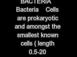
Size of a Bacterial Cell There are great variations in size of bacteria They measure from 075 µ to 15 µ but on an average each cell of bacterium measures about 125 µ to 2 µ in diameter ID: 1039033

cell bacteria gram membrane bacteria cell membrane gram granules wall acid acids negative nucleoid proteins positive present called lipid

Share:

Link:

Embed:

Download Presentation from below link

Download Presentation The PPT/PDF document "STRUCTURE AND CHEMICAL COMPOSITION OF BA..." is the property of its rightful owner. Permission is granted to download and print the materials on this web site for personal, non-commercial use only, and to display it on your personal computer provided you do not modify the materials and that you retain all copyright notices contained in the materials. By downloading content from our website, you accept the terms of this agreement.


Presentation Transcript

1. STRUCTURE AND CHEMICAL COMPOSITION OF BACTERIA

2. Size of a Bacterial Cell: There are great variations in size of bacteria. They measure from 0.75 µ to 1.5 µ but on an average each cell of bacterium measures about 1.25 µ to 2 µ ( in diameter). The smallest rod shaped eubacteria is Dialister sps and measures between 0.15 µm to 3.0 µm size. Sulphur bacteria Thiophysa volutans is the largest amongst all bacteria.

3. Shape and Arrangement of Bacterial Cell

4.

5. 1. Cell Envelope:It is the outer covering of protoplasm of bacterial cell. Cell envelope consists of 3 components— glycocalyx, cell wall and cell membrane. Glycocalyx (Mucilage Sheath):It is the outermost mucilage layer of the cell envelope which consists of non-cellulosic polysaccharides with or without proteins. Glycocalyx may occur in the form of loose sheath when it is called slime layer. If thick and tough, the mucilage covering is called capsule. Glycocalyx gives sticky character to the cell. It is not absolutely essential for survival of bacteria.It has several secondary functions: (a) Prevention of desiccation, (b) Protection from phagocytes(c) Protection from toxic chemi­cals and drugs, (d) Protection from viruses, (e) Attachment, (f) Immunogenicity and (g) Virulence.

6. Structure of Cell Membrane:Cell membrane is a thin structure that completely surrounds the cell only about 8 nm thick. This structure is a critical barrier separating the inside of cell from environment. The cell membrane is also highly selective barrier enabling the cell to concentrate a specific metabolite and excrete waste material. Mostly biological membrane is composed primarily of Phospholipids (about 20 to 30 percent) and proteins (about 60 to 70 percent). The phospholipids form a bilayer in which most of the proteins are strongly held (integral proteins) and these proteins can be removed only by destruction of the membranes, as with treatment by detergents. Other proteins are only loosely attached (Peripheral proteins) can be removed by mild treatment such as osmotic shock. The lipid matrix of membrane has fluidity, allowing the components to move around laterally. Fluidity is essential for various membrane functions and is dependent on factors such as temperature and on proportion of unsaturated fatty acids to saturated fatty acids present in phospholipids.

7. In eubacteria the phospholipids are phosphoglycerides in which straight chain fatty acids acids are ester linked to glycerol. In archaebacteria, the lipids are polyisoprenoid branched chain lipids, in which long chain branched alcohol (Phytanols) are ether linked to glycerol

8. (ii) Cell Wall: It is rigid solid covering which provides shape and structural support to the cell. Cell wall lies between plasma membrane and glycocalyx. Periplasmic space occurs between plasma membrane and cell wall. Cell wall protects the bacterial cells against bursting in hypotonic solution. In Gram positive bacteria wall is 20-80 nm thick, single layered and smooth. In Gram negative bacteria, wall is 8-12 nm thick, complex, wavy and two layered. The outer layer is also called outer membrane. It consists of lipopolysaccharides, lipids and proteins. The outer membrane has hydrophilic channels of 16-stranded (3-barrel proteins called porins. The single layered cell wall of Gram positive bacteria and inner wall layer of Gram negative is made up of pepidoglycan, proteins, non-cellulosic carbohydrates, lipids, amino acids, etc.

9. Cell Envelope of Prokaryote:Bacteria can be divided into two major groups called Gram positive bacteria and Gram negative bacteria, based on Gram staining. Gram positive bacteria and Gram negative bacteria differ in the appearance of cell wall. The cell wall of Gram negative bacteria is multilayered structure and quite complex.Gram positive bacteria contain primarily single type of molecule and is often much thicker.

10. Bacterial Cell Wall and Peptidoglycan Component: In the cell wall of bacteria, there is one rigid layer that is primarily responsible for strength of the wall. In Gram negative bacteria additional layer is present outside this rigid layer. The rigid layer of both Gram negative bacteria and Gram positive bacteria is very similar in chemical composition and is called Peptidoglycan (or murein). This layer is thin sheet composed of two sugar derivatives, N-acetyl glucosamine and N-acetyl muramic acid (NAM &NAG) and small number of amino acids consisting of L-alanine, D-alanine, D-Glutamic acid and either lysine or meso-diaminopimelic acid These constituents are connected to form a repeating structure, glycan tetrapeptide. Basic structure of peptidoglycan is a thin sheet in which the glycan chains formed by sugars are connected by peptide cross links formed by the amino acid. Glycosidic bonds connecting the sugars in the glycan chains are very strong but these chains alone cannot provide rigidity in all directions. In Gram-negative bacteria cross linkage usually occurs by direct peptide linkage of the amino group of diaminopimelic acid to the carboxyl group of the terminal D-alanine. In Gram positive bacteria cross linkage is usually by peptide inter bridge. The kinds and number of cross linking amino acids varies from org to org.

11. In Gram-negative bacteria cross linkage usually occurs by direct peptide linkage of the amino group of diaminopimelic acid to the carboxyl group of the terminal D-alanine.

12. Peptidoglycan chains connected through short tetrapeptide and pentagiycine bridge. The polysaccharide chain consisting of alternate residues of the amino acid N-acetyl glucosamine (NAG) and N-acetyl muramic acid (NAM) linked in β-1, 4 glycosidic linkage. Each NAM residue carries a short peptide chain of four amino acid residues (tetra peptide) which are L-alanine, D-Glutamic acid, L-Lysine and D-alanine.

13. Diversity of Peptidoglycon:Peptidoglycan is present only in bacteria.N Acetyl muramic acid and mesodiaminopimelic acid are never found in the cell walls of Archae and eukaryote. DAPA is present in all Gram negative bacteria and in some Gram positive bacteria but most Gram +ve cocci have lysine instead of DAP and a few other Gram positive bacteria have other amino acids. In peptidoglycan, The glycan portion is uniform with only the sugar NAG and NAM being present, and these sugars are always connected in β 1→ 4, linkage. Tetra peptide of the repeating unit shows major variation in only one amino acid, the Lysine, diaminopimelic acid alteration. More than 100 different peptidoglycan types are known and the greater variation among them occurs in inter-bridge. Peptidoglycan constitutes 70-80% of wall in Gram positive bacteria. Lipid content is little. 10-20% of wall in Gram negative bacteria is formed of peptidoglycan. Lipid content is 20-30%.

14. Teichoic Acids:Gram positive bacteria have acidic polysaccharide called teichoic acids attached to their cell wall. The term teichoic acid includes all wall, membrane or capsular polymers containing glycerophosphate or ribitol phosphate residues. Teichoic acids form receptor sites and surface antigens.These poly-alcohols are connected by phosphate ester bond to 6-OH group of NAM. Because they are negatively charged, Teicohic acids are partially responsible for the negative charge of cell surface and may function to effect passage of ions through the cell wall. Certain Glycerol containing acids are bound to membrane lipids of Gram positive bacteria, because these teicohic acids are intimately associated with lipid, they have been called lipo-teichoic acid.

15. In Gram negative bacteria outer membrane is covered by lipopolysaccharide. The lipopolysaccharide consists of complex polysaccharide covalently linked to lipid A. Polysaccharide consists two portions, the core polysaccharide and O-polysaccharide. Lipid portion of lipopolysaccharide, referred to as lipid A is not a glycerol lipid, but instead the fatty acids are connected by ester amine linkage to a disaccharide composed of N-acetyl glucosamine phosphate. The disaccharide is attached to core-o-polysaccharide through KDO. Fatty acid commonly found in lipid A : lauric, muristic, Palmitic and β-hydroxy meristic acid.

16. Cytoplasm:It is crystallo-colloidal complex that forms the protoplasm excluding its nucleoid. Cytoplasm is granular due to presence of a large number of ribosomes. Membrane bound cell organelles are absent. However, all biochemical pathways are found in prokaryotic cells. Cytoplasmic streaming is absent. Sap vacuoles are absent. Instead gas vacuoles are present

17. Various structures present in cytoplasm are as follows: (i) Mesosome: It is a characteristic circular to villi form specialisation of cell membrane of bacteria that develops as an ingrowth from the plasma membrane. It consists of vesicles, tubules and lamellae. Mesosme is of two types, septal and lateral. Septal mesosome It connects nucleoid with plasma membrane. It takes part in replication of nucleoid by providing points of attachment to the replicated ones. Septal mesosome is also believed to help in septum formation. At the time of cell division, plasma membrane grows in the region where the septal mesosme is present so that most probably it provides membranes for rapid elongation. Lateral mesosme It is not connected with nucleoid. It contains respi­ratory enzymes and is, therefore, often called chondrioid. It is believed to be equal to mitochondrion of eukaryotes. However, respiratory enzymes are also present over the plasma membrane

18.

19. (ii) Ribosomes: They are small, membrane less, submicroscopic ribo nucleoprotein en­tities having a size of 20 nm x 14-15 nm. Ribosomes are of two types, fixed and free. Fixed ribosomes are attached to the plasma membrane. Free ribosomes occur free in the cyto­plasmic matrix. The ribosomes are 70S in nature. (Here S denotes sedimentation coefficient or Svedberg number). Each ribosome has two subunits, larger 50S and smaller 30S. Ribo­somes take part in protein synthesis. Free or matrix ribosomes synthesize proteins for intracellular use while fixed ribosomes synthesize proteins for transport to outside. Ribo­somes generally occur in helical groups called polyribosomes or polysomes. In each polysome 4—8 ribosomes are attached to a single strand of messenger or mRNA. It is a mechanism to synthesize several copies of the same protein.

20. (iii) Chromatophores: They are internal membrane systems of photosynthetic forms which possess photosynthetic pigments. In purple bacteria the membranes are typical while in green bacteria they are non-unit, non-lipid and proteinaceous. Chromatophores of green algae contain photosynthetic pigments are bacteriochlorophyll, bacteriophaeophytin (bacterioviridin) and carotenoids.

21. Nucleoid:It represents the genetic material of prokaryotes. Several alternative names have been given to nucleoid— gonophore, prochromosome, incipient nucleus and chromoneme. Nucleoid consists of a single circular strand of DNA duplex which is super­coiled with the help of RNA and polyamines to form a nearly oval or spherical complex. The folding is 250-700 times. Polyamines or nucleoid proteins are different from histone proteins. DNA of prokaryotes is considered naked because of its non-association with histone pro­teins and absence of nuclear envelope around it. In E. coli, nucleoid has 1100 µm long DNA duplex with 4.6 x 10⁶ base pairs. Nucleoid is embedded freely in the cytoplasm. A cell can have 2 or more nucleoids but all are replicated copies of same nucleoid. It is equivalent to a single chromosome of eukaryotes because nucleoid consists of a single DNA double strand. Nucleoid may be directly attached to the plasma membrane or through the mesosome.

22. Plasmids:They are self-replicating, extra chromosomal segments of double stranded, circular, naked DNA. Plasmids provide unique phenotypic characters to bacteria. They are independent of main nucleoid. Some of them contain important genes like fertility factor, nif genes, resistance factors and colicinogenic factors. Plasmids can get associated temporarily with nucleoid are known as episomes. Plasmids are used as vectors in genetic engineering.

23. Inclusion Bodies:They are non-living structures present in the cytoplasm. The inclusion bodies may occur freely inside the cytoplasm (e.g., cyanophycean granules, volutin or phosphate granules, glycogen granules) or covered by 2-4 nm thick non-lipids, non-unit protein membrane (e.g., gas vacuoles, carboxysomes, sulphur granules, PHB granules). On the basis of their nature, the inclusion bodies are of 3 types— gas vacuoles, inorganic inclusions and food reserve.Gas Vacuoles: They are gas storing vacuoles found in cyanobacteria, purple and green bacteria and a few other planktonic forms. A gas vacuole is without any covering of its own. It consists of a variable number of hexagonal, hollow and cylindrical gas vesicles. Each gas vesicle is surrounded by a single non-unit, non-lipid protein membrane having ribs or folds. The membrane is impermeable to water but is permeable to atmospheric gases. Gas vacuoles protect the bacteria from harmful radiations. They also constitute buoyancy regu­lation mechanism for their proper positioning in water during daytime for photosynthesis.

24. (ii) Inorganic Inclusions: Several types of inorganic granules occur in bacteria. They include volutin granules, sulphur granules, iron granules, magnetite granules, etc. Because of the ability to pick up different colours with basic dyes, they are called metachromatic granules. Two common types of inorganic granules are volutin granules and sulphur gran­ules. Volutin granules are polymetaphosphates which function as storage reserve of phosphate. Sulphur granules occur in bacteria living in sulphur rich medium like the one which pick up hydrogen sulphide for obtaining reducing power in photosynthesis. Iron granules are simi­larly found in those bacteria which metabolise iron compounds for obtaining energy. Aquaspirillum magnetotacticum contains magnetosomes, which are vesicles having mag­netite. The granules help the bacteria to orientate themselves along geomagnetic lines

25. (iii)Food Reserve: Blue green algae have cyanophycean starch or α-granules, β- granules or lipid globules and cyanophycin or protein granules. In bacteria, starch is replaced by glycogen. Neutral fats are absent. Instead poly-beta-hydroxy-butyrate or PBH granules are present. A biodegradable plastic can be prepared from PBH. Protein granules are present. Carboxysomes occur in photosynthetic forms.

26. Flagella Bacterial flagella are unistranded, equivalent to a single micro- tubular fibre. It is about 20 nm (0.02 µm) in diameter and 1-7µm in length. Bacterial flagellum is made up of 3 parts— basal body, hook and filament. Basal body It is like a rod and is inserted in the cell envelope. The basal body bears ring-like swellings in the region of plasma mem­brane and cell wall. There are two pairs of rings (L and P ring in cell wall and S and M rings embedded in cell membrane) in Gram negative bacteria and only a single pair of rings (S and M rings embedded in cell membrane) in Gram positive bacteria.Hook It is curved tubular structure which connects the filament with the basal body. It is the thickest part of flagellum. Filament It is long tubular structure which causes turbulence in the liquid medium. It is made up of protein called flagellin. Protein molecules are globular. They are arranged in 3-8 spiral rows. It is believed that bacterial flagella perform rotation type movement that brings about backward pushing of the water. It results in the bacterium moving forward.

27.

28. Pili and Fimbriae:The two terms have been used interchangeably for bacterial appendages which are not involved in locomotion. Pili (singular-pilus) are longer, fewer and thicker tubular outgrowths which develop in response to F+ or fertility factor in Gram negative bacteria. They are made up of protein pilin. A donor bacterial cell having fertility factor develops 1-4 pili. Being long (18-20 (xm) they are helpful in attaching to recipient cell and forming conjugation tube. Fimbriae are small bristle-like fibres sprouting from cell surface in large number. There are 300-400 of them per cell. Diameter is 3-10 nm while length is 0.5-1.5 µm. Fimbriae are involved in attaching bacteria to solid surfaces (e.g., rock in water body) or host tissues (e.g., urinary tract in Neisseria gonorrhoeae). Some fimbriae cause agglutination of RBC. They also help in mutual clinging of bacteria