/
Root Cause Analysis(RCA) Root Cause Analysis(RCA)

Root Cause Analysis(RCA) - PowerPoint Presentation

hadley
hadley . @hadley
Follow
66 views
Uploaded On 2024-01-03

Root Cause Analysis(RCA) - PPT Presentation

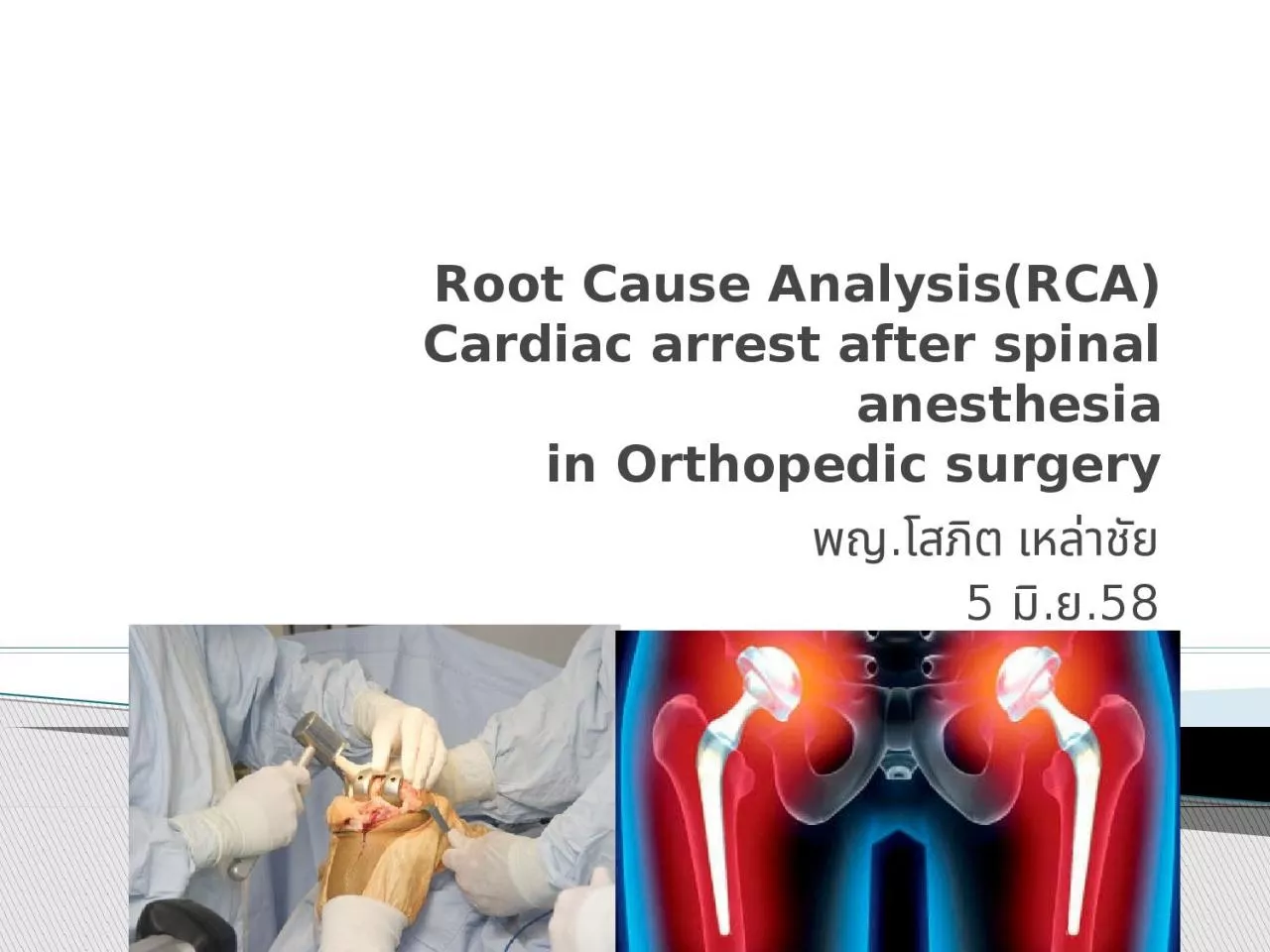
Cardiac arrest after spinal anesthesia in Orthopedic surgery พญโสภต เหลาชย 5 มย58 Hypotension Severe bradycardia Cardiac arrest Problems Cardiac Arrest During Spinal Anesthesia Common Mechanisms and Strategies for Prevention ID: 1038478

mechanism spinal arrest cardiac spinal mechanism cardiac arrest pulmonary dvt anesthesia high block embolism severe bradycardia risk heparin 000

Share:

Link:

Embed:

Download Presentation from below link

Download Presentation The PPT/PDF document "Root Cause Analysis(RCA)" is the property of its rightful owner. Permission is granted to download and print the materials on this web site for personal, non-commercial use only, and to display it on your personal computer provided you do not modify the materials and that you retain all copyright notices contained in the materials. By downloading content from our website, you accept the terms of this agreement.


Presentation Transcript

1. Root Cause Analysis(RCA)Cardiac arrest after spinal anesthesiain Orthopedic surgeryพญ.โสภิต เหล่าชัย5 มิ.ย.58

2. HypotensionSevere bradycardiaCardiac arrestProblems

3. Cardiac Arrest During Spinal Anesthesia: Common Mechanisms and Strategies for PreventionPollard, John B. MDAnesthesia & Analgesia:January 2001 - Volume 92 - Issue 1 - p 252–256Sudden cardiorespiratory arrest following spinalanesthesiaPERIODICUM BIOLOGORUMVOL. 115, No 2, 283–288, 2013

4. Cardiac arrests during spinal anesthesia พบได้ไม่บ่อย overall incidence 0.07% (7:10,000) spinal anestheticsA review of approximately 4000 regional anesthetics พบว่า 6 cases of severe bradycardia (pulse of 20 to 40 bpm) 6 others (0.15%) with cardiac arrest after spinal anesthesia Introduction

5. 0.03% reported for patients undergoing noncardiac surgery The incidence of cardiac arrest spinal anesthesia พบบ่อยกว่า epidural anesthesiaIntroduction

6.

7. CIRCULATORY MECHANISMCEREBRAL MECHANISMRESPIRATORY MECHANISMMECHANISM

8. หลังจากฉีดยาชา Spinal block motor somatic sensory fiber(test by pinprick) sympathetic block(test by temperature) 2 - 6 segment higher > somatic levelsThe cardiac sympathetic outflow emerges from C5 to T5 levelsmain supply to the ventricles from T1 to T4 ถ้า block > T4 จะทำให้เกิด severe bradycardia ได้CIRCULATORY MECHANISM

9. คือ การ ลดลงอย่างมากของ venous return เนื่องจาก การ dilatation ของ artery and venous vessels สามารถลด right atrial pressure 36% in low spinal block53% in high spinal blockทำให้มีการลดลงของ preload ทำให้เกิด reflexes ที่เป็นสาเหตุทำให้เกิด severe bradycardiaCIRCULATORY MECHANISM

10. ลดลงของ preload กระตุ้น Reflexes : cause severe bradycardia1. The pacemaker stretch reflex : Right atrium2. Low pressure baroreceptors : Right atrium and vena cava3. The Bezold-Jarisch reflex : mechanoreceptors in the left ventricleAcute coronary syndromePre existing coronary artery diseaseCIRCULATORY MECHANISM

11. 1. Proper selection of patient and operation2. Fluid support : Preload before SB 500-1000 ml crystalliods(10 ml/kg)3. Pharmacological supportIV atropine (0.4–0.6 mg) thenephedrine (25–50 mg) thenepinephrine (0.2–0.3 mg) in stepwise escalation of therapy when bradycardia develops following spinal anesthesiaPossible prevention of cardiac arrest

12. Treatment for Hypotension after Spinal Anesthesia (www.nysora.com).

13. In orthopedic surgeryBone cement : Made of polymethylmethacrylateThe process of cement hardening causes intramedullary HTN that may cause fat embolization, cement embolizationMay cause systemic vasodilation, platelet aggregation, and microthrombus that lead to hemodynamic instability, hypoxia, dysrhythmias, and pulmonary HTNPrevention : Increase FiO 2 and maintain euvolemia or slight hypervolemia during cementingCIRCULATORY MECHANISM

14. Tourniquet problems Physiologic changes include Application>60 mintourniquet pain and HTNApplication>2 hr muscle dysfunction and potential postoperative neurapraxia Metabolic acidosis after releasing the tourniquet, with transient increase in ETCO2, vasodilatation , hypotensionCIRCULATORY MECHANISM

15. spinal anesthesiaไม่ใช่สาเหตุโดยตรง แต่อาจทำให้เกิด การรบกวน cerebral oxygen balanceConsciousness: probably secondary to decreased afferent stimulation of the reticular activating systemลดการ sedation ลงCEREBRAL MECHANISM

16. oversedationอาจเป็นสาเหตุทางอ้อมที่ทำให้เกิด cardiac arrest after spinal anesthesiaเพราะว่า ผู้ป่วยส่วนใหญ่ที่ได้รับ sedative drug จะทำให้เกิด inadequate ventilation ขึ้น เพิ่ม risk ในการเกิด hypoxiaการ oversedation อาจเพิ่ม risk of bradycardia และผู้ป่วยไม่สามารถสื่อสารกับเราได้ทำให้ไม่ทราบอาการของ early warning of a vasovagal attack : sweating, nausea, and syncopeLateral decubitus position :THAPotential ventilation-perfusion mismatchresultant hypoxemiaRESPIRATORY MECHANISM

17. In orthopedic surgeryPulmonary embolismFat embolism : Rare but very serious complication (mortality up to 20%). Within 72 hr after long bone or pelvic fractureClassic presentation. Dyspnea, petechiae, and Increased fatty acids in plasma release of vasoactive substances. ARDS-like syndrome. Capillary damage to cerebral circulation. During GA. Decline in E TCO 2 and PaO2. Treatment. Supportive.Blood clot : DVT : immobilizationAir : in spine surgery (C-spine, Lumbar prone position , shoulder in sitting position (site of surgery above heart level)RESPIRATORY MECHANISM

18.

19.

20. stasis, hypercoagulability ทั้งชนิด congenital และacquired และ vessel wall injury 1. การผ่าตัดในระยะ 12 สัปดาห์ที่ผ่านมา 2. มีโรคมะเร็ง 3. เคยเป็น deep vein thrombosis (DVT) หรือ PE มาก่อน 4. immobilization นานเกิน 3 วัน ใน 4 สัปดาห์ที่ผ่านมา 5. ระยะหลังคลอด 3 สัปดาห์หรือการใช้ estrogen 6. ประวัติครอบครัวเป็น DVT หรือ PE 7. กระดูกหักบริเวณขาใน 12 สัปดาห์ที่ผ่านมา ภาวะ hypercoagulability ชนิด acquired อื่นอาจพบได้ใน antiphospholipid syndrome, nephrotic syndrome, และ thrombocytosis และ paroxysmal nocturnal hemoglobinuria ภาวะ hypercoagulability ทั้งชนิด congenital และacquired มักจะทำให้เกิด recurrent DVT/PE ปัจจัยเสี่ยงคือการเกิด pulmonary embolism (PE)

21. ใช้ Modified Wells Criteria ในผู้ป่วยที่มีอาการที่สงสัย PE ได้แก่ sudden dyspnea หรือ pleuritic chest pain ร่วมกับอาการและอาการแสดงของ DVT (ขาบวม, ปวด) 3 คะแนน ไม่มีสาเหตุอื่นๆ อธิบายได้ 3 คะแนน ชีพจรเร็วกว่า 100 ครั้ง/นาที 1.5 คะแนน Immobilization (> 3 วัน) หรือประวัติผ่าตัดในช่วง 4 สัปดาห์ก่อน 1.5 คะแนน ประวัติ DVT/PE 1.5 คะแนน ไอเป็นเลือด 1 คะแนน มะเร็ง 1 คะแนน > 6 จัดเป็นกลุ่มที่มีความน่าจะเป็นทางคลินิกสูง (high clinical probability) 2 -6 คะแนน จัดเป็นกลุ่มที่มีความน่าจะเป็นทางคลินิกปานกลาง <2 คะแนน จัดเป็นกลุ่มที่มีความน่าจะเป็นทางคลินิกต่ำ (low clinical probability) แนวทางการวินิจฉัยผู้ป่วยที่สงสัย pulmonary embolism

22. Arterial blood gas มี PaO2 ต่ำ และ PaCO2 ต่ำ ร่วมกับการมี respiratory alkalosis เล็กน้อยChest X-ray มักจะไม่พบความผิดปกติมากนัก อาจมีเงาเส้นเลือดลดลงบริเวณฐานของปอดและเพิ่มขึ้นบริเวณยอดปอดและขณะเดียวกันมี pulmonary vein โป่งพองเล็กน้อยร่วมกันกับมี wedge – shaped infarctInvestigation

23. EKG มักไม่ช่วยเท่าใดนัก ความผิดปกติที่อาจพบได้แก่ sinus tachycardia S wave in Lead I , Q – wave and T-wave inversion ใน lead III แต่สิ่งนี้สามารถพบได้ 20% ของผู้ป่วย(right ventricular strain, new incomplete right bundle branch block) S1Q3T3

24.

25.

26. Pulmonary angiogram : gold standard but invasive Echocardiography CT angiographyVentilation – perfusion scanCompression ultrasound ของเส้นเลือดดำที่ขา การตรวจพิเศษ

27. Anticoagulation แนะนำว่าขนาดของ unfractionated heparin ในการรักษา unstable PE นั้น ควรใช้ในขนาดสูง โดย bolus ทาง intravenous 15,000-20,000 unit และให้ต่อด้วย continuous infusion โดยปรับให้ระดับ aPTT ถึงเกณฑ์ภายใน 24 ชั่วโมงแรก เนื่องจากมีหลักฐานพบว่าหลังจากเกิด massive emboli จะมีการกระตุ้น platelet aggregation และการสร้าง thrombin ซึ่งมีผลทำให้ต้องใช้ขนาด heparin สูงกว่าปกติ การรักษา

28. Thrombolytic therapy มักจะเก็บไว้ในผู้ป่วยที่มีกรณี massive pulmonary emboli ที่มีระบบหัวใจและปอดทำงานผิดปกติมีผลกับ hemodynamic อย่างรุนแรงการรักษา

29. Caval filterคือการใส่ตะแกรงกรอง embolism ใน inferior vena cava ตัวกรองเหล่านี้จะเป็นตัวเก็บก้อนเลือดซึ่งมาจากขาหรือ iliac vein วิธีการนี้จะทำในผู้ป่วยที่มี recurrent PE ทั้ง ๆ ที่ให้ยา anticoagulation อย่างเพียงพอ หรืออาจใช้ในกรณีที่ผู้ป่วยไม่สามารถให้ยา anticoagulationได้ ก็จะพิจารณาอาจจะต้องใส่ caval filter ชั่วคราวSurgical embolectomyทำเฉพาะ High risk PE massive PE ที่มีข้อห้ามของการใช้ยาสลายลิ่มเลือด การรักษา

30. Prevention : PE prophylaxisunfractionated heparin 5000 U SC every 12 h begun preoperatively or immediately postoperatively in high-risk patients

31. Prevention : PE prophylaxis

32. Thank you