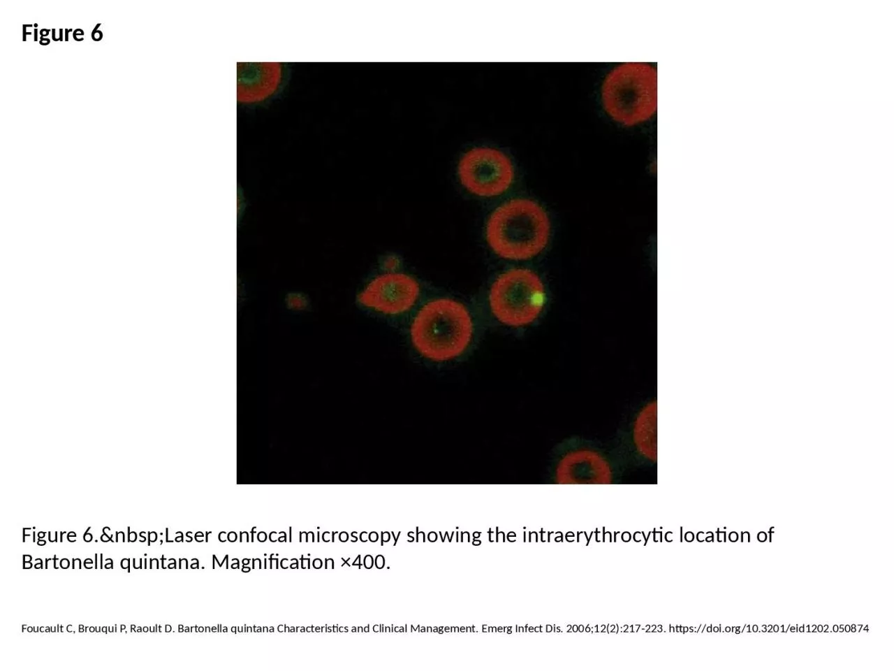
PPT-Figure 6 Figure 6. Laser confocal microscopy showing the intraerythrocytic location

Author : hadly | Published Date : 2023-07-08

Foucault C Brouqui P Raoult D Bartonella quintana Characteristics and Clinical Management Emerg Infect Dis 2006122217223 httpsdoiorg103201eid1202050874

Presentation Embed Code

Download Presentation

Download Presentation The PPT/PDF document "Figure 6 Figure 6. Laser confoc..." is the property of its rightful owner. Permission is granted to download and print the materials on this website for personal, non-commercial use only, and to display it on your personal computer provided you do not modify the materials and that you retain all copyright notices contained in the materials. By downloading content from our website, you accept the terms of this agreement.

Figure 6 Figure 6. Laser confocal microscopy showing the intraerythrocytic location: Transcript


Foucault C Brouqui P Raoult D Bartonella quintana Characteristics and Clinical Management Emerg Infect Dis 2006122217223 httpsdoiorg103201eid1202050874. Lecture . 07: . Confocal Microscopy. Adding the Third Dimension. Lecture . 7: Confocal Microscopy. Optical Sectioning: adding the third dimension. Wide-field . Imaging. Point . Spread Function. Deconvolution. lecture. 16. November 2011. Fourier. plane. Point . object. Image. f. f. f. f. a. a. ’. Magnification. : m=1. Angles. : sin(. a. ’)=sin(. a) .  . a. ’=. a. Magnification and resolution: the Abbe limit. Lecture . 07: . Confocal Microscopy. Adding the Third Dimension. Andres Collazo, Director Biological Imaging Facility. Wan-. Rong. (Sandy) Wong, Graduate Student, TA. Lecture . 7: Confocal Microscopy. Outline. Introduction. Optical Microscopy. Types of Optical Microscopes. Confocal Microscopy. Laser Scanning Confocal Microscopy. Examples. Optical Microscopy. Optical microscopy is the oldest type of microscope and uses visible light and a system of lenses to create an image of the sample. Jernigan JA, Stephens DS, Ashford DA, Omenaca C, Topiel MS, Galbraith M, et al. Bioterrorism-Related Inhalational Anthrax: The First 10 Cases Reported in the United States. Emerg Infect Dis. 2001;7(6):933-944. https://doi.org/10.3201/eid0706.010604. Kee AC, Yang S, Tambyah P. Atypical Chikungunya Virus Infections in Immunocompromised Patients. Emerg Infect Dis. 2010;16(6):1038-1040. https://doi.org/10.3201/eid1606.091115. Haapasalo K, Suomalainen P, Sukura A, Siikamäki H, Jokiranta TS. Fatal Babesiosis in Man, Finland, 2004. Emerg Infect Dis. 2010;16(7):1116-1118. https://doi.org/10.3201/eid1607.091905. Harris JR, Blaney DD, Lindsley MD, Zaki SR, Paddock CD, Drew CP, et al. Blastomycosis in Man after Kinkajou Bite. Emerg Infect Dis. 2011;17(2):268-270. https://doi.org/10.3201/eid1702.101046. Brent AJ, Matthews PC, Dance DA, Pitt TL, Handy R. Misdiagnosing Melioidosis. Emerg Infect Dis. 2007;13(2):349. https://doi.org/10.3201/eid1302.061290. Rolain J, Mathai E, Lepidi H, Somashekar HR, Mathew LG, Prakash J, et al. "Candidatus Rickettsia kellyi," India. Emerg Infect Dis. 2006;12(3):483-485. https://doi.org/10.3201/eid1203.050853. Song D, Kang B, Lee C, Saif LJ, Ha G, Kang D, et al. Transmission of Avian Influenza Virus (H3N2) to Dogs. Emerg Infect Dis. 2008;14(5):741-746. https://doi.org/10.3201/eid1405.071471. Shirwadkar CG, Samant R, Sankhe M, Deshpande R, Yagi S, Schuster FL, et al. Acanthamoeba Encephalitis in Patient with Systemic Lupus, India. Emerg Infect Dis. 2006;12(6):984-986. https://doi.org/10.3201/eid1206.060087. Aiki-Raji CO, Aguilar PV, Kwon Y, Goetz S, Suarez DL, Jethra AI, et al. Phylogenetics and Pathogenesis of Early Avian Influenza Viruses (H5N1), Nigeria. Emerg Infect Dis. 2008;14(11):1753-1755. https://doi.org/10.3201/eid1411.080557. Lee J, Chung I, Shin D, Cho S, Cho D, Ryang D, et al. Hemorrhagic Fever with Renal Syndrome Presenting with Hemophagocytic Lymphohistiocytosis. Emerg Infect Dis. 2002;8(2):209-210. https://doi.org/10.3201/eid0802.010299.

Download Document

Here is the link to download the presentation.
"Figure 6 Figure 6. Laser confocal microscopy showing the intraerythrocytic location"The content belongs to its owner. You may download and print it for personal use, without modification, and keep all copyright notices. By downloading, you agree to these terms.

Related Documents