/
For the first part of my legal career as a personal injury lawyer, we didn’t have MRIs—and, For the first part of my legal career as a personal injury lawyer, we didn’t have MRIs—and,

For the first part of my legal career as a personal injury lawyer, we didn’t have MRIs—and, - PowerPoint Presentation

holly
holly . @holly
Follow
342 views
Uploaded On 2022-06-07

For the first part of my legal career as a personal injury lawyer, we didn’t have MRIs—and, - PPT Presentation

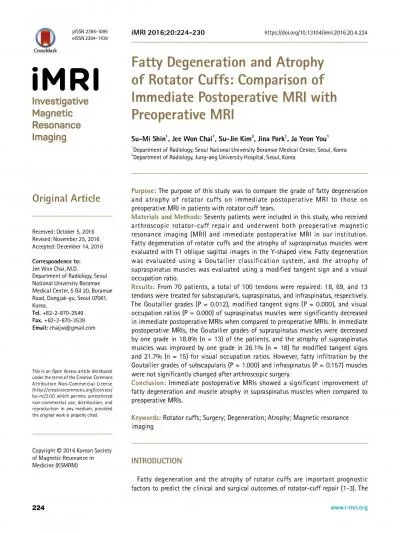
When I started seeing MRI scans proving the existence of disc injuries I was happy for the new technology but sad for all of those past clients who didnt get justice or the best possible medical care because their disc injury couldnt be proven Many were labeled complainers ID: 914515

spine motion ligamentous disc motion spine disc ligamentous pain cervical ligaments injury posterior flexion dmx ligament laxity ama extension

Share:

Link:

Embed:

Download Presentation from below link

Download Presentation The PPT/PDF document "For the first part of my legal career as..." is the property of its rightful owner. Permission is granted to download and print the materials on this web site for personal, non-commercial use only, and to display it on your personal computer provided you do not modify the materials and that you retain all copyright notices contained in the materials. By downloading content from our website, you accept the terms of this agreement.


Presentation Transcript

Slide1

Slide2

For the first part of my legal career as a personal injury lawyer, we didn’t have MRIs—and, in turn, no imaginable proof of disc injuries.

When I started seeing MRI scans proving the existence of disc injuries, I was happy for the new technology but sad for all of those past clients who didn’t get justice, or the best possible medical care, because their disc injury couldn’t be proven. Many were labeled “complainers”.

Slide3

We have known for many years that ligamentous injury is at the core of all Cervical Acceleration/Deceleration injuries (CAD).

However, until recently there was no objective proof of ligamentous injuries. Modern imaging techniques, both DMX and still imaging coupled with CRMA, now provide that proof.

CRMA has opened the door to a new level of understanding of how ligamentous injury affects the structural integrity of the cervical spine, causing negative consequences to the intervertebral discs and supporting muscles.

Slide4

I am now seeing case after case where my clients’

ligamentous

injuries are now objectively proven.

I can’t help but reflect on all the victims of whiplash injuries that haven’t received the justice they deserved because there was no objective proof of ligamentous injuries.

Hopefully, CRMA will help the chiropractor to understand ligamentous injuries that previously were hypothetical-and provide for more effective treatment-and justice for the patient’s personal injury claim.

Slide5

Combined with CRMA analysis and multi-positional upright MRIs, we now have solid objective proof of ligamentous and disc injuries that couldn’t be objectively proved in the past.

Slide6

CRMA Allows For Objective Determination of Ligamentous Injury.

The ligaments don’t show on imaging. But the results of ligamentous injury do show on imaging, both still and motion imaging.

Slide7

CRMA can quantitatively analyze the extent of the ligamentous laxity.

CRMA is Computerized Radiographic Mensuration Analysis, or CRMA. It is also called “digitized x-ray.” The term “mensuration” simply means “the measurement of geometric quantities.” Medical and chiropractic doctors have, for nearly 100 years, been measuring the translational and angular

malpositioning

of vertebrae.


CRMA means to measure

radiographics

(x-ray) using a computer. Thus CRMA provides for accurate, computerized measurement of the

malpositioning

of vertebrae due to ligamentous laxity.

Slide8

Slide9

TOTAL= $743,188.95

Slide10

Spinal ligaments are "binders", the important part of the anatomy that binds the vertebral column together. They are integral in providing and preserving the structure of the spinal column.

They are uniaxial structures. They are most effective in carrying loads along the direction in which the fibers run. In this respect, they are much like rubber bands. They readily resist tensile (stretching) forces.

Nature has designed the spinal motion segments in such a way that when the motion segment is subjected to different complex force and torque vectors, the individual ligaments, providing's resistance to the loads, experience only tensile (stretching) forces.

BASIC FUNCTIONS OF LIGAMENTS: 

Slide11

CRMA is Computerized Radiographic Mensuration Analysis.

“Mensuration” is simply an computer engineering technical term for “measurement”. It is done by taking a still Xray or using still slices from a Digital Motion Xray to measure the amount of subluxation between intervertebral discs.

Ligaments do not show up on imaging. However, when the spine is placed in a position of extreme flexion, extension, or other range of motion, the inability of the ligaments to hold the vertebral in

prper

position becomes apparent-and can be measured.

The amount of impairment, using AMA Guides, can be measured.

WHAT IS CRMA?

Slide12

1. The ligaments must allow adequate physiological motion and fixed postural attitudes between vertebrae, with a minimum expenditure of muscle energy.

2. They must protect the spinal cord by resisting the motions within well-defined limits.

3. They must protect the spinal cord in traumatic situations where high loads are applied at fast speeds. In these highly dynamic situations, not only is the displacement to be restricted within safe limits, but large amounts of energy that are suddenly applied to the spine must be absorbed.

Source: White & Panjabi.

Spinal ligaments have many different functions, some of which seem to be in opposition

to others.

Slide13

Slide14

Slide15

Slide16

The Anterior Longitudinal Ligament is a fibrous tissue structure which arises from the anterior aspect of the basioccipital and is attached to the atlas and the anterior surfaces of all the vertebrae, down to and including a part of the sacrum.

It attaches firmly to the edges of the vertebral bodies but it is not so firmly affixed to the annular fibers of the intervertebral disc.

The width of the anterior longitudinal ligament is diminished at the level of the disc. It is narrower and thicker in the thoracic region

.

Anterior Longitudinal Ligament

Slide17

Anterior posterior longitudinal ligament arises from the posterior aspect of the basioccipital, covers the dens and the transverse ligament (where it is called the

membrana

tectoria

), and runs over the posterior surfaces of all the vertebral bodies down to the coccyx. It too is thicker in the thoracic region. It has an interwoven connection with the intervertebral disc.

In contradistinction of the anterior longitudinal ligament, it is wider at the disc level and narrower at the vertebral body level.

Anterior Posterior Longitudinal Ligament

Slide18

The

ligamenta

flava extend from the anterior inferior border of the laminae above to the posterior superior border of the laminae below. They connect the borders of adjacent laminae from the second cervical vertebra to the first sacral vertebra.

They are thicker in the thoracic region. Although they seem to be paired due to a midline cleavage, each is rather like a single structure that extends from the roots of the articular process on one side to the corresponding process on the other.

The ligament is composed of a large amount of elastic fibers and represents the most pure elastic tissue in the human body. It has been noted.

Ligamenta

Flava

Slide19

Because the ligamentum flavum is located posterior to the axes of rotation of flexion/extension, it contracts with extension of the spine and elongates with flexion of the spine. Calculations show that with full extension of the spine from the neutral position, there is decreased in the length of ligamentum flavum by 10 percent.

Because the ligamentum flavum has 15 percent of pretension, it does not buckle into the spinal canal with extension.

Full flexion of the spine from the neutral position results in a 35 percent increase in its length. This is the total physiologic range. An additional 20 percent lengthening of the ligament, due to further flexion of the spine during trauma, results in failure.

Functional Biomechanics of the Ligamentum Flavum

Slide20

The supraspinous ligament originates in the ligamentum nuchae and continues along the tips of the spinous process as a round, slender strand down to the sacrum.

It is thicker and broader in the lumbar region than in the thoracic region.

Supraspinous Ligament

Slide21

The interspinous ligaments connect adjacent spines, and their attachments extend from the root to the apex of each process. They are narrow and elongated in the thoracic region, broader and thicker in the lumbar region, and only slightly developed in the neck.

Interspinous Ligaments

Slide22

The intertransverse ligaments pass between the transverse process in the thoracic region and are characterized as rounded cords, intimately connected with the deep muscles of the back.

Intertransverse Ligaments

Slide23

By the sharp increase in stiffness as the curve leaves the physiologic range. This is shown by the average stiffness in the trauma range, which is 75 times that in the flexion range. Thus , the two functions required of ligaments in the physiologic range are accomplished by the design of the load- deformations curve. When the large flexion loads are applied to the spine so that a traumatic situation exits, the design of the curve is such that large amounts of energy are absorbed before failure. Nearly seven time more energy is absorbed in the trauma range as compared to the flexion range.

Ligamentous Injury From CAD Trauma.

Slide24

Ligamentous injury is at the core of most CAD (Cervical Acceleration/Deceleration) injuries.

Slide25

Spinal ligaments are designed to stretch and return to their normal shape (elastic deformity).

When spinal ligaments are stretched beyond their intended anatomical limit, they are permanently stretched and unable to return to their nature shape (plastic deformity)

.

Plastic deformity reduces or destroys the ability of the ligaments to provide support to the vertebral column, placing increased stress on the muscle and intervertebral discs. The result is accelerated degeneration of the intervertebral discs and the development of myofascial strain (Myofascial Pain Syndrome).

OVERSTRETCHING OF LIGAMENTS CAUSES PLASTIC DEFORMITY.

Slide26

“Sub-failure” (stretch only-no tear) Ligamentous Injuries Are Serious!

1. Stretch (distension) damage resulted in abnormal laxity equal to having “no ligamentous support” at all!!!


2. Partial injury to these ligaments resulted in laxity equivalent to “completely compromised” ligaments.

Slide27

Plastic deformity reduces or destroys the ability of the ligaments to provide support to the vertebral column, placing increased stress on the muscles. The result is both ligamentous “sprain” and myofascial “sprain”. The result Myofascial Pain Disorder (Croft) or Myofascial Pain Syndrome .

Abnormal muscle stretch, i.e. “strain”. Croft says that the term “chronic strain” is “incongruous” because “strains heal in a few weeks”. The “wild card” is that the ligamentous “sprain” places continuous stress on the musculature, resulting in “Myofascial Pain Disorder”.

MYOFASCIAL PAIN DISORDER/SYNDROME

Slide28

Croft says that Myofascial Pain Disorder has two major components:

Trigger Points;

The referred zone of pain felt when palpating the trigger points.

The Clinical Review of MPD published in the Journal of PMR states that MPS is diagnosed in nearly one-third of patients with musculoskeletal disorders.

MYOFASCIAL PAIN DISORDER/SYNDROME

Slide29

TRAVELL & SIMONS

Slide30

Simons outlines the following criteria for a diagnosis of

myofascitis

:

Localized spontaneous pain;

Spontaneous pain or altered sensation in the referred area;

Taut, palpable bands in the accessible muscle;

Exquisite, localized tenderness in the tender band;

Some degree of restricted range of motion.

TRAVELL & SIMONS

Slide31

MYOFASCITIS/

MYOFASCIAL

PAIN SYNDROME

Dr. Jack Hubbard, “Myofascial Trigger Points: What Physicians Should Know about these Neurological Imitators”, Minnesota Medicine Journal, May 2010:

Definition of MPS:

Myofascial trigger points

are pathologic changes within muscle segments that are usually caused by trauma such as a motor vehicle accident…

identified on examination as painful

knots and taut bands within the affected muscle

, trigger points are capable of producing neurological symptoms including headache, dizziness, and sensory disturbances.

Trigger Point Examination

:

MFTPs can be objectively identified during a careful examination of the

patient

.

Any skeletal muscle can develop trigger points, which can be identified by careful examination of the affected muscle groups for knots that are 2 mm to 5 mm in diameter and tight bands that are painful when palpated. Diagnosis is based on the patient’s history and examination; there are no laboratory tests or imaging studies that can confirm the presence of MFTPs

.

Slide32

AMA-Myofascial Pain Syndrome

Myofascial Pain Syndrome is a condition of mild to severe muscle pain associated with trigger points

, the pain is typically localized, often to a single muscle. The syndrome commonly follows “muscle overload,” such as may occur with acute injury (e.g., following a near fall or intense exertion). Patients complain of a deep, aching pain that is worsened by activity. Postural muscles are often affected. The pain may wax and wane, but it is usually always present and at times can be severe.

Assessment and Diagnosis of Myofascial Pain

Trigger points are a diagnostic feature of this syndrome. On physical exam there will be muscle tenderness and limited range of motion, and these may be associated with palpable trigger points or “taut bands.” Palpating the trigger point produces a local twitch (a visible shortening of the muscle) and referred pain most often involves the posterior neck, low back, shoulders, chest. Chronic pain in the muscles of the posterior neck can refer to muscles in the head and cause persistent headaches; trigger points in the lower back muscles can cause referred pain to the leg that mimics sciatica.

Source: AMA Pain Management, An American Medical Association Continuing Medical Education Program for Primary Care Physicians.

Slide33

Managing Myofascial Pain

“The primary treatment for myofascial pain involves releasing the trigger points, which can be accomplished by stretching the affected muscle. One treatment, the “spray and stretch technique” involves stretching the muscle in conjunction with topical application of

vapo

-coolant sprays. A sudden drop in the skin temperature is thought to be associated with a localized anesthetic effect, which allows the affected muscle to be stretched. Massage of the trigger points may also provide pain relief. Provoking factors such as poor posture and repetitive work should be avoided.

Nonpharmacologic

management. In addition to “spray and stretch” and massage, other

nonpharmacologic

approaches include osteopathic manipulation, application of heat or ice, ultrasound exercise, TENS, acupuncture, and biofeedback. Reports of treatment effectiveness are based more on empirical clinical experience than data form controlled trials.”

Source: AMA Pain Management, An American Medical Association Continuing Medical Education Program for Primary Care Physicians

Slide34

Slide35

AMA EATS CROW REGARDING SUBLUXATION

Since 1993 the American Medical Association now recognizes ligamentous laxity to be an important cause of permanent disability. The AMA uses the terminology of:

AOMSI is Alteration of Motion Segment Integrity

.

(Does that sound like “subluxation”?)

LOMSI is Loss of Motion Segment Integrity.

The AMA Guides to the Evaluation of Permanent Impairment, 4th Edition, 5th Edition and 6th Edition both recognize AOMSI as a basis for the assessment of permanent impairment of the spine when verified by flexion/extension

Xrays

:

The term “alteration of motion segment integrity” was first used in the Fourth Edition of the Guides to describe loss of motion segment integrity, identified on flexion/extension X -rays (following specific protocols) and related to either instability or fusion, regardless of the cause.

Slide36

SURE SOUNDS LIKE A SUBLUXATION!!!

For all of 118 years since Daniel David Palmer founded the discipline of Chiropractic Medicine, the medical community has scoffed at the concept of “subluxations”!! The American Medical Association publicly declared chiropractic to be an “unscientific cult”. They finally figured it out-but refuse to use the terminology of “subluxation” that the DC community has known and appreciated for decades.

Slide37

SCLEROTOME-DERMATOME

CHART

Slide38

The

plasctic

deformity of the cervical ligaments results in permanent damage.

Slide39


The

Golden Triangle

now provides objective proof of

ligamentous

injury and the devastating consequences of the accelerated degeneration that follows:


-Digital Motion X-ray (DMX)-or still

Xrays

;


-Computerized Radiographic

Mensuration

Analysis (

CRMA

);


-Motion MRI.

Slide40

Virtually 100% of the DMX’s that have been done on my clients, over 100, have revealed objective proof of ligamentous laxity. (This is a carefully selected group, i.e. the majority being clients with significant symptoms lasting more than 1 year after the car accident.)

Over 80% have produced results that have allowed for a impairment (disability) rating using the AMA Guides.

Slide41

It is well established in the medical literature that flexion-extension trauma results in accelerated degeneration of the spine including the intervertebral discs. Until recently, that has only been a theoretical concept based on clinical observations. Recently, I have “circled” back and sent a number of my client back for repeat MRIs. The results have been astounding, showing clear proof of shockingly accelerated degeneration of the intervertebral discs in a matter of just a few years. See Appendixes A and B for case examples.

Slide42

Ligamentous Laxity Causes Accelerated Degeneration of the Intervertebral Discs.

the lack of motion at one level will be compensated for by hypermobility at adjacent levels, which in turn usually will result in degenerative disc disease and osteoarthritis sometime in the future

.”

Delayed instability of the spine due to ligamentous laxity is recognized in the medical literature. CRMA is a valuable tool in detecting delayed instability of the

ligments

. See Cusic, Clinical Biomechanics, Vol. 17, No 1, pp. 1-20 (2002)

Slide43

Deterioration of Intervertebral Discs Due to Ligamentous Injury.

Injury to the ligaments destroys their ability to support the discs. The result is accelerated deterioration of the discs.

In several of our injury cases where ligamentous damage was established, repeat MRIs done several years later, have revealed

accerated

disc degeneration.

Slide44

Lynn’s Case

Lynn’s Case: Lynn was 14 years old when she suffered a CAD injury. MRI studies done 2 months post accident were normal. CRMA studies revealed significant ligamentous injury. Follow up MRI, done at SUMA 2 years later, revealed major degeneration. Her Physical Medicine physician opined that she had a spine of a 40 year old. As the result of her ligamentous injury, she had suffered the equivalent of nearly 30 years of aging in only 2!!!

Slide45

Bruce’s Case-Major Disc Degeneration in only 2 years.

In another case where we had established major ligamentous injury, follow-up MRIs revealed major degeneration of the intervertebral discs in only slightly more than 2 years.

Slide46

DISC INJURIES CAUSE PAIN EVEN WITHOUT IMPINGEMENT OF A NERVE ROOT

Foreman & Croft-

Four ways that an anterior extrusion of an intervertebral disc can convey pain:

(a) via direct compression or irritation of the sympathetic chain ganglia;

(b) via the anastomosis between the recurrent meningeal nerve and its sympathetic connection;

(c) through the direct route into the ventral ramus of the spinal nerve via the white rami

communicantes

; and

(d) via nociceptive afferent activity (recurrent meningeal nerve) in the anterior

anulus

.

Both produce pain (direct and referred), muscle spasm, trigger points, further causing abnormal function and movement of the spine.

Slide47

DISC PAIN WITHOUT IMPINGEMENT

Medical literature universally establishes that disc injury causes “

vertebrogenic

pain” without nerve root impingement.

Jinkins

, et. al,

“The Anatomic Basis of

Vertebrogenic

Pain and the Autonomic Syndrome Associated with Lumbar Disc Extrusion,”

American Journal of

Roentgenology

, June 1989, p. 1277 et. seq. identifies

“the

vertebrogenic

symptom complex which involves ‘referred pain’ emanating from the injured disc through the autonomic nervous system.”

Slide48

Freemont, et. al,

“Nerve Growth into Diseased Intervertebral Disc in Chronic Back Pain,”

The Lancet 1997; 350; 178-81 explains that injuries to the outer third of the

annullus

fibrosus

of the disc can cause the ingrowth of painful nerve fibers which extrude chemical substance causing pain plays an “important role in the pathogenesis of chronic low back pain.”

Bogduk

et. al,

“The Innervation of the Cervical

Intrvertebral

Disc,”

Spine, Vol. 13, No. 1, pp. 1-8 (1987) states that “there has been no doubt that lumbar intervertebral discs are innervated…(and) the innervation is not just in their superficial layers but can be found as deep as the outer third of the annulus

fibrosus

” so that disc injuries that do not impinge on a nerve root or the spinal cord can be expected to be painful. This article states that studies have shown that the same is true of cervical discs, i.e. that injury to the disc can cause pain.

Slide49

FACET JOINT INJURIES

Present in 60% of cases with chronic pain lasting more than 1 year.

Ligament laxity can be detected by a careful clinical examination of the laxity of each vertebral joint in all 8 planes of motion (flexion-extension, rotation, side bending, and protraction/retraction. Findings of ligamentous laxity should be carefully documented in the record

Palpation of the facet joint in pillar examination will often produce radiation of symptoms in a well-established dermatome region. (See next slide) Radiation of symptoms within the dermatome region should be carefully documented

Facet joint damage is linked to myofascial trigger points. Myofascial trigger points can be detected by a careful examination. The location of each trigger point, naming the muscle group, should be carefully documented

Gold standard of DX: Medial Branch Block Injections

Treatment: Radiofrequency Neurotomy. (Useful in conjunction with chiropractic!

Slide50

The evidence it provides to a jury in the court room setting is also amazing. In the very first case in which

DMX

was introduced as evidence in a court trial, the jury was obviously fascinated. They “

scooched

” forward on their seat, craned their necks forward, and stared “buggy-eyed” at the screen as the

DMX

film played out. The result was a jury verdict of $743,188.95 in a case where the last formal offer before trial was only $10,000.00.

The evidence that DMX videos provides to the Doctor of Chiropractic and the patient is amazing.

Slide51

It is well recognized and respected in the medical literature—but the large majority of the medical community has virtually no knowledge of it.

It has been accepted as reliable evidence in court in a number of cases. The courts and arbitrators have accepted DMX/CRMA in over 50 cases where the Schmidt Law Firm has represented the injury victim without a single case where it has been rejected.

DMX is a respected technological development.

Slide52

As early as 1993, the AMA Guides stated:

AMA Guide to Evaluation of Permanent Impairment 4

th

Edition:

The loss of (motion segment) integrity

is defined as:

-an

antero-posterior motion

or slipping of one vertebrae over another greater than 3.5 mm for a cervical vertebrae are greater than 5 mm for a vertebrae in the thoracic or lumbar spine (“translation”); or

-a

difference in the

angular motion

of two adjacent motion segments greater than 11° in response to spine flexion and extension. Motion of the spine segments is evaluated with flexion and extension. (Loss of integrity of the lumbosacral joint is defined as an angular motion between L5, and S1 that is 15° greater than the motion at the L4, L5 level.)”

Slide53

Slide54

The AMA Guides acknowledge that ligament injuries produce two types of “motion segment” impairment- translational and angular.

Slide55

Foreman/Croft used a similar illustration of the same concept in 1995:

Slide56

It’s all about motion!!!

Motion of the individual spine segments cannot be determined by physical examination, but is evaluated with flexion and extension roentgenograms (see Figures 15-3a through 15-3c)

AMA Guides,

(5

th

Ed. 2001)

Slide57

How does DMX work?

Digital Motion X-ray utilizes x-ray technology, and couples it with new digital and optic technology in the image intensifier to create high-resolution images of the spine and skeletal system in real-time motion.

DMX can produce 2700 still x-rays with the same radiation dose as the seven (7) view Cervical Davis series.

Slide58

DMX can detect ligamentous laxity that static x-rays cannot see.

Most of these (static x-ray) studies…fail to define movement in a dynamic sense…deviation from the normal

biokinetics

may occur somewhere between these arcs of motion, which would not be visualized by static radiographic techniques.

Slide59

DMX is respected by the chiropractic community.

DMX has been approved by the Council on Chiropractic Practice:

Vertebral Subluxation in Chiropractic Practice Guidelines

, 3rd Ed, Council on Chiropractic Practice, 2008, p 318:


Videofluoroscopy

may be employed to provide motion views of the spine when abnormal patterns are clinically suspected.

Videofluoroscopy

may be valuable in detecting and characterizing spinal

kinesiopathy

associated with vertebral subluxation.”

Slide60

DMX is respected by the U.S Government.

The AHQR (Agency for Healthcare Research & Quality of the U.S. Department of Health & Human Services) recommends DMX in two separate practice guidelines. The

Guideline: Vertebral Subluxation in

ChiropracticPractice

states:

Videofluoroscopy

may be employed to provide motion views of the spine when abnormal motion patterns are clinically suspected.

Videofluoroscopy

may be valuable in detecting and characterizing spinal

kinesiopathology

associated with vertebral subluxation.”

Slide61

DMX is respected by the medical community.

The

American Academy of

Pain Management’s Practical Guide to Clinicians

, 5th Edition states:

"...digital motion radiography is currently a valuable diagnostic method in evaluating painful hyper- mobility and instability of capsular and axial ligaments in the cervical spine."

Slide62

DMX has been accepted as reliable evidence by the courts.

Actual Appeals Court decision:

Appellant argues that DMX technology does not meet the Daubert standard because it has not been proven to aid in diagnosing or treating any injury and that DMX gives no more information than standard x-rays. Appellant also asserts that the scientific community has not generally accepted the use of DMX is for diagnosing or treating any injury or ailment. We disagree.

Slide63

CRMA can be used to establish a percentage impairment rating using the AMA Guides rating system.

Motion of the individual spine segments cannot be determined by physical examination, but is evaluated with flexion and extension roentgenograms (see Figures 15-3a through 15-3c) AMA Guides

, (5

th

Ed. 2001), pg. 379.

Slide64

CRMA can be used to provide an AMA-based disability rating.

The AMA Guides to the Evaluation of Permanent Impairment, 5

th

Ed. (2000), p. 392, states that Alteration of Motion Segment

Intergrity

(AOMSI) results in a 25-28% impairment rating in each of the following cases:


Alteration of Motion Segment Integrity or

bilateral or multilevel radiculopathy

; alteration of motion segment integrity is defined from flexion and extension radiographs as at least

3.5 mm of translation

or one vertebrae on another, or

angular motion of more than 11 degrees greater than at each adjacent level

.

Slide65

How do we know that the ligamentous laxity was caused by CAD trauma?

When routine x-rays are normal and severe trauma is absent, motion segment alteration is rare

; thus, flexion and extension x-rays are indicated

only

when the physician suspects motion segment alteration from history or findings on routine x-rays. AMA Guides, 5Ed. 2001, pg. 379.

The flip side of the coin is that when severe trauma is present and digital motion X-rays show laxity, motion segment alteration is likely present.

Slide66

When ligamentous laxity is demonstrated, repeat MRIs can, in some cases, objectively prove accelerated degeneration.

Slide67

DMX provides objective proof of the existence of both translational laxity and angular laxity in ligaments.

CRMA provides a scientifically reliable method of accurately measuring or quantifying the exact amount of laxity in each category.

Motion MRI provides a method of proving the existence of accelerated degeneration.

Together, they form the “Golden Pyramid” of Objective Proof of ligamentous injury.

The combination of DMX, CRMA, and Motion MRI can be extremely valuable in proving major impairment in cases that previously were dismissed as minor “soft tissue” injuries.

Slide68

Motion MRIs provide a method of detecting herniations that are not observable in conventional MRIs.

SUMA MRI has developed a new technology that may be as revolutionary as Digital Motion X-rays, namely “Motion MRIs”. This procedure involves taking MRIs are a number of positions, or stations, throughout the flexion-extension cycle. Remarkably, it has been shown that some herniations will be revealed at one station but not others. Some will be revealed at one station, then disappear at other stations, and reappear at yet others.

Slide69

DMX provides proof of the cause of Facet Joint Injuries.

The posterior ligamentous complex acts to stabilize the vertebral column and hold the facet joints in the neighboring vertebrae in fixed relation with each other.

Each facet joint has capsular ligaments that are often damages when the posterior ligaments are excessively stretched.

Slide70

Gina’s Case

-There is a high degree of correlation between the specific

location of the ligamentous laxity and the specific location of

the symptoms.


-There is a high degree of correlation between the specific

location of the

ligamentous

laxity and the specific location of

the disc pathology.


-Patients with

ligamentous

laxity have demonstrated grossly

accelerated disc degeneration on repeat MRIs.


-There is a high degree of correlation between laxity of the

Atlas and occipital symptoms/occipital headaches.

Our series of DMX results has allowed us to do some additional research that has produced astounding results:

Slide71

-Over 90% of our clients who have been

DMX’d

have evidence of abnormal

ligamentous

laxity.

-35% have sufficient laxity to qualify for a 25% Impairment rating using the AMA Guides.

-40% have abnormal shifting of the Atlas that corresponds to high cervical and occipital pain and headaches. (Those with bilateral shifting have bilateral headaches. Those with unilateral shifting have unilateral symptoms.

New Data From

Schmidt Salita Law Team cases:

Slide72


Serious ligamentous injuries have been dismissed as “soft tissue sprain/strain” injuries of minimal consequence because of the lack of objective proof.


We now have that proof of the ligamentous damage!!! DMX provides that objective proof.


We also now have objective proof of accelerated degeneration resulting from ligamentous laxity!!!


Gina’s case is a classic example.

CLASSIC EXAMPLE OF HOW DMX & MRI CAN ESTABLISH PERMANENT LIGAMENTOUS DAMAGE

Slide73

Case study-“Gina’s case,” shows accelerated degeneration of intervertebral discs secondary to ligament damage.

Simply put, Gina’s ligamentous injury had produced laxity which imposed abnormal stresses on the intervertebral discs, causing grossly abnormal degeneration. In the course of only 2 years since her whiplash injury, Gina’s neck had “aged” or “degenerated” the equivalent of 30-40 years! Imagine what she must look forward to in her remaining 60-year life expectancy!!!

Slide74


-14 year old girl sustains a whiplash injury.


-After 2 years of treatment, by DC, MD, PT and Massage ($32,000.00), she continues to experience major pain and disability.


-The MRI is “unremarkable” in all aspects.


-The Chiropractic Rx is “sprain/strain”.


-The MD Rx is “ligamentous injury” with no objective proof.


-The offer of settlement is $2000.00 more than the medical bills.

BEFORE DMX:

Slide75

Ligamentous injury to a 14 year old girl, previously healthy, has resulted in two impairments of her cervical spine, each

rateable

pursuant to AMA Guides at 25% of the whole body.

Proof of accelerated degeneration of her spine.

Her spine has aged the equivalent of 30-40 years in 2 years!!!

BOTTOM LINE-MAJOR ACCELERATED DEGENERATION:

Slide76

The anterior vertebral line (AVL) should be visualized as a smooth, unbroken arc extending from C2 to C7 along the anterior border of the vertebral bodies (Fig. 4-l A). A patient who presents with either a straight or a kyphotic curve should also have a smooth line, and any disruption of this line should be noted.

Anterior Vertebral Line

Slide77

The posterior vertebral line (PVL), also known as George’s line, like its anterior counterpart, should also be visualized in a smooth, unbroken manner extending from C2 to C7 along the posterior border of the vertebral bodies. This line delineates the anterior wall of the spinal canal or the posterior longitudinal ligament, and its disruption may localize the source of a neu­rological deficit.

Posterior Vertebral Line

Slide78

The posterior canal line is visualized di­rectly over the

spinolaminar

line from lev­els Cl to C7. The

spinolaminar

line is the short cortex line produced by muon of the right and the left lamina to the spinous process. The absence of the

spinolaminar

line, usually seen at the level of the atlas, indicates a failure of fusion.

Posterior Canal Line

Slide79

This specialty radiology report is compiled upon evidence based objective biomechanical analysis for

Roentgenometric

Digitization of the spine. This evaluation will not include a pathological report. The report results

will

provide the referring physician with radiologic analysis necessary to help determine the ligamentous and alignment factors of the spine for a more accurate diagnosis and treatment plan. The Digital Radiographic Images used were of acceptable quality and in compliance with our protocols for X-ray imaging. This report follows the AMA Guides to The Evaluation of Permanent Impairment, 5th edition. This report is based on digitization printout and accepted consensus of what these numbers indicate. All findings in this report must be correlated clinically:

'Objective Spinal Motion Unit Assessment through AMA Precision Compliant Procedures'

Computer Aided Radiographic Mensuration Analysis-VS

Slide80

Lateral Cervical Spine: interruptions of the George's Line at C5/C6 are indicative of ligamentous instability or sub failure. Atlas lateral shift is 3.65 mm during left lateral bending and 5.81 mm during right lateral bending.

Cervical Motion Study: The angular motion segment integrity is abnormal which may be clinically significant and must be clinically correlated. The translational motion segment integrity is ratable at C3 and cs.

'Objective Spinal Motion Unit Assessment through AMA Precision Compliant Procedures'

Computer Aided Radiographic Mensuration Analysis-VS

Slide81

IMPRESSIONS:

l. Interruptions of the George's Line at C5/C6 are indicative of ligamentous instability or sub

failure.Ligamentous

instability is indicated in the cervical spine with the measurements in this report. All findings must be clinically correlated to the doctor's clinical findings.

Motion Segment Integrity, Angular variation is abnormal at CS. This patient's digital analysis reveals CS =7,53°.

Cervical motion study indicates Translational Motion Seg1i1ent Integrity change at C3, CS. The impairment of the cervical region is due to ratable Loss of Motion Segment Integrity and is ratable at 25% for cervical spine (AMA Guides, Fifth Edition, Errata). This patient's digital analysis reveals Loss of Motion Integrity at C3 = 3.70 mm, C5 = 3.66 mm yielding an impairment estimate based on plain film forensics at 25% whole person. Motion Segment Integrity, Translational variation is abnormal at C2, C4. This patient's digital analysis reveals C2 = 1.92 mm, C4 = 2.97 mm.

'Objective Spinal Motion Unit Assessment through AMA Precision Compliant Procedures'

Computer Aided Radiographic Mensuration Analysis-VS

Slide82

Cervical Notes: *Measurements over 1mm Translation and/ or over 7° Angular Variation are considered to be clinically significant and in excess of normal flexibility of the cervical spine. (SPINE 200l, February; 26(3): (256-261), Lin, Tsai, Chu and Chang.

**Abnormal measurements of more than 11° Angular Variation and/ or greater than or equal to 3.5mm Translation (Loss of Motion Segment Integrity) by definition constitutes ligament damage which results in instability and calculates a whole person impairment of 25% to 28%. (Guides to the Evaluation of Permanent Impairment, Fifth Edition, 2000.) DRE Category IV.

***Lateral shift of Atlas on Axis greater than 1.7mm is considered subluxation and associated with poor prognosis for whiplash injury.

Source:

Krakenes

J,

Kaale

BR, Moen G, Nordli H,

Gilhus

NE,

Rorvik

J. MRI assessment of the alar ligaments in the late stage of whiplash injury- structural abnormalities and observer agreement. Neuroradiology 2002 Jul;44(7):617-24.

'Objective Spinal Motion Unit Assessment through AMA Precision Compliant Procedures'

Computer Aided Radiographic Mensuration Analysis-VS

Slide83

C5-6: There is normal disc height, but mild disc desiccation. There is mild annular bulging with associated spondylosis and uncinate hypertrophy, right greater than left resulting in mild to moderate right foraminal compromise. The canal and left foramen are mildly stenotic and there is slight effacement but not impingement of the subjacent cord. Mild facet arthrosis is noted bilaterally. The posterior disc bulge becomes less prominent with overall extension motion and more prominent with overall extension motion with ti1e central spinal canal narrowing to approximately 9.5 mm. With overall flexion motion, the AP dimension measures approximately 12 mm.

LIFESCAN UPRIGHTMRI

MINNEAPOLIS ST. PAUL:

Slide84

C6-7: There is normal disc height, but mild disc desiccation There is relatively prominent annular bulging with associated spondylosis, somewhat eccentric to the left. along with mild uncinate hypertrophy resulting in mild to moderate overall canal and foraminal compromise, left greater than right without definite nerve root impingement. There is slight effacement of the ventral subjacent cord. The posterior disc bulging becomes somewhat less prominent with overall flexion motion and somewhat more prominent with overall extension motion. The AP dimension of the canal is 9mm with extension and 11mm with flexion. Mild facet arthrosis is no ted bilaterally.

LIFESCAN UPRIGHTMRI

MINNEAPOLIS ST. PAUL:

Slide85

At C7-Tl and Tl-2, there is normal disc height and hydration without disc bulge or herniation. There is mild to moderate facet arthrosis at these levels, greatest at C7-T1, left greater than right. This results in at least mild left foraminal compromise at C7-T l . The canal and right foramen are adequately patent. The canal and foramina are adequately patent at T1-2.

T2-3: There is mild loss of disc height and disc desiccation . There is mild annular bulging. The canal and foramina are adequately patent. There is mild facet arthrosis noted bilaterally, right greater than left.

T3-4: There is normal disc height and hydration without disc bulge or herniation. The canal and foramina are adequately patent. The facet structures arc unremarkable.

LIFESCAN UPRIGHTMRI

MINNEAPOLIS ST. PAUL:

Slide86

CONCLUSION:

1) Multilevel degenerative disc changes, disc bulging and facet arthrosis throughout the cervical spine, as described. The disc bulging and spondylosis is most prominent at C5-6 and C6-T becoming more prominent with overall extension motion and less prominent with overall flexion motion. At C5-6, the uncovertebral arthrosis is greatest on the right resulting in at least moderate right foraminal compromise, that may affect the right

cxiting

. C6 nerve root. At C6-7, the disc bulging and spondylosis is somewhat eccentric

posterolaterally

to the left resulting in at least mild left foraminal compromise. Clinical correlation for left C7 nerve root symptoms is recommended in this regard.

BRUCE’S FIRST MRI

Slide87

1. Left paracentral posterior disc herniation at C3/4.

2. Broad-based posterior disc herniation at CS/6 and C6/7.

3. Annular bulge at C2/3 and C4/5.

4. Dehydration of the discs as noted above.

5. Mild loss of disc height.

6. Straightening of the normal lordosis.

7. Moderate stenosis of the bilateral intervertebral neural foramina at C5/6 and C6/7 with impingement of the bilateral C6 and C7 nerve roots.

8. Moderate stenosis of the left

intervertcbral

neural foramen at C3/4 with impingement of the left C4 nerve roots.

9. Mild central canal stenosis at C3/4, CS/6 and C6/7.

BRUCE’S MRI-4 YEARS LATER.

Slide88

1. In the neutral lateral projection: Shows straightening of the cervical lordosis. There is spurring at C3-C4 and C4-C5.

The integrity of the cervical lordosis and overall condition of the cervical spine is evaluated. The loss of the cervical lordosis may be a result of damage to the posterior longitudinal, capsular or interspinous ligaments.

Digital Motion X-ray

Cervical Spine (AL)

Slide89

Widening of posterior disc space

Anterolisthesis

Damaged Interspinous Ligament

Digital Motion X-ray

Cervical Spine:

Slide90

Neutral lateral projection

Digital Motion X-ray

Cervical Spine (AL:

Slide91

2. Motion in the neutral lateral projection to full flexion: There is an anterolisthesis of C2 on C3 and C3 on C4.

This view examines the integrity of the posterior longitudinal ligament demonstrated by a forward (anterior) movement of one vertebrae over the vertebrae below or by the posterior widening of the intervertebral disc space (increased disc angle).

The integrity of the interspinous ligament is evaluated in the lateral flexion view. Damage to this ligament results in increased separation of the spinous processes in flexion.

Digital Motion X-ray

Cervical Spine:

Slide92

Full flexion projection

Digital Motion X-ray

Cervical Spine:

Slide93

3. Motion in the neutral lateral projection to full extension: Is restricted.

This view examines the integrity of the anterior longitudinal ligament demonstrated by a backward (posterior) movement of one vertebrae over the vertebrae below or by the anterior widening of the intervertebral disc space (increased disc angle).

Retrolisthesis

Widening of the anterior disc

Digital Motion X-ray

Cervical Spine:

Slide94

4. Motion in the oblique flexion projection: There is gapping of the facet joints at C4-C5 on the right.

This view examines the integrity of the capsular ligaments by observing gapping of the facet joints, located on the posterior cervical spine (C2-C7), there are five capsular ligaments on the right and the left.

Capsular ligament damage

Digital Motion X-ray

Cervical Spine:

Slide95

Left IVF flexion

Digital Motion X-ray

Cervical Spine:

Slide96

5. Motion in the oblique extension projection: There is intervertebral foraminal encroachment of the facet joint at C3-C4 on the right.

This view examines the integrity of the capsular ligament by encroachment into the intervertebral foramen, located on the posterior cervical spine (C2-C7), there are five capsular ligaments on the right and the left.

Right IVF oblique extension

Digital Motion X-ray

Cervical Spine:

Slide97

6. Motion in the A-P rotation projection: Is within normal limits.

This view examines the rotational range of motion between Occiput-C1-C2. Increased motion indicates damage to the alar and accessory ligaments.

7. Motion in the A-P open mouth lateral bending projection: Is restricted.

This view examines the integrity of the alar and accessory ligaments either by the lateral overhang of C1 on C2 or by the changes in the para-odontoid spaces.

Digital Motion X-ray

Cervical Spine:

Slide98

C1 lateral mass overhang

Change in Para-odontoid space

Digital Motion X-ray

Cervical Spine:

Slide99

IMPRESSION for patient (AL):

• Damage to the posterior longitudinal ligament is indicated by an anterolisthesis at C2 on C3 and C3 on C4.

• Damage to the capsular ligament is indicated by gapping of the facet joint at C4-C5 on the right.

• Damage to the capsular ligament is indicated by intervertebral foraminal encroachment of the facet joint at C3-C4 on the right.

Sincerely,

Chintan Desai, MD. Diplomate ABR

ACR Certified in

NeuroRadiology

Fellowship trained in MSK MRI.

Digital Motion X-ray

Cervical Spine: