/
Facial  nerve And Taste Pathway And its clinical Facial  nerve And Taste Pathway And its clinical

Facial nerve And Taste Pathway And its clinical - PowerPoint Presentation

isabella2
isabella2 . @isabella2
Follow
66 views
Uploaded On 2023-07-21

Facial nerve And Taste Pathway And its clinical - PPT Presentation

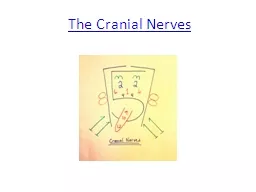
By DR ROBERTON GAUTAM SR JNMC ALIGARH Functional Components In addition to having similar somatic and visceral components as spinal nerves some cranial nerves also contain special sensory and motor components ID: 1009774

nerve amp motor facial amp nerve facial motor nucleus fibres side part muscles paralysis visceral affected taste root branch

Share:

Link:

Embed:

Download Presentation from below link

Download Presentation The PPT/PDF document "Facial nerve And Taste Pathway And its ..." is the property of its rightful owner. Permission is granted to download and print the materials on this web site for personal, non-commercial use only, and to display it on your personal computer provided you do not modify the materials and that you retain all copyright notices contained in the materials. By downloading content from our website, you accept the terms of this agreement.


Presentation Transcript

1. Facial nerve And Taste Pathway And its clinicalBy DR ROBERTON GAUTAM’SR, JNMC ALIGARH

2.

3.

4.

5.

6. Functional Components In addition to having similar somatic and visceral components as spinal nerves, some cranial nerves also contain special sensory and motor components

7. Functional ComponentAbbreviationGeneral FunctionCranial NervesGeneral Somatic AfferentGSAPerception of touch, pain, temperature Trigeminal, Facial, Vagus General Visceral AfferentGVASensory input from visceraGlossopharyngeal, VagusSpecial AfferentSASmell, Taste, Vision, Hearing and BalanceOlfactory, Optic, Facial, Vestibulocochlear, Glossopharyngeal, VagusGeneral Somatic EfferentGSEMotor innervations to skeletal musclesOculomotor, trochlear, Abducent, Accessory, HypoglossalGeneral Visceral EfferentGVEMotor innervations to smooth muscles, heart muscles and glandsOculomotor, facial, glossopharyngeal, vagusSpecial Visceral Efferent/ Branchial MotorBEMotor innervations to skeletal muscles derived from pharyngeal arch mesodermTrigeminal, Facial, Glossopharyngeal, Vagus

8. Innervations of the musculature derived from the five pharyngeal arches are

9. INTRODUCTION Facial nerve (7th cranial nerve) consists of a motor & sensory root.Sensory root is also known as nervous intermedius. It contains following functional components: (a).Branchio-motor fibres-to supply the muscles developed from the second branchial arch.(b).Preganglionic secreto-motor fibres for submandibular, sublingual, lacrimal glands & glands of soft palate & nasal cavity.(c).Taste fibres from the anterior 2/3rd of tongue & from the soft palate.(d).Cutaneous somato-sensory fibres from concha of auricle.

10. facial nerve nucleusMotor nucleus- it is situated in the caudal part of pons, below & in front of the abducent nucleus. The motor nucleus represents special visceral( branchial) efferent column. - The fibres from motor nucleus pass dorso-medially towards caudal end of abducent nucleus, & then run rostrally superficial to that nucleus forming facial colliculus of the floor of fourth ventricle. At cranial end of abducent nucleus the fibres bend abruptly downwards & forwards forming an internal genu & emerge at the lower border of pons through the motor root.

11.

12. To be cont.Superior salivatory nucleus- it is situated dorso-lateral to caudal part of the motor nucleus & represents general visceral efferent column. It gives origin to preganglionic secreto-motor fibres. upper part of nucleus of tractus solitarius- it represents special visceral afferent & general visceral afferent column. It receives taste sensation from anterior 2/3rd of tongue & from the soft palate.

13. upper part of spinal nucleus of trigeminal nerve- it receives cutaneous sensations from the auricle through the vagus.

14. Central connectionThe cortico-nuclear fibres(pyramidal) of the opposite side control the activities of that part of motor nucleus which supplies muscles of the lower part of the face.The part of motor nucleus which supplies muscles of forehead & eyelids is controlled by cortico-nuclear fibres of both sides.

15. Superficial origin both the roots of facial nerve are attached to the lower border of pons between olive & inferior cerebellar peduncle.The motor root is large & lies on the medial side of the sensory root. sensory root is also called as nervous intermedius.

16. Course & relationsIntracranial –intrapetrous part- from brainstem both roots of facial nerve, accompanied by vestibulo-cochlear nerve, pass laterally & forwards & enter the internal acoustic meatus . At bottom of the meatus the two roots of facial nerve unite to form a trunk which enters the bony facial canal. In the bony canal at first nerve passes laterally above the vestibule of the internal ear & reaches the medial wall of the epitympanic part of tympanic cavity where it bends backward forming a genu(external genu).At the genu facial nerve presents an asymmetrical swelling known as the genicular ganglion.Finally nerve passes vertically downward along the posterior wall of the tympanic cavity & leaves the temporal bone through stylomastoid foramen.

17. . In the facial canal (two branches): (a). Nerve to stapedius- it arises from the facial nerve opposite the pyramidal eminence & supplies the stapedius muscle.(b). Chorda tympani nerve- It arises from the facial nerve about 6mm. above the stylomastoid foramen. It convey taste fibres from the anterior 2/3rd of tongue except vallate papillae

18.

19. Extra cranial part From the stylomastoid foramen the facial nerve changes the direction , passes forward superficial to the styloid process of the temporal bone & pierces the postero-medial surface of the parotid gland. Within the gland the nerve runs forward for about 1cm. Superficial to retro-mandibular vein & external carotid artery & then subdivides into five terminal branches

20.

21. Below the stylomastoid foramen(a).posterior auricular nerve(b).nerve to the posterior belly of digastric(c).nerve to the stylohyoid muscleIn the face (five terminal branches)- goose-foot pattern (pes anserinus)(a).temporal branch(b).zygomatic branch(c).buccal branch(upper & lower)(d).marginal mandibular branch(e).cervical branch

22.

23.

24. secrto-motor fibresSecreto-motor supply of the lacrimal gland is derived from the parasympathetic nerves. The preganglionic fibres arises from lacimatory nucleus in the pons ,& pass successively through the nervous intermedius, trunk & genicular ganglion of the facial nerve , greater petrosal nerve & nerve to pterygoid canal & reach the pterygo-palatine ganglion where the fibres are relayed. The postganglionic fibres pass through the maxillary nerve, zygomatic nerve & its zygomaticotemporal branch & finally supply the gland via the lacrimal nerve

25. secreto-motor fibresFor lacrimal glands:

26.

27.

28. secretomotor to soft palate & nasal mucosa

29. secretomotor to submandibular & sublingual gland

30.

31. taste from anterior 2/3rd of the tongue

32.

33. Applied anatomy of facial nerveLesion of facial nerve may be supranuclear or infranuclear & paralysis is commonly unilateral . supranuclear paralysis- it involves upper motor neurons of the cortico-bulbar & cortico-reticular fibres to the facial nucleus. This results in impairment or loss of movements of the lower facial muscles of the contralateral side, but upper facial muscles are escaped. This is due to bilateral control of the motor cortex to the subgroups of motor nuclei which supply the upper facial muscles.

34. to be cont.Nuclear paralysis- a lesion in the pons may involve the motor nucleus of the facial nerve along with abducent nucleus around which motor root makes a loop . This results in lower motor neuron paralysis producing loss of movements of all facial muscles on the affected side, associated with internal strabismus due to involvement of the lateral rectus muscle of the eyeball.

35. Infranuclear ParalysisA lesion interrupting the peripheral part of facial nerve is known as Bell’s palasy, which when complete, produces lower motor neuron paralysis of all facial muscles on affected side with abolition of both voluntary & reflex movements. The manifestations of peripheral injury vary according to the site of involvement.Lesion in the internal acoustic meatus produces Bell’s palsy & deafness due to involvement of the vestibulo-cochlear nerve.A lesion at the genu produces diminished lacrimation & submandibular salivary secretion, reduced taste sensation on the anterior 2/3rd of the tongue, hyperacusis due to involvement of nerve to the stapedius, along with signs of Bell’s palsy on the affected side. Involvement of pinna in herpes zoster of geniculate ganglion (Ramsay Hunt syndrome):

36. to be cont.During recovery from an injury proximal to genicular ganglion, some regenerating salivary fibres may pass through greater petrosal nerve & reach the pterygo-palatine ganglion. This is manifested by paroxysmal lacrimation during eating & is known as crocodile tear syndrome.3.A lesion of the facial nerve between genu & pyramidal eminence produces all manifestations without disturbance of lacrimation & taste sensation from the soft palate.

37. to be cont.4. An injury of the facial nerve below the stylomastoid foramen produces Bell’s palsy without affecting other functions. In a typical paralysis the face become asymmetrical & following manifestations are observed on the affected side:(a). Transverse wrinkles of the forehead disappear & eyebrow droops.(b). Palpebral fissure is wider than that of normal side, due to unopposed action of levator palpebrae superioris. The pt. Is unable to close his eyelids & tear roll over the cheek. Corneal reflex is disturbed which may culminate into corneal ulcers & blindness. When an attempt to close eyelids ,eyeball on the affected side may be seen to roll upward this is known as Bell’s phenomenon.

38. to be cont.(c). Nasolabial fold disappears, ala nasi does not move & tip of the nose is deviated to the sound side.(d). During smiling the angle of the mouth remains motionless on the affected side, whereas the other angle moves upward & laterally.(e). Due to paralysis of buccinator , food accumulates in the vestibule of the mouth, & occasionally dribbles out between paralysed lips.(f). Lips on the affected side cannot move, pursing of the whistle is disturbed & labial speech is affected.(g).Bell’s palsy belongs to the flaccid type of lower motor neuron paralysis & caused by sudden exposure to cold, middle ear infection, fractures, tumours etc.

39.

40.

41. • Trigeminal neuralgia (tic douloureux): It is a clinicalcondition characterized by sudden paroxysmal attacks oflancinating pain lasting from few hours to several days,confined to distribution of one or more divisions oftrigeminal nerve. It commonly starts in the maxillaryterritory and more frequently on the right side.• Herpes zoster ophthalmicus: It is a viral infection involvingthe ophthalmic nerve. It presents as severe pain andedema in the ophthalmic territory and is characterized bythe appearance of vesicles along the course of cutaneousbranches of the ophthalmic nerve.