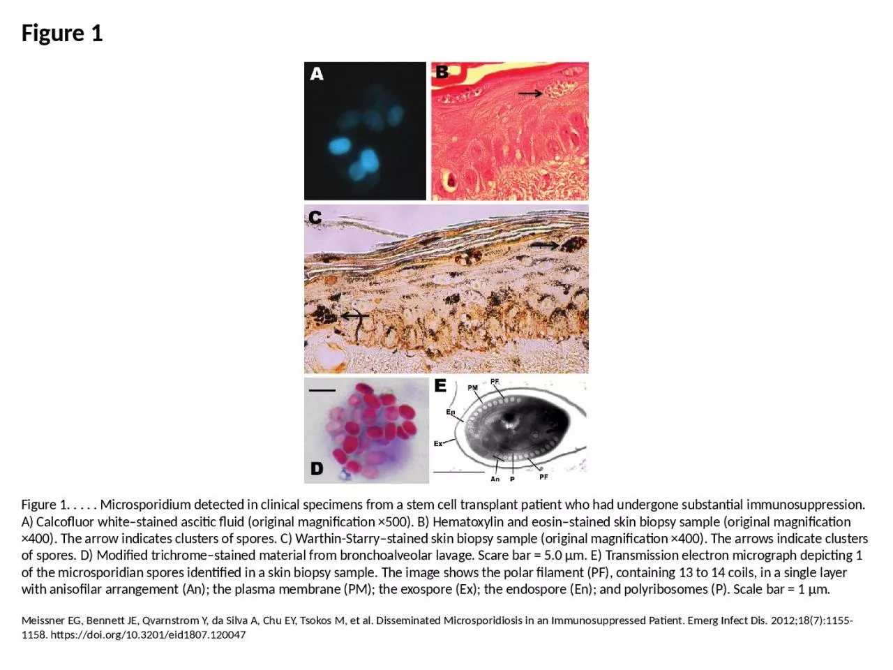
PPT-Figure 1 Figure 1. . . . . Microsporidium detected in clinical specimens from a stem cell

Author : isabella2 | Published Date : 2023-07-19

Meissner EG Bennett JE Qvarnstrom Y da Silva A Chu EY Tsokos M et al Disseminated Microsporidiosis in an Immunosuppressed Patient Emerg Infect Dis 201218711551158

Presentation Embed Code

Download Presentation

Download Presentation The PPT/PDF document "Figure 1 Figure 1. . . . . Microsporidiu..." is the property of its rightful owner. Permission is granted to download and print the materials on this website for personal, non-commercial use only, and to display it on your personal computer provided you do not modify the materials and that you retain all copyright notices contained in the materials. By downloading content from our website, you accept the terms of this agreement.

Figure 1 Figure 1. . . . . Microsporidium detected in clinical specimens from a stem cell: Transcript


Meissner EG Bennett JE Qvarnstrom Y da Silva A Chu EY Tsokos M et al Disseminated Microsporidiosis in an Immunosuppressed Patient Emerg Infect Dis 201218711551158 httpsdoiorg103201eid1807120047. Figure 12.1a The functions of cell division: Reproduction. Figure 12.1b The functions of cell division: Growth and development. Figure 12.1c The functions of cell division: Tissue renewal. Figure 12.2 Eukaryotic chomosomes. -1- Figure 1 -2- Figure 3 Figure 4 Figure 5loss resistance is zero, and that the radiation resistance is the textbook figure of 35 ohms. The feedpointimpedance would then be 15+0+35 = 50 ohms, and th Ia. Study to Assess the Safety and Immunogenicity of New Malaria Vaccine Candidates ChAd63 CS administered alone and with MVA CS. Eoghan. F. de Barra. 1. , Susanne H. Sheehy. 2. , Katie J. Ewer. 2. Figure 23.3. Human Microbiota – Respiratory Tract. The lungs and trachea are usually sterile.. The ciliated mucous lining of the trachea, bronchi, and bronchioles makes up the . mucociliary escalator. At Life’s first dawn,. God chose for us,. This magic blessed place.. That we might together share,. The joys of Time’s embrace. Cellular Components,. Cytokines and Growth Factors. of the. Immune Response.. A cell is the basic structural and functional unit of living organisms.. The activity of an organism depends on the collective activities of its cells.. Continuity of life has a cellular basis.. Chemical Components of Cells. Figure 12.1b The functions of cell division: Growth and development. Figure 12.1c The functions of cell division: Tissue renewal. Figure 12.2 Eukaryotic chomosomes. Figure 19.0 Chromatin in a developing salamander ovum. expression. Feedback. inhibition. Precursor. Genes that . encode enzymes. 1, 2, and 3. Enzyme 1. Enzyme 2. Enzyme 3. trp. E. trpD. trpC. trpB. trpA. Tryptophan. Regulation of enzyme. activity. (b) Regulation of . Simões RO, Monteiro FA, Sánchez E, Thiengo SC, Garcia JS, Costa-Neto SF, et al. Endemic Angiostrongyliasis, Rio de Janeiro, Brazil. Emerg Infect Dis. 2011;17(7):1331-1333. https://doi.org/10.3201/eid1707.101822. ! epidermotropic lymphocytes displayed irregularity in nuclear size and shape (Figure E) with CD4 to CD8-positive T-lymphocyte ratio of approximately 3:1 and loss of CD7 staining (Figure F). A Peri Suankratay C, Thiansukhon E, Nilaratanakul V, Putaporntip C, Jongwutiwes S. Disseminated Infection Caused by Novel Species of Microsporidium, Thailand. Emerg Infect Dis. 2012;18(2):302-304. https://doi.org/10.3201/eid1802.111319. Consort flow chart summarizing the number of patients screened, enrolled, treated and followed on the study. *one patient was re-assessed for eligibility at recurrence and re-enrolled on the study. Created with BioRender.com.. Kirschner DE, Webb G. Resistance, Remission, and Qualitative Differences in HIV Chemotherapy. Emerg Infect Dis. 1997;3(3):273-283. https://doi.org/10.3201/eid0303.970303. Outline. Review of Photosynthesis. Finish up Cam plants . Cell-Cell Communication. Local vs Long distance. Three Stages of Signaling. Reception. Transduction. Response. Photosynthesis - Overview. “Fixing” Carbon.

Download Document

Here is the link to download the presentation.
"Figure 1 Figure 1. . . . . Microsporidium detected in clinical specimens from a stem cell"The content belongs to its owner. You may download and print it for personal use, without modification, and keep all copyright notices. By downloading, you agree to these terms.

Related Documents