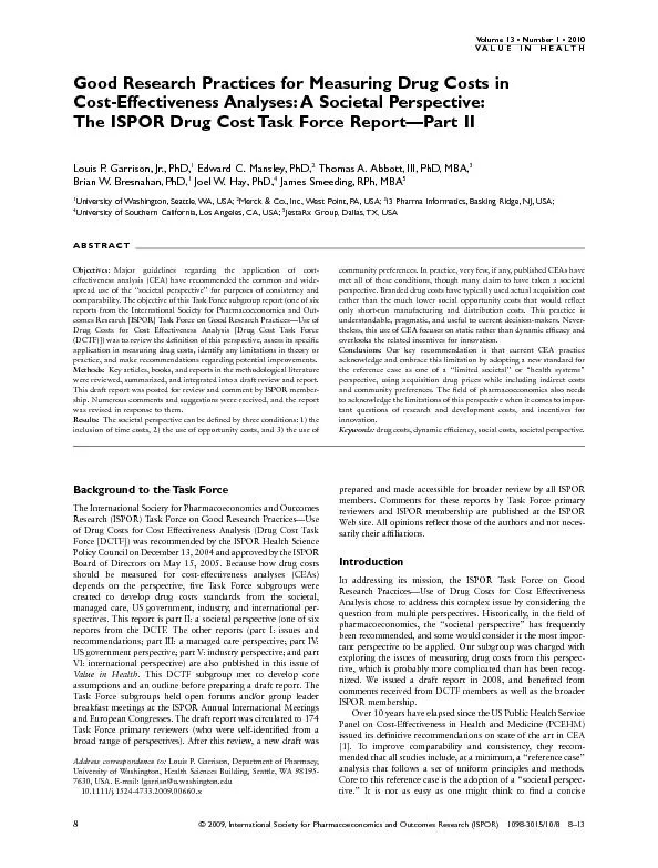
/
View Wesley E Shankland DDS MS PhD The Journal of Craniomand View Wesley E Shankland DDS MS PhD The Journal of Craniomand

View Wesley E Shankland DDS MS PhD The Journal of Craniomand - PDF document

isabella2
isabella2 . @isabella2
Follow
342 views
Uploaded On 2022-08-25

View Wesley E Shankland DDS MS PhD The Journal of Craniomand - PPT Presentation

This fourpart series of articles will serve as indepth study guides and sources of information concerning the trigeminal nerve The first article is an overview each of the additional three artic ID: 941658

trigeminal nerve ganglion branches nerve trigeminal branches ganglion branch posterior superior anterior fibers nerves inferior division sensory maxillary lateral

Share:

Link:

Embed:

Download Presentation from below link

Download Pdf The PPT/PDF document "View Wesley E Shankland DDS MS PhD The J..." is the property of its rightful owner. Permission is granted to download and print the materials on this web site for personal, non-commercial use only, and to display it on your personal computer provided you do not modify the materials and that you retain all copyright notices contained in the materials. By downloading content from our website, you accept the terms of this agreement.


Presentation Transcript

View Wesley E. Shankland, D.D.S., M.S., PhD. The Journal of Craniomandibular Practice October 2000, VOL. 18, NO. 4 ABSTRACT: The trigeminal nerve is the largest and most complex of twelve cranial nerves. Its vas sizand influence are greatly appreciated when one attempts to diagnose and treat patients suffering from orofacial pain and temporomandibular joint disorders. Without a thorough knowledge of the trigeminal nerve, the efficacy of diagnostic and therapeutic procedures will be very disappointing. This is the first of a four-part series of articles about the trigeminal nerve, a basic over-view of both the gross and neuroanatomical structures is presented. As practitioners from various backgrounds and specialties, we are all charged with evaluation patients suffering with orofacial, temporomandibular joint, head and neck pain. The finest service we can offer our patients is to discover an accurate diagnosis or diagnoses, as the case may be. Due to the great anatomical complexity of the head and neck regions, diagnostic procedures are often difficult at best. This four-part series of articles will serve as in-depth study guides and sources of information concerning the trigeminal nerve. The first article is an over-view; each of the additional three articles describes and discusses the individual divisions of the trigeminal nerve. Cranial Origin of the Trigeminal Nerve The trigeminal, or fifth cranial nerve, is comprised of three major divisions: the ophthalmic, the maxillary, and the mandibular. The largest cranial nerve is the great afferent nerve of the face, of the mucous membranes of the head, the afferent nerve (The term afferent, Òto bring to,Ó is interchanged with the term sensory in this article.) of internal cranial structures, the afferent nerve of the teeth and temporomandibular joints, and efferent (motor) nerve of the first branchial arch (Table 1). The trigeminal nerve has its origin at the lateral border of the pons through two roots, the sensory Anton B. R. Hirsch in 1765 in admiration of his mentor and professor, Johann Ludwig Gasser.2 This size of the cavernous area which contains the Gasserian ganglion varies from person to person and even one side to the other in t

he same person3. The mass of this ganglion is basically flat, semilunar in shape, and measures approximately 1 x 2 centimeters4. The central cellular processes (i.e., the portio major) exit the concavity of the ganglion, passing under the superior petrosal sinus of the temporal bone. The peripheral fivers leave the convexity of the crescent of the ganglion as the three divisions of the trigeminal nerve. The internal carotid artery and the posterior portion of the cavernous sinus are positioned on the medical aspect of the ganglion, from which sympathetic innervation is provided to the ganglion. The greater superficial petrosal nerve is situated between the ganglion and the petrous portion of the temporal bone. This ganglion is unique in that it is the only site in which primary afferent neuron cell bodies are derived from neural crest cells during development. In addition, it is the largest collection of cell bodies outside of the central nervous system in the body (Figure 1). The cells of the trigeminal ganglion are pseudo-unipolar, this making this structure analogous to the dorsal root ganglion of the spinal cord. Theses neurons are somatotopically (Somatotopic organization: an orderly representation of the various body parts within central nervous system structures that process sensory information such that neighborhood relation in the periphery are preserved in the central nervous system. This organizational representation in the central nervous system is maintained in the spinal cord and throughout the various areas of the brain.) organized from medial to lateral. Each neuron in the ganglion has two branches or processes. The peripheral processes of these ganglion cells form, from medial to lateral, the ophthalmic, maxillary, and mandibular divisions of the trigeminal nerve. The other branches of these ganglion neurons, the central processes, form the large sensory root which courses to the lateral border of the pons, through a groove in the petrous portion of the temporal bone, entering the pons and dividing into upper and lower roots. The upper roots synapse in the principle (chief sensory, main sensory, or pontine) nucleus positioned lateral in the pons. The lower roots descend a

s the spinal trigeminal root and become continuous with the substantia gelatinosa of Rolando of the spinal cord. Blood supply to the Gasserian ganglion is delivered from branches of the cavernous portion of the internal carotid artery as this vascular structure passes medially and superiorly to the ganglion through the carotid canal in the temporal bone. Table 1 Components of the Trigeminal Nerve _______________________________________________________________________________ Component Function ________________________________________________________________________________ Special visceral efferent (Branchial motor) To muscles of mastication, (Autonomics) tensor tympani and tensor veli palatine muscles,zygomandibularis muscle, mylohoid muscle, and the anterior belly of the digastric muscle. General somatic afferent From the face, scalp, conjunctiva, bulbus of eye; portion of the mandibular branchial arch; similarly, the mandibular nerve supplies superficial and deep structures which were derived from the ventral portion of the mandibular branchial arch). However, all three divisions have distributions of their branches corresponding to facial structures that develop from one of three fetal structures; namely, the frontonasal (ophthalmic nerve distribution), maxillary portion of the first branchial arch ( maxillary nerve distribution), and the mandibular portion of t horseradish peroxidase tracer techniques15 have demonstrated that a significant number of both ophthalmic and maxillary axons are responsible for innervation of the cornea. The dental pulp receptors of the trigeminal nerve are also unique. Both small myelinated (A-delta fibers) and unmyelinated fibers ( C fibers) provided afferent innervation to the pulps of the teeth. Until recently, it was thought that these fibers carried only pain information concerning dental diseases. diameter in response to pulpal inflammation as well as immunoregulatory functions, rather than simply conveying pain transmission information. Motor Portion of the Trigeminal Nerve (Portico Minor) Determined by embryological development, the motor fibers (portico minor) of the trigeminal nerve supply all the muscles of

the first branchial arch, usually termed the muscles of mastication (Table 2). This category of muscles also includes the tensor veli palatine, the tensor tympani, the anterior belly of the digastric, the mylohyoid, and the zygomandibularis, a newly discovered muscle of mastication.17 Traditionally it has been taught that the anterior motor root is comprised of six to eight rounded filaments of Vulpian and emerges from the pons slightly superior to the larger posterior sensory root. However, this multistranded This inferior root is also termed the intermediate root by some authors. The exact function of this intermediate root or inferior motor root is unknown and there is no actual anatomical evidence for the specific functions motor or sensory. Yet, Pelleteir and colleagues19 contend that the physiological characteristics of this intermediate root were identical to that of the portico minor fibers and subserved either a motor or proprioceptive function father than a sensory function. Young contends that the fibers of the portico intermedia or inferior motor rootlet simply are randomly displaced fibers which provide accessory pathways into or out of the pons or cross communications between the portico major and minor for either sensory or motor fiber. Obviously, further research is needed to determine the actual physiological function of this inferior portico intermedia. Table 2 Muscles of Mastication and Associated Innervations __________________________________________________________________________________ Muscle Nerve branch of motor division V3 __________________________________________________________________________________ Temporalis Posterior deep temporal Ð usually two (from Anterior division V3) Zygomandibularis Superior belly of lateral pterygoid Lateral pterygoid (from anterior division of V3) Inferior belly of lateral pterygoid Lateral pterygoid (from anterior division of V3) Medial pterygoidMedial pterygoid (from main trunk of V3) Tensor veli palatine* Branch of the medial pterygoid nerve Tensor tympani* Branch of the medial pterygoid nerve Mylohyoid* Mylohyoid nerve (from posterior of division V3) Anterior belly of digastricMylohyoid nerv

e ____________________________________________________________________________________ * Although not true muscles of mastication, these muscles are traditionally included with the masticatory muscles due to the common innervation of branches of V3 In addition to motor fibers of the third division of the trigeminal nerve, proprioceptive fibers, which originate in the mesencephalic nucleus in the pons, accompany the motor root to be distributed throughout the third division20. Paradoxically, May and Horsley21 demonstrated early in this century that afferent fibers to the mesencephalic nucleus ran through the motor root as well. After the motor and mandibular sensory divisions of the trigeminal nerve exit the middle cranial fossa through the foramen ovale, they unite to form the mandibular trunk which further divides into anterior and posterior divisions to provide innervation to specific structures which will be discussed in the fourth of this series of articles (The Mandibular Division). This union of the motor and sensory nerves forms a mixed nerve in contrast to the ophthalmic and maxillary which are purely sensory. Autonomics of the Trigeminal Nerve The autonomic ganglia associated with the trigeminal nerve belong to the general systemic autonomic nervous system. Although totally separate and independent from the central and peripheral nervous systems, the autonomics are intimately affiliated with them by communicating branches. In the head, the smooth muscles of the orbit, the lacrimal and salivary glands, the ciliary body of the eye, and secretory cells all come under the dual control of the sympathetic and parasympathetic divisions of the autonomic nervous system. Traditionally, all nerves which entered one of the ganglia of the head were called roots of the ganglion. However, since only true roots of a ganglion are nerves which synapse on cells of a ganglion, only parasympathetic fibers are true roots. Sympathetic roots pass t This internal carotid plexus sends filament to several cranial nerves. In the carotid canal two nerves, the caroticotympanic nerves, arise and enter the tympanic cavity to form the tympanic plexus. In the cavernous sinus, the deep petrosal nerve originates. T

his nerve joins the parasympathetic greater petrosal nerve (from the facial nerve) in the pterygoid canal of the sphenoid bone to form the pterygoid or Vidian nerve which continues to the sphenopalatine ganglion. The sympathetic fibers pass through the These fine filaments from the internal carotid plexus supply postganglionic fibers through the ciliary. A plexus associated with the maxillary and middle meningeal arteries provides postganglionic The Sphenopalatine Ganglion (pterygopalatine; ganglion of Meckel) The largest of the parasympathetic ganglia of the trigeminal nerve, the sphenopalatine ganglion is placed deeply within the pterygopalatine fossa. It is a triangular or heart-shaped, reddish-gray collection of cells located below the maxillary nerve (V2) after the nerve enters the pterygopalatine fossa through the foramen rotundum. The nerve fibers which synapse in the sphenopalatine ganglion are derived from the superior salivary nucleus in the pons and leave the pons with the facial nerve as two nerves; namely, the chorda tympani and the greater superficial petrosal. After coursing through the middle cranial fossa, exiting through the foramen lacerum, the greater superficial petrosal is joined by the deep petrosal ( from the internal carotid plexus) in the pterygoid canal to form the Vidian nerve. This mixed nerve leaves the pterygoid canal and enters the sphenopalatine ganglion. Here, the sympathetic fibers pass through the ganglion and terminate in the glands and blood vessels of the palate, nasal fossa and pharynx. The parasympathetic fibers of the greater superficial petrosal nerve synapse on ganglion cell bodies. From the sphenopalatine ganglion the parasympathetic fibers leave through the zygomatic, nasal branches, and pharyngeal nerve to innervate the lacrimal gland, the glands of the nasal fossa, and pharynx. In addition, The Submandibular Ganglion The chorda tympani branch of the seventh cranial nerve, after coursing through the temporal bone and middle ear, emerges through the petro-tympanic fissure of the temporal bone and descends into the infratemporal fossa to join the lingual branch of the mandibular division of the trigeminal nerve. With the lingual nerve, the chor

da tympani travels to the submandibular ganglion and the parasympathetic fibers synapse. From the ganglion, some fine filaments are provided to the submandibular gland and others join the lingual nerve to travel to the sublingual gland and ultimately, to the lingual glands, or taste buds, of the tongue. Sympathetic fibers from the plexus around the facial artery pass through the submandibular gland to reach the submandibular and lingual glands. The Otic Ganglion The Otic Ganglion (ganglion oticum or ArnoldÕs ganglion25) is a very small, oval-shaped , flat and reddish-gray cluster of cells situated immediately below the foramen ovale and associated with the third division of the trigeminal nerve. The ganglion lies on the medial surface of the mandibular nerveÕs main trunk near the origin of the medial pterygoid and tensor veli palatini nerves. The fibers of the otic ganglion originate in the inferior salivary nucleus of the glossopharyngeal or ninth cranial nerve in the medulla oblongata. These parasympathetic fibers leave the brain stem in the glossopharyngeal nerve and enter the tympanic branch, which passes through a small canal in the occipital bone between the jugular foramen and the carotid canal to reach the tympanic cavity, passes through the tympanic plexus, and emerges through the roof of the tympanic cavity of the temporal bone as the lesser superficial petrosal nerve. After a few sympathetic fibers from the facial nerve join the lesser superficial petrosal, the nerve proceeds to the otic ganglion. These sympathetic fibers are really derived from the plexus of the middle meningeal artery and do not synapse in the otic ganglion. The parasympathetic fibers, after synapsing on cells in the otic ganglion, have a wide distribution through the mandibular division of the trigeminal nerve; specifically, trigeminal motor fibers to the medial pterygoid and tensor tympani and tensor veli palatini nerves. But the real contribution of the glossopharyngeal nerve is through the auriculotemporal nerve to provide secretomotor fibers to the parotid gland. Some of the sympathetic fibers which traveled with the lesser superficial petrosal terminate also in the parotid gland. Table 3 Parasympath

etic Ganglia ______________________________________________________________________________________ GangliaCN*Cell Origins Specific nerve Trigeminal division Target organ ______________________________________________________________________________________ Ciliary III Motor root from V1 Ciliary muscles Edinger-Westphal Nucleus of CN III Sphenopalatine VII Superficial petrosal V2 a e sensory and one motor) and is located lateral and slightly inferior to the motor nucleus in the mid-portion of the pons. It is considered to be analogous to the posterior dorsal column of the spinal cord and being so, the function of the principle nucleus is to process all types of somatosensory information. This large collection of neurons receives large fiber, heavily myelinated afferents. The nucleus is specifically located in the dorsal Ðlateral area of the pontine tegmentum at the level of entry of the afferent fibers into the pons. It is continuous with the inferiorly placed spinal nucleus. The principle nucleus is composed of two sub-nuclei: 1. a ventrolateral; and 2. dorsomedial subnucleus. The ventrolateral subnucleus extends from the level of the motor nucleus inferiorly to the subnucleus oralis of the spinal trigeminal tract. The dorsomedial extends more caudally (inferiorly) than its ventrolateral counterpart.27 The ventrolateral subnucleus is primarily composed of uniformly medium-size cells with small multipolar cells scattered between the larger cells. the trigeminal nerve superiorly to the uppermost area of the midbrain. The neurons resemble those of the dorsal root ganglion cells of the spinal cord. The central processes from the mesencephalic tract of the trigeminal nerve which descends to the level of the motor nucleus of the trigeminal nerve. There is recent evidence,31 at least in the cat, that some of these neuronal cells are bipolar or multipolar with one to nine dendrites, making these cells distinctly different from those of the Gasserian ganglion. Like the principle nucleus, the mesencephalic is composed of two regions. The majority of the pseudounipolar neurons are located in the inferior or caudal portion of the nucleus whose perip

heral processes primarily innervate the periodontal ligament fibers. These fibers may also innervate the teeth32,33 and the ocular muscles.32 The multipolar cells are mostly located in the superior or rostral portion of the mesencephalic nucleus. According to Hinrichsen and Larramendi,34 the heterogenous organization may reflect the dual embryologic origin of the mesencephalic neurons, some from neural crests cells and others from the alar plate. The peripheral fibers of the mesencephalic nucleus are distributed through the peripheral processes of the three divisions of the trigeminal nerve. Some central fibers terminate in both the motor nucleus and the principle nucleus. Some collateral fibers give off to the reticular formation and on the thalamus through the trigeminal lemnisci. Also, the mesencephalic nerve send fibers to the cerebellum through the superior cerebellar peduncle. The Spinal Trigeminal Tract Nucleus The third sensory nucleus, termed the spinal trigeminal tract nucleus, is perhaps the most influential systemically of all three and is actually composed of three subnuclei. This complex extends from the principle nucleus inferiorly to merge with the substantia gelatinosa of the cervical spinal cord. Therefore, this trigeminal nuclear complex is actually located not in the pons only, but within the medulla and spinal cord, too. Since at lease 191838 anatomists have taught that the spinal trigeminal tract descended caudally to the level of the first or second cervical nerve. However, recently several researchers have demonstrated in the cat that the spinal trigeminal tract conveys fibers through the substantia gelatinosa of the spinal cord as far caudally as T-9 and in some cases as far as L-3, or the very end of the spinal cord. There are definite histological differences among these three subnuclei; they are distinctly separate and each processes different types of information. Afferent fibers of all three divisions of the trigeminal nerve enter the pons in a distinct Subnucleus Oralis The subnucleus oralis (pars oralis) of the spinal trigeminal tract is the most superior of the three subnuclei and itself is subdivided into three subdivisions. It extends from the

caudal pole of the motor nucleus of the trigeminal nerve (see below) inferiorly to the rostral or superior pole of the nucleus of the facial nerve. The most rostral division is interdigitated with the ventrolateral subnucleus of the principle trigeminal nucleus, lying medial to this sub nucleus. The area of these two subnuclei which overlap is termed either the oralis gamma subdivision 29 or the rostrodorsomedial subdivision. 27 Characteristic to the subnucleus oralis is the vast numbers of multipolar neurons of various sizes. Subnucleus Interpolaris Situated between the subnucleus oralis rostrally and subnucleus caudalis caudally is the subnucleus interpolaris. This second of the three subdivisions of the spinal trigeminal nucleus extends rostrally from the rostral pole of the hypoglossal nucleus caudally to the obex (The obex is a ÒVÓ shaped area in the midline which is the caudal boundary of the fourth ventricle in the medulla oblongata.). There appears to be an equal mixture of predominately small and medium-sized neurons in the subnucleus interpolaris, 75% of which are low threshold mechanoreceptors, although all classes of neurons 5. Dodd J, Kelly JP: The trigeminal system. In; Kandel ER, Schwartz JH, Jessel TM, eds. Principles of neural sciences. 3rd ed. 1991. 6. Young RF: The trigeminal nerve and its central pathways. In Rovit RL, Murali R, Jannetta PJ, eds. Trigeminal neuralgia, 1990:27-51. 7. Moore KL: The developing human, 4th ed. Philadelphia: WB Saunders co. 1988: 396-416. 8. Romer AS: The vertebrate body. 4th ed. Philadelphia: WB Saunders Co, 1971: 365-366. 9. Sarnat HB, Netsky MG: Evolution of the nervous system. London: Oxford University Press, 1974:7710. Weichert CK: Anatomy of the chordates. New York: McGraw-Hill Book Co. 1971:644-645. 11. Beck F, Moffit DB, Davies DP: Human embryology ed. Oxford: Blackwell Scientific Publications, 1985:125. 12. Brash JC, Jamieson EB eds.: CunninghamÕs textbook of anatomy. 8th ed. London: Oxford University Press, 1947:982. 13. Romanes GJ. Ed: CunninghamÕs textbook of anatomy. 11th ed. London: Oxford University Press, 1972. 14. Tervo T., Joo R, Huikuri KT, Toth I, Palkama A: Fine structure of sensory nerves in the rat cornea: an e

xperimental nerve degeneration study. Pain 1979; 6:57.70 15. Morgan C, Jannetta PJ, de Groat WC: Organization of corneal afferent axons in the trigeminal nerve root entry zone in the cat. Exp Brain Res 1987; 68: 411-416. 16. Silverman JD, Kruger L: An interpretation of dental innervation based upon the patterns of calcitonin gene-related peptide (CGRP)- immunoreative thin sensory axons. Somatosensory Res 1987; 5:157-175. 17. Shankland WE, Negulesco JA, OÕBrian B: The Òpre-anterior bellyÓ of the temporalis muscle: a preliminary study of a newly described structure. J Craniomandib Pract 1996: 14: 106-112. 18. Saunders RL, Sachs E: Relation of the accessory rootlet of the trigeminal nerve to its motor root. J Neurosurg 1970; 33: 317-324. 19. Pelleteir VA, Poulos DA, Lende RA: Functional localization in the trigeminal root. J Neurosurg 1974; 40:504-513. 20. Corbin KB, Harrison R: Function of mesencephalic root of the 5th cranial nerve. 22. Barr ML, Kierman JA: The human nervous system. 5th ed. Philadelphia: JB Lippincott, 1988: 353-358 23.DuBrul L: SicherÕs oral anatomy. 8th ed. St. Louis: CV Mosby Co., 1988:246. 24. DuBrul L: SicherÕs oral anatomy. 8th ed. St. Louis: CV Mosby Co., 1988:1100. 25. Gray H: Anatomy, descriptive and surgical. 2 30. Johnson LR, Westrum LE: Structure of the dorsomedial subdivision of the feline trigeminal main sensory nucleus (abstract). Anat Rec 1981; 199:130A.31. Walberg F: On the morphology of the mesencephalic trigeminal cells. New data based on tracer studies. 32. Corbin KB, Harrison R: Function of mesencephalic root of the 5th cranial nerve. Hinrichsen CFL, Larramendi LMH: Features of trigeminal mesencephalic nucleus structure and organization. 1. Light microscopy. Am J Anat 1969; 126:497-506. 35. Hinrichsen CFL, Larramendi LMH: The trigeminal nucleus, II. Electron microscopy. Am J Anat 1970; 127:303-320. 36. Lucchi ML, Bortolami R, Callegar E: Ultrastructural features of mesencephalic trigeminal nucleus in cat, rabbit ad pig. Submicro Cytol 1972; 4:7-18. 37. Hinrichsen CFL: Electronic coupling between cells in the mesencephalic nucleus. In : Andersen DJ, Matthews B, eds. Mastication. Bristol: Wright, 1976. 38. Robinson R, ed.: Cunni

nghamÕs textbook of anatomy. 5th ed. New York: Wm Wood and Co., 1918. 39.Matsushita M. Okado N, Ikeda M, Hosoya Y: Descending projection from the spinal and mesencephalic nuclei of the trigeminal nerve to the spi trigeminal region. Amsterdam: Elserier/North-Holand Biomedical Press, 1977: 443-453. 48. Johnson LR, Westrum LE, Henry MA: Anatomic organization of the trigeminal system and the effects of deafferentation. In The Journal of Craniomandibular PracticeJanuary 2001, VOL. 19, NO. 1 ABSTRACT: The ophthalmic, or first division (V1) of the trigeminal nerve, is the smallest of the three divisions and is purely sensory or afferent I function. It supplies sensory branches to the ciliary body, the cornea, and the iris; to the lacrimal gland and conjunctiva; to portions of the mucous membrane of the nasal cavity, sphenoidal sinus, and frontal sinus; to the skin of the eyebrow, eyelids, forehead, and nose; and to the tentorium cerebelli, dura mater, and the posterior area of the falx cerebri, At first glance, one might not expect one interested in the diagnosis and treatment of orofacial pain and temporomandibular joint disorders to have a need to be concerned with the ophthalmic division. Although much of this divisionÕs influence is dedicated to structures within the orbit, nose, and cranium, still, the ophthalmic division may be afflicted with a lesion or structural disorder which can cause all sorts of orofacial pain. Ignorance of this or any portion of the trigeminal nerve will lead to diagnostic and therapeutic failures. In this, the second of four (4) articles concerning the trigeminal nerve, the first division of this vast cranial nerve will be described in detail. The ophthalmic, or first division (V1) of the trigeminal nerve is the smallest of the three and is purely sensory or afferent in function (Figure 1). It supplies sensory branches of the ciliary body, the cornea, and the iris; to the lacrimal gland and conjunctiva; to portions of the mucous membrane of the nasal cavity, sphenoidal sinus, and frontal sinus; to the skin of the eyebrow, eyelids, forehead, and nose; and to the tentorium cerebelli, dura mater, and the posterior area of the falx cerebri (Table 1). Never leaving the

protection of the cranium, the ophthalmic nerve exits the Gasserian g The Lacrimal Nerve The lacrimal nerve (n. lacrimalis) is the smallest of the three divisions of the ophthalmic nerve, Traveling forward in a separate tube of dura mater after leaving the ophthalmic nerve just prior to the superior orbital fissure of the sphenoid bone, the lacrimal nerve enters the orbit through the narrowest portion of the superior orbital fissure and travels above and parallel to the lateral rectus muscle and receives a communicating branch from the maxillary or second division of the trigeminal nerve, which provides postganglionic parasympathetic motor fibers for the lacrimal gland. According to Goss,4 These parasympathetic fibers, carried by the zygomaticotemporal nerve, have their cells bodies of origin in the sphenopalatine ganglion. They travel from the sphenopalatine ganglion through the zygomatic nerve, which divides into the zygomaticofacial and zygomaticotemporal nerves, thus bringing secretomotor fibers to the lacrimal gland, Parasympathetic and sympathetic innervation are also provided to the lacrimal gland by the mixed greater superficial petrosal nerve, a branch of the seventh cranial nerve.5 Further, additional sympathetic fibers, also from the carotid plexus, after running with the sixth cranial or trochlear nerve, are given to the lacrimal gland.5 As the lacrimal nerve enters the lacrimal gland with the lacrimal artery, it also gives off branches The Nasociliary Nerve The nasociliary nerve (nasal nerve; n. nasociliaris) is intermediate in size when compared to the lacrimal and the frontal nerves. It runs deeper within the orbital contents than the former two nerves. The nasociliary enters the orbital cavity through the superior orbital fissure of the sphenoid bone between the two heads of the rectus lateralis muscle, between the superior and inferior rami of the oculomotor nerve, and with the abducent nerve (cranial nerve VI) within the tendinous ring from which the four rectus muscles (superior rectus; inferior rectus; medial rectus; and lateral rectus) take their origin to lie within the cone formed b the extrinsic ocular muscles.7 From there, the nasociliary curves anteromedially an

d travels superior to the ophthalmic artery and the optic nerve to leave the muscular cone, travels between the 2. The lateral branch: and 3. The anterior or superficial branch. The internal or septal branch of the ethmoidal nerve travels inferiorly and anteriorly , supplying the anterior portion of the nasal septum of the nose with afferent fibers. The lateral branch of the ethmoidal nerve actually is comprised of two or three filaments which are distributed to the anterior portions of the lateral walls of the nasal fossa, which includes the middle and inferior conchae. 1 The anterior or superficial branch of the ethmoidal nerve is the largest of the three branches and is actually considered the continuation of the ethmoidal nerve. It passes inferiorly in a longitudinal canal in the nasal bone until the bone reaches the nasal cartilage where the canal ends. From there, the anterior branch travels under the nasalis muscle and terminates in the wing, spine, and tip of the nose1 and is named the external nasal nerve. Table 1 Branches of the Ophthalmic Division of the Trigeminal Nerve Division Branches Area of distribution ____________________________________________________________________________________ Frontal nerve Supraorbital, medial branch Forehead, medial; conjunctiva; skin of upper eyelid; frontal sinusSupraorbital lateral Forehead, later; scalp Supratrochlear Medial upper eyelid; forehead Nasociliary nerve Long Ciliary nn*Sclera Long root of the ciliary ganglion Ciliary ganglionCiliary nerves to sclera Sclera Anterior ethmoid * nn: nerve Prior to entering the anterior ethmoidal foramen and subsequently becoming the anterior ethmoid nerve, the nasociliary nerve gives off at least four (4) branches: Viz, 1. The long root of the ciliary ganglion;2. The long ciliary; 3. The infratrochlear; and 4. The posterior ethmoid. The long root of the ciliary ganglion (radix longa ganglii ciliaris) generally originates from the nasociliary nerve between the two heads of the lateral rectus muscle.3 This long but slender nerve travels forward on the lateral side of the optic nerve and enters the ciliary ganglion but does not synapse with any

cell bodies. This sensory nerve, after leaving the ciliary ganglion, divides into several fine filaments and supplies the bulb of the eye. These fine filaments are known as the short ciliary nerves or ciliary nerves to the sclera. Most likely, the long root of the ciliary ganglion is joined in the ciliary ganglion by a filament from the internal carotid plexus, which, the later being derived form the superior cervical ganglion, provides sympathetic innervation. The long ciliary nerves (nn. ciliares Frequently the nasociliary gives off small branches to the superior ad inferior recti muscles.1 Also, a branch to the levator palpebrae superioris has been reported. The posterior ethmoidal nerve (n. posterior ethmoidalis; the sphenoethmoidal) comes off the nasociliary nerve in the anterior medial corner of the eye and enters the posterior ethmoidal foramen. From there, the nerve provides afferent innervation to the posterior ethmoidal air cells, the mucous membranes of the sphenoidal sinuses, and the dura mater. The Frontal Nerve The frontal nerve (n. frontalis) is the largest division or branch of the ophthalmic nerve. It should be regarded as the continuation of the ophthalmic past its entrance to the orbit through the superior orbital fissure. From this large fissure in the sphenoid bone, the nerve runs forward between the levator palpebrae superioris muscle and the periosteum of the sphenoid bone. The frontal then divides into two branches, the larger supraorbital and smaller supratrochlear nerves. The supraorbital nerve continues anteriorly through the orbit and exits through the supraorbi Cryer The supraorbital nerve also innervates the upper eyelid, the mucous membrane of the frontal sinus of the frontal bone, the galea aponeurosis (Latin: a leather helmet; the fascial tissue which extends between the frontalis and occipitalis muscles of the skull.), and the orbicularis oculi. In addition, these branches anastomose with the temporal branches of the facial, or seventh, cranial nerve as well as send small twigs to the pericranium (the periosteum of the skull) to innervate the frontal and parietal bones. The supratrochlear nerve (n. supratrochlearis) branch is the smaller of the t

wo terminal branches of the frontal nerve. It travels anterior and medially, over the trochlear muscle to the pulley of the superior oblique muscle and gives off a descending filament or branch which anastomoses with the infratrochlear nerve branch of the nasociliary nerve. The supratrochlear nerve then turns superiorly on the medial portion of the supraorbital margin of the frontal bone and supplies sensory innervation to the medial portion of the upper eyelid, the conjunctiva, and substance of the forehead. In the next article, the larger and very influential maxillary nerve or second division of the trigeminal will be presented in detail. References 1. Cryer MJ, The Internal Anatomy of the Face. 2nd ed. Philadelphia: Lea & Febiger, 1916. 2. Williams PL, Warwick R, Dyson M, Bannister LH, eds. Grays Anatomy. 37th ed. Edinburgh: Churchill Livingstone, 1989. 3. Sutherland S, Hughes ERS: The pupilloconstrictor pathway and nerves to the ocular muscles in main. Brain 1946; 69:301. 4. Goss CM (ed). Grays Anatomy. 26th ed. Philadelphia: Lea & Febiger, 1954. 5. Sachs E: The role of the nervus intermedius in facial neuralgia. J Neurosurg 1968; 28:54-60. 6. Parkinson D, Johnson J, Chaudhuri A: Sympathetic connections to the fifth and sixth crania Wesley E. Shankland, II, D.D.S., M.S., PhD. The Journal of Craniomandibular Practice January 2001, VOL. 19, NO. 2 ABSTRACT: The maxillary nerve gives sensory innervation to all structures in and around the maxillary bone and the midfacial region including the skin of the midfacial regions, the lower eyelid, side of nose, and upper lip; the mucous membrane of the nasopharynx, maxillary sinus, soft palate, palatine tonsil, roof of the mouth, the maxillary gingivae, and maxillary teeth. This vast and complex division of the trigeminal nerve is intimately associated with many sources of orofacial pain, often mimicking maxillary sinus and /or temporomandibular disorders, knowledge of this nerve must be second nature. Just providing the difficult services of a general dental practice should be stimulus enough to understand this trigeminal division, but if one hopes to correctly diagnose and treat orofacial pain disorders, dedication to understanding this

nerve cannot be overstated. In this, the third of a four part series of articles concerning the trigeminal nerve, the second or maxillary division will be described and discussed in detail. The maxillary nerve (n. maxillaries; superior maxillary nerve), or second division of the trigeminal nerve, is intermediate in size in comparison to smaller ophthalmic and largest mandibular divisions. Designated as V2, this large nerve with multiple branches, like the ophthalmic nerve (V1), is purely sensory in function. The maxillary nerve gives sensory innervation to all structures in and around the maxillary bone and the midfacial region including the skin of the midfacial regions, and lower eyelid, side of nose and upper lip; the mucous membrane of the nasopharynx, maxillary sinus, soft palate, palatine Table 1 Branches of the V2 ___________________________________________________________________________________ Origin of branchNerve or branch ___________________________________________________________________________________ In the cranium Middle meningeal Zygomatic Orbital Orbital branches Pharyngeal Posterior palatine Middle palatine Anterior palatine (greater palatine) Lateral posterior superior nasal branches Medial posterior superior nasal branches Posterior Inferior nasal branches Nasopalatine ZygomandibularisPosterior superior alveolar In the infraorbital Middle Superior alveolar Canal Anterior superior alveolar On the face Inferior palpebral Lateral nasal Superior labial ______________________________________________________________________________________ The ganglionic branches. After the maxillary nerve exits the middle cranial fossa through the foramen rotundum of the sphenoid bone and enters the pterygopalatine fossa, two nerves, providing sensory innervation to the sphenopalatine ganglion, These two small nerves thereafter branch themselves. A few of their branches provide sensory innervation to the ganglion itself, with other branches passing directly to the palatine nerves. The zygomatic nerve. The zygomatic nerve (n. zygomaticus; orbital nerve; temporomalar nerve)

arises from the maxillary nerve in the pterygopalatine fossa and travels anteriorly and superiorly to enter the orbit through the inferior orbital fissure. After entering the orbit, the zygomatic nerve divides in two branches: a. the zygomaticotemporal and b. zygomaticofacial nerves. The zygomaticotemporal nerve (ramus zygomaticotemparalis; temporal branch) travels along the infer-lateral angle of the orbit in a groove in the zygomatic or malar bone. It supplies a communicating branch to the lacrimal nerve of the ophthalmic nerve (V1), or first division of the trigeminal nerve. At times, when the lacrimal nerve is lacking, the zygomaticotemporal becomes its replacement and innervates the lacrimal gland. Exiting the zygomatic bone through the zygomaticotemporal foramen, the nerve travels within the substance of the temporalis muscle superior to the zygomatic arch, ascends to the surface of the muscle and pierces the temporal fascia, and branches to areas of the skin in the lateral forehead and anterior temporal areas of the skull. The zygomaticotemporal nerve produces a small branch which runs in fascia to the lateral corner of the eye and provides a communicating branch to the auriculotemporal nerve, a major branch of the mandibular division (V3) of the trigeminal. Lastly, this nerve also communicates with the facial nerve. The zygomaticofacial nerve (ramus zygomaticofacialis; malar nerve) is the second branch of the zygomatic nerve. This sensory nerve passes along the inferior and lateral edge of the orbit and appears on the face as two branches after passing through the zygomaticofacial foramen of the zygomatic portion of the zygomatic arch, anterior to the zygomaticotemporal foramen. It perforates the orbicularis oculi muscle After leaving the main trunk the maxillary nerve in the pterygopalatine fossa, the orbital nerve arises anteriorly and superiorly, passes through the inferior orbital fissure, passes through the ciliary ganglion without synapsing, and pierces the globe of the eye. The orbital branches Different from the orbital nerve, there are two or three small rami which originate from the sphenopalatine ganglion, pass through the inferior orbital fissure, and provide sympath

etic innervation to the orbitalis muscle and orbital periosteum; the sympathetic filament being supplied by the internal carotid plexus. The pharyngeal nerve (pterygopalatine nerve). The pharyngeal nerve is a small maxillary branch arising from the posterior portion of the sphenopalatine ganglion. Being purely afferent, this small filament travels with the pharyngeal branch of the maxillary artery in the pharyngeal canal (palatino-vaginal canal) in the roof of the nasal fossa, placed between the body of the sphenoid bone and the sphenoidal process of the palatine bone. It brings innervation to the mucous membrane of the pharynx and the sphenoidal sinus.2 The posterior palatine (n. palatinus posterior; least palatine nerve) nerve. The posterior palatine nerve descends posteriorly and inferiorly from the sphenopalatine ganglion through the pterygopalatine below the opening of the sphenoidal sinus to provide innervation to the mucosa of the posterior portion of the roof and the nasal septum.The posterior inferior nasal nerves. These nerves, which vary in number, arise from the anterior palatine nerve while the latter courses through the greater palatine canal. These branches travel through the perpendicular plate of the palatine bone through unnamed foramina to provide sensory innervation to the inferior conchae of the nose and the walls of the middle and inferior meatuses and to the soft palate via a palatine branch of this group.The nasopalatine nerve. The largest nerve emerging from the sphenopalatine ganglion, the nasopalatine nerve travels through the sphenopalatine foramen to enter the nasal fossa just below the orifice to the sphenoidal sinus to reach the nasal septum. From there, it runs in an oblique direction anteriorly and inferiorly between the periosteum and mucous membrane of the nasal septum. Continuing inferiorly and after anastomosing with the opposite nasopalatine, it exits the maxilla through the nasopalatine foramen (incisive foramen), posterior and between the maxillary central incisor teeth, and is thereafter called the incisive nerve. According to Williams, et al.1 at times both the posterior and anterior incisive foramina may exist in the nasal fossa, with the left nas

opalatine nerve leaving the fossa through the anterior foramen and the right nerve through the posterior foramen before uniting as the incisive nerve. The incisive nerve communicates with both anterior palatine (greater palatine) nerves to provide senso The pre-temporal or zygomandibularis nerve. Recently described by Shankland, et al.the pre-temporal nerve seems to arise from the sphenopalatine ganglion and travel anterior and laterally to innervate the zygomandibularis muscle, possibly conveying parasympathetic fibers and somatosensory innervation. The posterior superior alveolar nerves. Traditionally taught as just one nerve, this group of branches (rami alveolares superiors poateriores; superior alveolar nerve) arise from the maxillary nerve in the pterygogpalatine fossa just prior to the maxillaryÕs entrance to the inferior orbital fissure in the maxilla. Cryer3 reported the posterior superior nerve leaves the maxillary just after the former provides its two branches to the sphenopalatine ganglion. These nerves descend on the maxillary tuberosity to enter the posterior portion of the maxilla. This is true, but in addition, the present writer has noted on numerouoccasions during anatomical dissections that the posterior superior alveolar nerve generally enters the maxilla laterally and not posteriorly, just superior to the apices of the second and third molar teeth. Mercuri6 agreed with this finding and reported that the posterior superior alveolar nerve divided into an external gingival branch (corresponding to the lateral foramina observed) and an internal dental branch. This fact is demonstrated by examining dry human skulls and reviewing photographs like those demonstrated by White and Folkens.7 After entering the maxilla in numerous unnamed foramina, the posterior superior alveolar nerves travel forward under the mucosa of the maxillary sinus, supplying afferent innervation to these membranes. Continuing anteriorly, the posterior superior alveolar nerves communicate with the middle superior alveolar nerve and give branches which form a dental plexus and innervate the second and third molars as well as the palatal and distobuccal roots of the maxillary first molar. In addition, th

e posterior superior alveolar nerve provides innervation to the maxillary posterior gingivae and mucous membranes of the buccal mucosa. Nerves Originating in the Infraorbital Canal After the maxillary nerve enters the maxilla through the inferior orbital fissure and becomes the infraorbital, the latter travels through the medial and superior area of the maxillary sinus, traveling below the orbit to emerge on the face through the infraorbital foramen. In the maxillary bone, two main branches arise from the infraorbital. The middle superior alveolar nerve (ramus alveolaris medius; middle superior dental nerve) leaves the infraorbital nerve in the posterior portion of the infraorbital canal. This nerve descends downward and runs forward in a canal in the lateral wall of the maxillary sinus to produce three terminal branches which enter the mesiobuccal root of the first molar and the apices of each of the premolar teeth. The middle superior alveolar also anastomoses with the posterior superior alveolar. This nerve is not without controversy. Wood8 denied that there was a frequent occurrence of the middle superior alveolar nerve. But Fitzgerald9 disagreed, demonstrating that in his dissections, a middle superior alveolar nerve was present in 82% of his specimens and absent in only 18%, being replaced by the anterior superior alveolar branch. Yet, Loetscher and Walton (1988) reported that in such cases, the posterior superior alveolar supplied innervation when the middle branch was missing. The anterior superior alveolar nerve (ramus alveolaris superior anteriores; superior dental nerve) is the other branch which arises from the infraorbital in the infraorbital canal. This nerve, being larger than the middle superior alveolar, leaves the infraorbital just prior to the infraorbitalÕs exit through the infraorbital foramen. It descends inferiorly in a canal in the anterior wall of the maxillary sinus and divides into branches which innervate the central and lateral incisor teeth as well as the canine. After communication with the middle superior alveolar nerve, a nasal branch is give off which travels through a small, unnamed foramen in the lateral wall of the inferior meatus of the nasal foss

a, thus supplying sensory innervation to the mucous membrane of the anterior portion of the inferior meatus and floor of the nasal cavity. This nasal branch continues further to anastomose with the nasal branches of the sphenopalatine The inferior palpebral branches (rami palpebrales inferiors), which are usually two or three in number, ascend posterior to the orbicularis oculi muscle to supply afferent innervation to the skin and conjunctiva of the lower eyelid. These branches then communicate with the zygomatic branch of the facial nerve near the lateral canthus of the eye. A branch of the inferior palpebral nerve also anastomoses with the external nasal branch of the ophthalmic (V1) division of the trigeminal nerve. The lateral nasal nerve, usually two or three in number, supply the skin of the nose and the movable portion of the nasal septum and also join the external nasal branch of the ophthalmic nerve. In addition, the lateral nerve innervates the mucous membrane lining of the nostril and communicates with the nasociliary of the ophthalmic nerve. The superior labial nerve is the largest of the three branches, both in area of distribution and numbers of branches. After exiting the infraorbital foramen, the superior labial nerve descends inferiorly beneath the quadratus labia superioris muscle, divides into several branches, and provides afferent innervation to the upper lip and its skin, labial mucous glands, and the mucous membrane of the upper lip and buccal vestibule. These superior labial branches anastomose with the zygomatic branch of the facial nerve to form the infraorbital plexus. In the fourth and last article, the third division or mandibular nerve will be discussed in detail. References 1. Williams PL, Warwick R, Dyson M, Bannister LH, eds.: GrayÕs anatomy. 37th ed. Edinburgh: Churchill Livingstone, 1989. 2. Brash JC, Jamieson EB, eds. : CunninghamÕs textbook of anatomy. 8th ed. London: Oxford University Press, 1947. 3. Cryer MH: The internal anatomy of the face. 2nd ed. Philadelphia: Lea & Febiger, 1916. 4. Ruskell GL: Form of the choroidocapillaris. The Trigeminal Nerve. Part IV: The Mandibular Division Unlike muscles of the viscera, these muscles are voluntary

and striated.1 These visceral muscles, better referred to as branchial muscles, are innervated by cranial nerves; chiefly, the fifth, seventh, ninth, and tenth cranial nerves. However, only the fifth or trigeminal nerve also contains somatic sensory fibers, which also provide afferent processing of most of the afferent information for the latter three cranial nerves. The posterior extent of the infratemporal fossa is the articular tubercle of the temporal bone (including the temporomandibular joint), the carotid sheath, the styloid Nerve branch of motor division of V3 _________________________________________________________________________________________ Temporalis Posterior deep temporal Ðusually 2 (from anterior division of V3) Zygomandibularis Anterior deep temporal (from anterior division of V3) Masseter Masseteric (from anterior division of V3) Superior belly of lateral pterygoid Lateral pterygoid (from anterior division of V3) Inferior belly of lateral pterygoid Lateral pterygoid (from the anterior division of V3) Medial pterygoidMedial pterygoid (from main trunk of V3) Tensor veli palatine* Branch of the pterygoid n. Tensor tympani* Branch of the medial pterygoid n. Mylohyoid* Mylohyoid n. (from posterior division of V3) Anterior belly of digastric Mylohyoid n. _________________________________________________________________________________ *Although not considered muscles of mastication, these muscles are traditionally included with the masticatory muscles due to the common innervation of the branches of V3. Table 2 Branches of V3 ___________________________________________________________________________________________ Division NerveSensory Neurological component Prop ___________________________________________________________________motor___________________ Undivided trunk Meningeal x Medial pterygoidTensor veli palatine x x x Tensor tympani x x x Anterior division BuccalLateral pterygoid x x x Massetric x x x Anterior deep temporal x x x Posterior deep temporal x x x Posterior division AuriculotemporalLingual x x*** Inferior alveolar x

Mylohyoid x x ____________________________________________________________________________________________ * There are differences of opinions concerning motor fibers in the buccal nerve. Carries postganglionic parasympathetic fibers to the parotid gland from the glossopharyngeal nerve v The Undivided Trunk After the mandibular nerve, both sensory and motor portions, leaves the middle cranial fossa through the foramen ovale, just prior to the main trunk dividing into anterior and posterior divisions, four nerve branches leave the undivided trunk. The recurrent meningeal nerve. The first branch off the undivided trunk is the recurrent meningeal or nervous spinosus. This small nerve re-enters the cranium through the foramen spinosum with the middle meningeal artery from the maxillary artery. The nerve, bringing sensory innervation to the dura mater of the middle cranial fossa, divides into anterior and posterior divisions. The anterior branch communicates with the meningeal branch of the maxillary nerve and the posterior branch sends filaments to the mucous membrane of the mastoid air cells.2 The medial pterygoid nerve (n. pterygoideus medius; n. pterygoideus internus) exits the undivided trunk below the foramen ovale, travels through the otic ganglion with some synapsing, and provides sensory, motor, and proprioceptive innervation to the medical pterygoid muscle. The tensor veli palatine nerve. The nerve to the tensor veli palatine muscle is given off the undivided mandibular trunk and like the medial pterygoid nerve, travels through the otic ganglion to enter the tensor veli palatine near its origin at the base of the medial pterygoid plate of the sphenoid bone. This short nerve provides sensory and motor innervation to the muscle. The tensor tympani nerve may arise separately or in common with the tensor veli palatine nerve from the undivided trunk. Like the medial pterygoid and tensor veli palatine nerves, it too, passes through the otic ganglion and yet, does not synapse with any cell bodies. The nerve pierces the cartilage of the auditory canal and enters the tensor tympani muscle to supply motor and sensory innervation. At times the buccal nerve is the main trunk fo

r the anterior division. If this occurs, the buccal nerve gives off at least five branches as it courses form deep within the infratemporal fossa to its destination in the buccinator muscle: 1.0 two or three lateral pterygoid branches; 2. an anterior deep This makes sense academically. Even though the anterior deep temporal nerve is listed in many anatomy texts, its destined structure to innervate is not listed. The posterior deep temporal nerves (nn. Temporalis profundus posteriors) arise with their anterior mate and basically travel together with the anterior branch as well. The posterior deep temporal nerves, generally two or three in number, after arising to the level of the temporal fossa, enter the temporalis muscle on its deep surface about an inch above the zygomatic arch. From there, the nerves branch, course upward and somewhat posteriorly to communicate with the auriculotemporal and facial nerves. These nerves also give sensory branches to the medial portion of the temporomandibular joint. Like the anterior deep temporal, te posterior deep temporal nerves carry afferent, efferent, but mostly proprioceptive fibers according to Mahan and Alling.8 The Posterior Division The larger posterior division of the mandibular nerve, although still classified as a mixed nerve, is primarily sensory. It does carry some motor fibers, but even at this time, there are questions as to what extent the motor fibers exert influence. The auriculotemporal nerve (n. auriculotemporalis) is a large nerve, innervating a vast area. Throughout its course, the auriculotemporal itself produces five terminal branches. The common representation of the origin of the auriculotemporal is either that of two short roots that tightly envelop the middle meningeal artery and then combine or of a large, posterior branch of the mandibular nerve that splits to enclose the artery, only to recombine again. For more than a century this was the accepted and undisputed anatomy of the nerve. However, Baumel et al.9 reported that the roots of the auriculotemporal nerve do not, contrary to accepted anatomical dogma, form a loop around the middle meningeal artery. Rather, they discovered that the nerve was formed by two roo

ts, both of which sprung from the undivided trunk, and not the posterior division, of the trigeminal nerve. Further, they wrote: ÒThe upper, larger root lies lateral to the middle meningeal artery; the lower, smaller root is medial to the artery.Ó In the 85 cadavers measured, the vertical mean distance between these two roots was six mm and the roots were relatively long (mean average of 15mm). Lastly, they reported, contrary to former understanding, that the auriculotemporal nerve consistently formed six, not five, terminal branches. The main trunk of the auriculotemporal nerve passes out of the infratemporal fossa posteriorly to the lateral pterygoid muscle, proceeds between the neck of the mandibular condyle and the sphenomandibular ligament. It then ascends posterior to the temporomandibular joint and anterior to the cartilaginous tragus of the ear and deep to the parotid gland to rise superiorly, traveling with but deep to the superficial temporal artery to provide distribution in a similar fashion to that of the superficial temporal artery on the lateral aspect of the skull. The auriculotemporal nerve is closely associated with the seventh cranial or facial nerve, but rarely were actual communications mentioned in anatomical textbooks. The facial nerve provides motor innervation for the muscles of facial expression and parasympathetic secretomotor fibers to the buccal and labial glands, the lacrimal gland, mucous membranes of the nose, the paranasal air sinuses, the submandibular and sublingual glands, and glands in the hard and soft palate. This branchial cranial nerve also carries neurons which originate within the geniculate ganglion to provide taste sensation to the anterior two-thirds of the tongue. After exiting the sylomastoid foramen of the temporal bone, the facial nerve divides into two major branches in the parotid gland, the upper (temporofacial) and the lower (cervicofacial) divisions. It was not until a report published in 1994 that these communicating branches were known. Namking, et al.10 classified the various types of anastomosing of these two nerves. They reported that in 60.4% of their 55 cadaver specimens, the facial and auriculotemporal nerves communicated via

two separate branches only between the upper division of the facial nerve posteriorly at the posterior border of the masseter muscle. They also noted one communicating branch in 20.7% of the specimen and three branches in 15.1% of the cadavers. Classically, it has been accepted that the auriculotemporal nerve provides five branches alo travel with the auriculotemporal nerve to be distributed to the parotid gland. 2. Communicating branches with the facial nerve (rami communicates n. auriculotemporalis cum n. faciali). These are the communicating nerves described by Namking, et al.10and briefly discussed above. 3. The anterior auricular nerve (n. auricularis anteriores). This branch of the auriculotemporal usually divides to become two in number. They course laterally, turn posteriorly and run with the superficial temporal artery and vein. The anterior auricular nerves then pierce the interval between the lamina of the tragus of the ear and crus of the helix to supply the skin above and below the point of entry.10 4. The external acoustic meatus nerve (n. meatus acusticus externus). Like the auricular nerve, the external acoustic meatus nerve generally is duplicate, one being superior and the other inferior. Forming a neurovascular plexus with fine twigs of the auricular artery, they both pass between the temporal bone and cartilage to enter the external acoustic meatus. Both branches enter the acoustic canal at is osseocartilaginous junction. The superior branch pierces the wall of the external acoustic meatus at the squamotympanic fissure just posterior to the postglenoid tubercle,9 thus supplying one or two twigs to the temporomandibular joint and the tympanic membrane. The inferior branch enters the anterior and inferior area of the bony canal. Both the superior and inferior branches of the external acoustic meatus nerves penetrate deep into the cutaneous lining and divide numerous times. 5. The articular nerve (n. articularis). This branch of the auriculotemporal nerve enters the posteriolateral aspect of the joint capsule of the temporomandibular joint and divides into an anterior and posterior division. These nerves provide most of the jointÕs innervation, evidenced by the fact

that anesthesia of the joint can easily be obtained by administering a local anesthetic block of the auriculotemporal nerve or the articular nerve itself. 6. The parotid branches (nn. Parotidel rami). This may be a single nerve arising from the auriculotemporal which, upon entering the parotid gland, divides into numerous filaments or twigs. Or, there may be several of these rami which arise from the main trunk of the auriculotemporal nerve and enter the parotid. In either case, sensory and secretomotor innervation is brought to the parotid gland. 7. Vascular rami ( rami vasculares). Apparently present infrequently, Bramel, et al.9 reported communications between the peri-arterial nerve plexes of the maxillary, middle meningeal, superficial Wadu, et al.17 recently reported that the vein was anterior to the artery which was anterior to the inferior alveolar nerve. However, Fawcett18 described, in his excellent description of the mandibular canal, that: At the entrance into the canal, the artery lies behind the nerve; beyond that point , and so long as the canal descends, the artery usually lies below and behind the nerve; subsequently the artery in nearly all cases lies on the outer side of the nerve. The inferior alveolar nerve is configured as a slightly S-shaped curve between its origin and the entrance into the mandibular foramen. This shape provides the necessary slack for the straightening of the nerve during wide opening of the mandible. The Creator used this simple yet ingenious technique of urves in nerves and vessels to provide necessary slack when required (e.g., the facial artery and retromandibular vein). There have been well-documented studies which have demonstrated the existence of well-defined foramina in the retromandibular fossa of the mandible (the area just posterior to the third molar). These foramina appear to be passages for accessory innervation of the mandible. DuBrul20 reported an aberrant inferior alveolar branch which is sometimes released superior to the mandibular foramen and enters an anterosuperior foramen on the ramus of the mandible which courses through a separate bony canal to innervate, along with the normal inferior alveolar nerve, the third molar.

The intramandibular course of the inferior alveolar verve may be quite varied. Carter and Keen21 classified three types of distribution of the nerve: Type 1: A single nerve running in a bony canal close to the root apices of the mandibular teeth: Type 2: Was positioned more inferiorly in the body of the mandible th inferior alveolar nerve is quite varied. The mental nerve (n. mentalis) exits the mandible through the mental foramen and divides beneath the triangularis muscle generally into three branches; one branch descends to the skin of the chin and two branches ascend to the skin and mucous membrane of the lower lip. However, this present writer has witnessed as many as five branches of the mental nerve. These branches all anastomose with branches of the mandibular branch of the facial nerve. The mental nerve is purely sensory, with no motor fibers. The incisive nerve, the smallest terminal branch of the inferior alveolar nerve, generally consists of a plexus instead of a nerve trunk. Olivier27 described the incisive canal as the canal housing the incisive nerve. Barr and Stephens28 reported that the mandibular canal divided into two small canals, both of which run forward and cease to exist in the bone beneath the mandibular incisor teeth. However, Denissen, et al.29 did not find and incisive canal when they studied cadaver mandibles. It has been this authorÕs experience that the incisive nerve should be termed the incisive plexus and this agrees with Starkie and Stewart. the auriculotemporal nerve has been identified as another source of mandibular tooth innervation,34 in addition tot the inferior alveolar nerve. Above the inferior alveolar nerve, the dental branches form a plexus of nerves which form a plexus of nerves which form fine filaments that pass through the apical foramina in the roots of the teeth to supply innervation to both the external and internal portions of the teeth. The mylohyoid nerve (n. mylohoideus), after leaving the trunk of the inferior alveolar, descends and pierces the sphenomandibular ligament, and runs in a small groove on the medical surface of the mandible. Traveling forward below the mylohyoid line of the mandible, the nerve is joined by the subme

ntal artery and vein in the submandibular fossa and reaches the inferior surface of the mylohyoid muscle, releasing several branches which supply the muscle with both sensory and motor innervation. There appears to be good evidence that along its course to the mylohyoid muscle, the mylohyoid nerve frequently provides small branches which enter unnamed medial (lingual) mandibular foramina of the References 1. Hyman LH. Comparative vertebrate anatomy. Chicago: University of Chicago Press, 1944. 2. Goss CM (ed): GrayÕs anatomy. 26th ed. Philadelphia: Lea & Febiger, 1954. 3. Crafts RC: A textbook of human anatomy. New York: The Ronald Press Co, 1966. 4. Davies DV, Copeland E (eds): GrayÕs anatomy. 34th ed. London: Longmans 1967. 18. Fawcett E: The structure of the inferior maxilla with special reference to the position of the inferior dental canal. J Anat Lond 1895; 29:355-366. 19. DuBrul L : 20. DuBrul L : SicherÕs oral anatomy. 7th ed. St. Louis: CV Mosby Co. 1980. 21. Carter RB, Keen EN: The intramandibular course of the inferior alveolar nerve. J Anat 1971; 108:433-440. 22. Cryer, MH: Cribiform tube, In: Studies of the internal anatomy of the face. Philadelphia: SS White Dental MFG Co, 1901:11-13 23. Oliver E: The inferior dental canal and its nerve in the adult. Br Dent J 1928; 49:356-358. 24. Lindh C, Petersson A: Radiographic examination for the location of the mandibular canal: a comparison between panoramic radiography and conventional tomography. Int J Oral Maxillfac Implants 1989; 4:249-253. 25. Gowgiel JM: The position and course of the mandibular canal. J Oral Inplantol 1992; 18:383-385. 26. Anderson LC, Kosinski TF, Mentag PJ: A review of the intraosseous course of the nerve of the mandible. J Oral Implant 1991; 17:394-403. 27. Olivier E: Le canal dentaire inferieur et son nerf chez lÕadulte. Ann Anat Path (Paris) 1927; 4:975-987. 28. Barr JH, Stephens RG: Radiographic anatomy. In: Dental radiograph.. Philadelphia: WB Saunders Co; 1980:230-258. 29. Denissen HW, Veldhuis HA, van Faassen F: Implant placement in the atrophic mandible: an anatomic study. J Prosthet Dent 1984; 52:260-263. 30. Starkie C, Stewart D: The intra-mandibular course of the inferior de