/
Lymphoma of cheek a case report Lymphoma of cheek a case report

Lymphoma of cheek a case report - PDF document

isla
isla . @isla
Follow
343 views
Uploaded On 2022-08-22

Lymphoma of cheek a case report - PPT Presentation

L FILIPPAP MOWAN Primary nasal paranasaloropharyngeal lymphoma in the pediatric agegroup Cancer 1990 65 14381444YP SYWXF ZXH LRadiotherapy As Primary Treatment for stage IEand IIE nasal na ID: 939900

oral lymphoma cheek hodgkin lymphoma oral hodgkin cheek therapy case cell patient malignant med lymphomas cases diagnosis mass review

Share:

Link:

Embed:

Download Presentation from below link

Download Pdf The PPT/PDF document "Lymphoma of cheek a case report" is the property of its rightful owner. Permission is granted to download and print the materials on this web site for personal, non-commercial use only, and to display it on your personal computer provided you do not modify the materials and that you retain all copyright notices contained in the materials. By downloading content from our website, you accept the terms of this agreement.
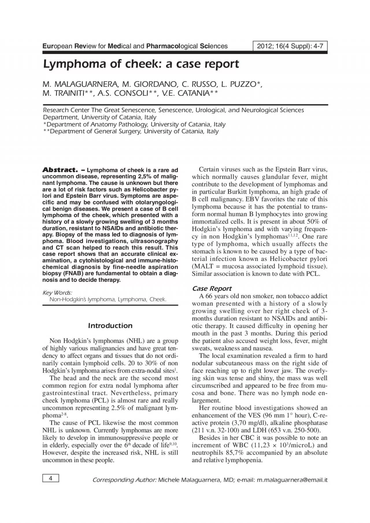

Presentation Transcript

Lymphoma of cheek: a case report L, FILIPPAP, MOWAN. Primary nasal paranasaloropharyngeal lymphoma in the pediatric agegroup. Cancer 1990; 65: 1438-1444.YP, SYW,XF, ZXH, LRadiotherapy As Primary Treatment for stage IEand IIE nasal natural Killer/T-cell lymphoma. JClin Oncol 2006; 245: 181-189.S, GG, KS, NV, O'HOral non- Hodgkin's lymphoma: review of the litera-ture and World Health Organization classificationwith reference to 40 cases. Oral Surg Oral PatholOral Radiol Endod 2008; 105: 194-201.ITALITALA, JENRYP, BARATM, RJ, BPeripheral neuropathies and lymphoma withoutmonoclonal gammopathy: a new classification. JNeurol 1990; 237: 177-185.M, AS, SBilateral facial paralysis secondary to lymphoma.ORL J Otorhinolaryngol Relat Spec 1993; 55:OMATSUDAM, NT, A. An autopsycase of malignant lymphoma associated with re-markable infiltration in skeletal muscle. RinshoKetsueki 1981; 22: 891-895.YC, PKP, LLym-phoma. Presenting as an intramuscular small cellmalignant tumor. Cancer 1984; 53: 1586-1589.The use of flow cytometry in the diag-nosis and monitoring of malignant hematologicaldisorders. Pathology 1999; 31: 382-392.I, IE, ES, SM, E. Primary lymphoma of thehead and neck: two case reports and review ofthe literature. Cases J 2008; 1: 426.TP, DS, CM, CE, F. Chemotherapy alone com-pared with chemotherapy plus radiotherapy forlocalized intermediate- and high-grade non-Hodgkin's lymphoma. N Engl J Med 1998; 339:JY, CKH, YWK, HJP, TJH, C. Radiation therapy versus chemotherapyas initial treatment for localized nasal natural killer(NK)/T-cell lymphoma: A single institute survey inTaiwan. Ann Oncol 2004; 15: 618-625.M, FK, KR, MEILLYS, SJJ, SJ, W. Introduction of combined CHOP plus rit-uximab therapy dramatically improved outcome ofdiffuse large B-cell lymphoma in British Columbia.J Clin Oncol 2005; 23: 5027-5033. DELAG, BS, A. Dedifferentiat-ed liposarcoma of the cheek. Ann Diagn Pathol2004; 8: 352-357. FC also has limited capability in classifyinglymphomas into different, well recognized subcate-gories. This is made possible by studies of surfacemarker expression and is best applicable for low. Through this technique is pos-sible to distinguish malignant lymphomas fromnon-lymphoid neoplasm, evaluating the presenceof positive staining for leukocyte common antigen(LCA). Diffuse large B-cell lymphoma is mostcommonly positive for CD20 and CD 79a and lesscommonly positive for germinal centre cell mark-A correct and early diagnosis is important tobegin on time therapy. It is easy to change a non-Hodgkin’s lymphoma of the cheek or oral cavitywith other benign disease. This causes concernabout the performance status and the progressionof the cancer. Generally the standard treatmentfor patients with early stage diffuse large celllymphoma consists in chemo-therapy followedby involved field radio-therapyNon Hodgkin’s lymphoma well responds to ra-diotherapy and the cheek is a region that can beeasy treated. However, this therapy is responsibleof different side effects like dry mouth, pharynxpain, dysphagia etc. The patient decided to stopthe therapy because of the aggravation of perfor-mance status also caused by an aggressive therapy. Patients with advanced stage (bulky stage II,stage III and IV) have to be treated by combinedchemotherapy. At this stage the standard treat-ment should be constituted by 7 cycles ofRCHOP (cyclophosphamide, doxorubicin, vin-Our patient received three cycles of chemother-apy (fludarabine 3.75 mg e.v. days 1-3 + mytox-antron 15 mg e.v. day 1), fo

llowed by 18 cycles oflocal radiotherapy on the interested area by 6MeV linear accelerator for a total dose of 36 Gybut she decide to stopped the therapy for the ap-parition of side effects depending of radiotherapy.In conclusion, non Hodgkin’s lymphomashould be considered in the differential diagnosisof various benign and malignant lesions becausethe treatment and prognosis are very different inthe several cases. However, according to the in-ternational prognostic index (IPI), established forpatients aged less than 60 years, the outcome ofpatients with extranodal DLBCL is similar to thatof patients with nodal DLBCL. Between malig-nant diseases we have to consider the presence ofdifferent histopathological cancers such as li-posarcoma, carcinoma, Sarcoma of Kaposi andHodgkin’s lymphomaFinally, an accurate clinical examination, a cy-nosis by FNAB and flow cytometry became fun-damental steps to decide a proper therapeuticReferencesE, RE, BERTONIF, CAVALLI. Primaryextranodal non-Hodgkin’s lymphoma. Part 1: Gas-trointestinal, cutaneous and genito urinary lym-phomas. Ann Oncol 1997; 8: 727-737.L, SR, SOral pre-sentations in non-Hodgkin’s lymphoma: a reviewof thirty-one cases. Part I Data analysis. OralSurg Oral Med oral Path 1983; 56: 151-156.. A review of non-Hodgkin’s lymphoma ofthe oral cavity 1950-1980. J Oral Med 1986; 41:Y, IT, FM, UT, AMalignant lymphoma of the oral cavity: Clinico-pathologic analysis of 20 cases. J Oral Pathol1987; 16: 8-12.F, OPrimarynon-Hodgkin’s lymphoma of the oral region. JOral Path Med 1989; 18: 84-91. I, G. A non-Hodgkin'slymphoma in the buccal mucosa. A case report.Oral Surg Oral Med Oral Pathol 1992; 74: 340-342.OLVIUSVANDERP, VANDERVANPJ, HVANDERI, S. Pri-mary extranodal non-Hodgkin lymphoma of theoral cavity. An analysis of 34 case. Eur J CancerB Oral Oncol 1994; 30: 121-125.. Charac-teristics of oral and paraoral malignant lym-phoma: a population-based review of 361 cases.Oral Surg Oral Med Oral Pathol Oral Radiol En-dod 2001; 92: 519-525.L, CRISTALDIE, Mrole of immunità in elderly cancer. Crit Rev OncolHematol 2010; 74: 40-60. L, FL, DS, I. Immunosenescenceand cancer, a review. Arch Gerontol Geriatr 2001;32: 77-93.K, BDP, B. Burkitt lymphoma in Papua, New Guinea.Br J Cancer 1967; 21: 657-664.E, K. Burkitt lymphoma. Semin CancerBiol 2009; 19: 345-346.Primary malignant lymphoma of theoral cavity and paranasal sinus. Radiology 1971;100: 151-153.K-J, FG, KClinical utility of lymphocytesurface markers combined with the Lukes-Collinshistologic classification in adult lymphoma. N EnglJ Med 1979; 301: 512-518.M. Malaguarnera, M. Giordano, C. Russo, L. Puzzo, M. Trainiti, A.S. Consoli, V.E. Catania Ultrasonography revealed a hypoechoic wellcircumscribed mass with the maxima diameter of4 cm and a laterocervical and submandibularlymphadenopathy. Therefore, patient was sub-jected to fine needle aspiration biopsy (FNAB)that showed a lymphomatous swelling. CT scan confirmed the presence of a solid, iso-dense, hypervascolarizated mass of 2,5 cm diame-ter in her right cheek in absence of laterocervicalnode involvement and metastasis. A percutaneousbiopsy was performed in order to obtain anhistopathological typing that revealed a small dif-fuse B cell CD20+ non-Hodgkin’s lymphoma.The immunophenotyping showed B-cell typeCD20 and CD79a positive and CD3, CD10,CD15, CD30, CD68 negative. It was also foundsmall lymphocytes with round or cleaved nucleus,minor component of blasts and plasmacytoid cells,compatible with a lymphocyte prolifer

ation. The patient was considered no operable.Therefore, she received three cycles ofchemotherapy (Fludarabine 3.75 mg e.v. days 1-3+ Mytoxantron 15 mg e.v. day 1). This was fol-lowed by 18 cycles of local radiotherapy to thediseased area on 6 MeV linear accelerator. Shereceived a total dose of 36 Gy. Following the treatment there was a partial re-gression of the tumor but after two months of theregression, patient refused to continue the thera-py because of the worsening of performance sta-tus caused by pharyngeal infiltration and side ef-fects of radiotherapy (dysphagia, asthenia, pares-thesias). Subsequently the patient had a ischio-pubic fracture after a long period of bone painsThe tumors of oral cavity include several typesof malignancies in correspondence of lip, tongue,cheek and pharynx. In the USA, the annual inci-dence is 19 cases on 100,000 people and the lifeexpectative to five years is about 52%. Non Hodgkin’s lymphoma is one of the possi-ble cancers in the head and neck region and, be-tween extra nodal non Hodgkin’s lymphomas,this is the second most common site after gas-. In the head and neck,Waldeyer’s ring is the most common site of ori-gin and may be accompanied by cervical nodeinvolvement. Nose, paranasal sinus, orbits, sali-vary gland are other possible organs affected indecreasing order of frequency, with rare spreadto the regional lymph nodes. Particularly,nasal non Hodgkin’s lymphoma is rare in West-ern countries but it is very common in East AsianThe diffuse large B-cell lymphoma (DLBCL)appears to be the most common type of primaryThere are no characteristics clinical features oflymphoma of the oral region and they depend bythe site of the swelling, the lymph node involve-ment and/or the presence of metastasis. The mostcommon beginning symptoms are local mass,pain or discomfort, dysphagia or sensation of aforeign body in the throat, in the case of tonsillarNHL. It is frequent that a NHL may be confusedwith a benign disease. Therefore, is useful oto-laryngology examination. Sometimes, it is also possible found facial hemi-plegia and parestesia because of a neural involve-ment. However, peripheral neuropathy is an unusu-al complication of lymphomalike so distinctmuscular involvement. Our patient presented aslowly growing swelling resistant to antibiotics andNSAIDs, weight loss, fever, might sweats, weak-ness and nausea. Therefore, the symptoms wereThe first step, to make a correct diagnosis, isan inspection and palpation of oral cavity and re-A part of that instrumental techniques, percu-taneous ultrasound (US) and computed tomogra-phy (CT) scan are well established procedure.Cytohistological diagnosis is mandatory for di-agnosis and treatment with an accurate fine nee-dle aspiration diagnosis of PCL is critical to berealized in time to avoid surgical managementand obviates the need for an exploratory la-paratomy. FNAB is considered a safe, rapid andeasy procedure with high diagnostic accuracy.Percutaneous biopsy should be performed to es-Flow cytometry (FC) has significantly en-hanced the diagnostic role of FNAB, particularlyin the case of hematolymphoid malignancies. FCis extremely sensitive in the detection of antigenexpression and identifies small clonal population.FC analysis distinguishes lymphomas fromclonality based in surface of Ig light chain ex-pression studies. In lymphomas, IG light chainexpression is usually restricted to either kappa orlambda, whereas inflammatory processes reveal amixed expression of kappa and

lambda light Lymphoma of cheek: a case report Review for Lymphoma of cheek is a rare aduncommon disease, representing 2,5% of malig-nant lymphoma. The cause is unknown but thereare a lot of risk factors such as Helicobacter py-lori and Epstein Barr virus. Symptoms are aspe-cific and may be confused with otolaryngologi-cal benign diseases. We present a case of B celllymphoma of the cheek, which presented with ahistory of a slowly growing swelling of 3 monthsduration, resistant to NSAIDs and antibiotic ther-apy. Biopsy of the mass led to diagnosis of lym-phoma. Blood investigations, ultrasonographyand CT scan helped to reach this result. Thiscase report shows that an accurate clinical ex-amination, a cytohistological and immune-histo-chemical diagnosis by fine-needle aspirationbiopsy (FNAB) are fundamental to obtain a diag-nosis and to decide therapy. Non-Hodgkin’s lymphoma, Lymphoma, Cheek.IntroductionNon Hodgkin’s lymphomas (NHL) are a groupof highly various malignancies and have great ten-dency to affect organs and tissues that do not ordi-narily contain lymphoid cells. 20 to 30% of nonHodgkin’s lymphoma arises from extra-nodal sitesThe head and the neck are the second mostcommon region for extra nodal lymphoma aftergastrointestinal tract. Nevertheless, primarycheek lymphoma (PCL) is almost rare and reallyuncommon representing 2.5% of malignant lym-The cause of PCL likewise the most commonNHL is unknown. Currently lymphomas are morelikely to develop in immunosuppressive people orin elderly, especially over the 6decade of lifeHowever, despite the increased risk, NHL is still Certain viruses such as the Epstein Barr virus,which normally causes glandular fever, mightcontribute to the development of lymphomas andin particular Burkitt lymphoma, an high grade ofB cell malignancy. EBV favorites the rate of thislymphoma because it has the potential to trans-form normal human B lymphocytes into growingimmortalized cells. It is present in about 50% ofHodgkin’s lymphoma and with varying frequen-cy in non Hodgkin’s lymphomastype of lymphoma, which usually affects thestomach is known to be caused by a type of bac-terial infection known as Helicobacter pylori(MALT = mucosa associated lymphoid tissue).Similar association is known to date with PCL.A 66 years old non smoker, non tobacco addictwoman presented with a history of a slowlygrowing swelling over her right cheek of 3-months duration resistant to NSAIDs and antibi-otic therapy. It caused difficulty in opening hermouth in the past 3 months. During this periodthe patient also accused weight loss, fever, mightThe local examination revealed a firm to hardnodular subcutaneous mass on the right side offace reaching up to right lower jaw. The overly-ing skin was tense and shiny, the mass was wellcircumscribed and appeared to be free from mu-cosa and bone. There was no lymph node en-largement. Her routine blood investigations showed anenhancement of the VES (96 mm 1°hour), C-re-active protein (3,70 mg/dl), alkaline phosphatase(211 v.n. 32-100) and LDH (653 v.n. 250-500).Besides in her CBC it was possible to note anincrement of WBC (11,23 neutrophils 85,7% accompanied by an absoluteand relative lymphopenia. 2012; 16(4 Suppl): 4-7Lymphoma of cheek: a case report M. TRAINITI**, A.S. CONSOLI**, V.E. CATANIA**Research Center The Great Senescence, Senescence, Urological, and Neurological Sciences*Department of Anatomy Pathology, University of Catania, Italy**Department of General Surgery, University of Catania, Italy