/
KoleoptRdsch 75 2005 tissues of those specimens which were fixed in KoleoptRdsch 75 2005 tissues of those specimens which were fixed in

KoleoptRdsch 75 2005 tissues of those specimens which were fixed in - PDF document

jade
jade . @jade
Follow
342 views
Uploaded On 2022-10-11

KoleoptRdsch 75 2005 tissues of those specimens which were fixed in - PPT Presentation

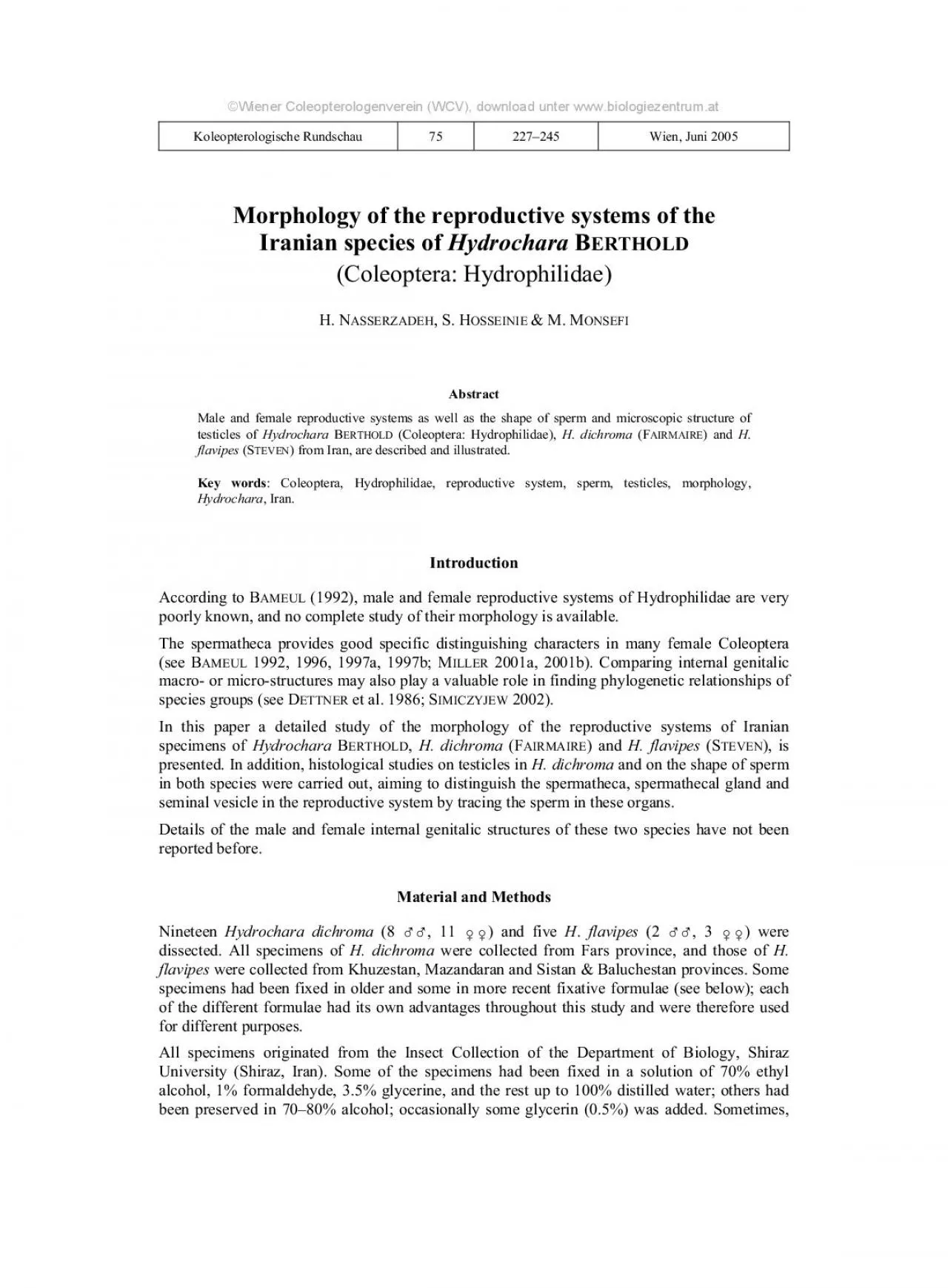
ASSERZADEH et al Reproductive systems of the Iranian species of Hydrochara HYDROPHILIDAE copulatrix was cleaned of muscles and connective tissues washing with distilled water and stored in glyce ID: 958547

reproductive 133 genitalia species 133 reproductive species genitalia bursa duct copulatrix hydrophilidae male view female seg spermatheca hydrochara 144

Share:

Link:

Embed:

Download Presentation from below link

Download Pdf The PPT/PDF document "KoleoptRdsch 75 2005 tissues of those sp..." is the property of its rightful owner. Permission is granted to download and print the materials on this web site for personal, non-commercial use only, and to display it on your personal computer provided you do not modify the materials and that you retain all copyright notices contained in the materials. By downloading content from our website, you accept the terms of this agreement.


Presentation Transcript

KoleoptRdsch. 75 (2005) tissues of those specimens which were fixed in the solution without formaldehyde, were found to be softer and more tender, which helped to find ducts and junctions under the stereo microscope in situ. Also, to study the bursa copulatrix and spermatheca, the softer specimens were superior. However, in specimens fixed with formaldehyde, the genitalia became harder and thus were much easier to be extracted than the softer ones. Apparently, formaldehyde helped to remove the whole abdominal membranous covering fat, and the ovarioles, testes, accessory glands and ducts Terminology follows that of SNODGRASS (1935), IMMSExtracting male genitalia: To extract the reproductive system (in both sexes), the abdomen was opened dorsally by lifting the wings and removing the whole abdominal tergites. The looped and folded hind gut was extracted, exposing the testes and accessory glands located among fat, muscles and tracheae of the third and fourth abdominal segments,. Occasionally, few drops of diluted met

hylen blue in and around the insects abdomen were used during the operation to increase the contrast to distinguish the structures. re pulled out and put into distilled water. In the older specimens the genitalia could be extracted easier; however, to clean and clear them, they were put in 10% KOH (potassium hydroxide) for about 15…20 minutes to loosen excessive parts, relaxed them as well as made their ducts and junctions visible without unnecessary cutting or destroying the adjacent parts. The remaining fat and excessive tissues were then removed by use of a fine pin. During this procedure, care was taken that KOH did not harm the genitalic tissue. If this happened, the process was stopped and they were washed by distilled water, then placed in distilled water, again some methylen blue was added and cleaning continued. Finally, the cleaned and stained structures were washed with distilled water, kept in glycerine and studied.Preparing sperm smear: A voluminous seminal vesicle of both species was chose

n for sperm smear preparation. The vesicle was separated, masticated by a pin in one drop of distilled water and some smear slides were made. They were stained by Aniline Blue & Eosin (G134) for 8…9 minutes. Permanent slides were made using Canada balsam. After making block paraffin of the testis of the preparation was cut by a Leitz microtome in 5 µm thickness (B 1990). The sections were stained by Hematoxilin & Eosin method (GPermanent slides were prepared using Canada balsam. Preparing whole mount (w.m.) slides of testicles: carefully and then put into ethyl alcohol rinses of 50%, 75% and finally 96% (each about 2…3 minutes). Each testis was washed with distilled water and stained with Anilin blue & Eosin. Permanent slides were made using Canada balsam with the cover glass slightly pushed down to scatter the testicles. Separating female genitalia: The method of separating female genitalia is the same as described for the male, except for cleaning the ovarioles, these were kept in a mixture of warm w

ater, detergent powder and a small amount of bleaching agent for five minutes, and then The freezing method for dissecting the fresh specimens was applied as usual (BAMEUL 1992; Separation of the bursa copulatrix was easier than separation of ovaries. A few drops of 10% KOH were added to the dissected females reproductive system and left for about 15 minutes to remove surrounding tissues. The bursa ASSERZADEH et al.: Reproductive systems of the Iranian species of Hydrochara (HYDROPHILIDAE) copulatrix was cleaned of muscles and connective tissues, washing with distilled water, and stored in glycerine for study. Adding diluted methylen blue improved the contrast for examination of details. More tender specimens, that were kept in alcohol only were preferred to those which were killed and fixed with alcohol and formaldehyde because the latter had been kept in warm water for 15…20 minutes before treating with KOH, the membraneous wall of the bursa copulatrix as well as the common oviduct was always destroy

ed during the procedure.To study the whole reproductive system, the entire abdomen had to be opened dorsally, but to dissect the bursa copulatrix and the spermatheca, it was sufficient to open only the last two or To clean the spermatheca and its duct, they were kept in 10% KOH for about 10 hours to permit Male internal genitalia consist of a pair of testes, and each testis consists of short tubules or testicles. Each tubule leads to the vas deferens by a small stalk, the NODGRASS 1935: 507…508). Each vas deferens dilates distally, called seminal vesicle, and the two seminal vesicles join to form the median ejaculatory duct. On top of each seminal vesicle there is a large accessory gland, a blind tube that folds circularly and finally continues posteriorly as a tube to join the ejaculatory duct. (1964: 762), two general types of reproductive organs are recognized, based closely coiled, each being enclosed in a membrane, in Polyphaga [for example species] they are compound and divided into a number of s

eparate folliclesŽ. The accessory glands are very large (except in the immature stages or out of their reproductive cycle), and, according to SNODGRASS (1935: 573), have the function of secreting a mucous or viscid substance. This substance is either discharged as a liquid together with the spermatozoa, or, serves to form a hard cover or capsule known as a spermatophore. No work on distinguishing spermatophores or spermatozoa in et al. (1959…1995) described some secretory cells in Tenebrio molitor (mealworm integrated into a particular layer of the spermatophore secretion. The shape and volume of the accessory glands of this species are very similar to those of This may represent a similar evolutionary path for both, but this would need further studies. The male accessory glands generally arise from the short divergent anterior branches of the ejaculatory duct (S 1935: 573). The ejaculatory duct is ectodermal in origin, and is formed as a median ventral invagination of the ectoderm at the posterior end

of the ninth abdominal segment, therefore, it has a cuticular NODGRASSmiddle of ejaculatory duct, there is a swelling which is more sclerotized than the rest of the duct (Fig. 1a). This seems to be a pumping site for sperm to be pumped through the median lobe into the bursa copulatrix of the female by the force of a strong muscular sheath surrounding the epithelial wall of the ductŽ (S 1935: 572) (Fig. 1a, mscl). In both species, the sperm are thin and elongate with a long flagellum (Figs. 3, 4). KoleoptRdsch. 75 (2005) 7) reveals that they have a very typical cellular structure as in most other insects. Nomenclature as in MATHEWS (1991: The structure and the shape of the male internal genitalia of the two species examined are Male external genitalia in insects are similar in evolutionary origin as those of females (SMITH, male external genitalia (Figs. 8…11) are rather simple and primitive, of the trilobed type, and rest inside the abdomen in the primitive position, with the apical opening of the

median lobe facing ventrally, stemming from segment IX (see S 1980; NODGRASSThe male copulatory organ, or aedeagus, is of prime importance to distinguish species (Sinvaginated ninth and tenth abdominal segments. The capsule consists of: 1) a dorsal proctiger, tergite X, 2) two lateral paraprocts, the divided tergite IX, and 3) the spiculum gastrale, representing the modified sternite IX (MARSHALLThe median lobe (Figs. 8…11) is membraneous throughout the ventral surface and sclerotized on the dorsal surface; a fine longitudinal line arises from the apex and extends to the bases ATANABE 1975). The parameres inare significantly The general structure of female internal genitalia in both species examined (Figs. 12…17) agree well with those of other beetles (see NODGRASSMMS 2001a, b). They consist of a pair of ovaries, each composed of some separated ovarioles. All ovarioles from each side join to form a lateral oviduct, which, converges posteriorly; left and right lateral oviducts join to form the common ov

iduct posteriorly. Several accessory glands on top and below each ovary join the oviduct on each side (clearly seen in Figs. 14, 15). The common oviduct ventrally leads into the middle of a large, sac-like bursa copulatrix. A long spermatheca opens into the bursa copulatrix anteriorly with a slender and long duct. A globular spermathecal gland is connected with the spermatheca. The ovarioles consist of three parts (Fig. 14b): 1) a terminal filament; 2) an egg tube which contains the germ cells and their derivatives and at the end of some tubes there exist corpora shows this type of ovarioles (Fig. 14b), but histological investigation is needed ETTNER et al. (1986: 348) mentiones two theories with respect to the formation of corpora lutea: 1) follicular cell degenerates after egg deposition to remain as corpus luteum. The number of corpora lutea is correlated with the number of eggs deposited, 2) it may be due to egg resorption, in which no statement on completion of egg deposition would be ELTMATE (1994), egg coco

ons in Hydrophilidae are made from silk secreted by Malpighian tubules, and the function of accessory glands must be in attaching egg cases to stems or leaves of plants or on the mud directly. The spermatheca is primarily an invagination of the integument at the posterior end of the venter of the eighth abdominal segment (SNODGRASS 1935: 556). It is cylindrical, long and filled with spermatozoa (Fig. 16, spz); a globular spermathecal gland secrets a fluid into which the sperms ASSERZADEH et al.: Reproductive systems of the Iranian species of Hydrochara (HYDROPHILIDAE) are discharged (S 1935); the spermathecal duct enters the anterior portion of the bursa copulatrix. It seems the duct gradually merges into the wall of the bursa copulatrix (Fig. 16, fsd). The genital chamber, or bursa copulatrix, receives the common oviduct ventromedially. Just under the junction between the bursa copulatrix and common oviduct, the bursa copulatrix is slightly expanded forming a sac (= vagina) resembling the ventral pouch in Marsup

ials (Fig. 16, vg). On the wall of the bursa copulatrix four fine tube-like structures can be observed which seems to be the starting point for the entrance of the spermathecal duct, continuing along the wall (1964), these fine tubes are not mentioned in the literature. (1964) describes a second passage or canal of fecundation leading from the spermatheca ABRICIUS, 1792)(Carabidae). The tissue structure of these tubes seems to be different from the wall tissue of the bursa copulatrix. Further histological studies are needed to Female external genitalia (Figs. 18, 19) of laterotergite VIII (formerly ninth tergite), mediotergite VIII (formerly valvifer), coxostyle IX and gonostyle IX (formerly stylus) (B 1992). The tenth segment (proctiger) appears as a NODGRASSThere is a very fine membraneous part behind the gonostylus which we named median membranous pieceŽ. Under the compound microscope, a very fine hair-like cover can be observed, wrapping around the gonostylus on each side.The mal

e and female internal reproductive systems and sperm of similar to those of in regard to general shape and structure, with the exception of the parameres. Certainly, because of the size difference between these two species, all structures are smaller and finer in H. flavipes. The median lobe of is also smaller, narrower and more elongate (in relation to parameres) than the median lobe of ; the parameres of are more slender, and with the external margins more smoothly curved (Figs. 8…11). The external female genitalia of the two species show no significant differences, either, even for the spermatheca, just unlike other genera (B are generally slightly smaller, the size is not a very reliable distinguishing This preliminary study could be improved with analysis of additional species, and use of biometric comparisons. Acknowledgements The authors heartily thank Dr. F. Bameul (France) for his advice and encouragement. Thanks are also due to Mr. K. Elmi, technician at the Department of Biology, Shiraz Universi

ty for collecting the specimens and for offering the necessary technical assistance in the laboratory. We also appreciate the help of Miss M. Adnafi in preparing the paraffin blocks of testes, and also acknowledge the effort of Mr. Tahamtani, Mr. Hadayegh and Mr. Khaki, biology students, for the Photography and Xerox Units at the Department of Biology, Shiraz University. KoleoptRdsch. 75 (2005) ; male reproductive system; a) dorsal view; b) ejaculatory duct, enlarged; acgl … accessory glands terminal tube; cutl … cuticular line; ejd … ejaculatory duct; maf … mass of fat; mscl … covering muscle; parapt … paraproct (of seg. IX); spga … spiculum gastrale (of seg. IX), pt X … proctiger (of seg. X). Fars, Dasht-e-Arjan; coll. no. 453. ASSERZADEH et al.: Reproductive systems of the Iranian species of Hydrochara (HYDROPHILIDAE) ; male reproductive system, ventral view. Fars, Dasht-e-Arjan; coll. no. 453. KoleoptRdsch. 75 (2005) ; 3) photo micrograph of smear of spermatozoa

; acs … acrosome; 400 X; 4) spermatozoa. Fars, Bamoo, Darreh-Bisheh. ASSERZADEH et al.: Reproductive systems of the Iranian species of Hydrochara (HYDROPHILIDAE) Fig. 5: , male reproductive system, whole mount, photomicrograph; tescl … testicle; tew … testicular wall; vd … vas deferens; 32 X. KoleoptRdsch. 75 (2005) Fig. 6…7: Hydrochara dichroma;photomicrographs of 6) section of testicle; dsp … differentiated spermatid; rb … residual body; psp … primary spermatocyte; spd … spermatid; spg … spermatogonium; ssp … secondary spermatocyte; 160 X; 7)clusters of spermatozoa and residual bodies; 200 X. ASSERZADEH et al.: Reproductive systems of the Iranian species of Hydrochara (HYDROPHILIDAE) ; 8) male external genitalia (aedeagus); a) dorsal view; b) ventral view; ; 10) male external genitalia (aedeagus); a) dorsal view; b) ventral view; 11) penis; a) dorsal view, b) right lateral view. Abbreviations: aop … apical opening of internal sac; bp … basal pie

ce (phallobase) (of seg. IX); pen … penis (of seg. IX); pm … paramere (periphalic organ) (of seg. IX); scr … sclerotized ring. KoleoptRdsch. 75 (2005) ; female reproductive system, dorsal view; acgl … accessory gland; bc … bursa copulatrix; et … egg tube; grm … germarium; gsty … gonostylus (of seg. IX); ltg … laterotergite (of seg. VIII); mscl … covering muscle; ov … ovary; ovl … ovariol; pdcl … pedicel; sth … spermatheca; sthd … spermathecal duct; sthg … spermathecal gland; tf … terminal filament. Fars, Bamoo, Cheshmeh-ye- ASSERZADEH et al.: Reproductive systems of the Iranian species of Hydrochara (HYDROPHILIDAE) Hydrochara dichroma; female reproductive system, ventral view; covi … common oviduct; ovi … oviduct. Fars, Bamoo, Cheshmeh-ye-Ghanbari. KoleoptRdsch. 75 (2005) ; female reproductive system; a) dorsal view; b) ovariole, enlarged; cax … calyx; clt … corpus luteum. Khuzestan, Ahvaz to Khorramshahr; co

ll. no. 2245. ASSERZADEH et al.: Reproductive systems of the Iranian species of Hydrochara (HYDROPHILIDAE) ; female reproductive system, ventral view. Khuzestan, Ahvaz to Khorramshahr; coll. no. 2245. KoleoptRdsch. 75 (2005) ; 16) external genitalia, bursa copulatrix and spermatheca, ventral view. Fars, Dasht-e-Arjan; 17) spermatheca and spermathecal gland. Abbreviations: covi … common oviduct; fsd … fusing spermathecal duct; spz … spermatozoa; sth … spermatheca; sthd … spermathecal duct; sthg … spermathecal gland; tstr … tube-like structure (fecundation ASSERZADEH et al.: Reproductive systems of the Iranian species of Hydrochara (HYDROPHILIDAE) Fig. 18…19: Female external genitalia; a) dorsal view; b) ventral view of 18) and H. flavipesAbbreviations: cxsty … coxostylus (of seg. IX); gsty … gonostylus (of seg. IX); ltg … laterotergite (of seg. VIII); mmp … memberanous median piece; mtg … mediotergite (of seg. VIII); prg … proctiger (scleroti

zed ring of seg. X). KoleoptRdsch. 75 (2005) ROWNE, F. 1958: British water beetles, Vol. III. … London: Ray Society, 162 pp. , F. 1992: Revision of the genus dOrchymont from southern India and Sri Lanka (Coleoptera, Hydrophilidae, Omicrini). … Systematic Entomology 17: 1…20, F. 1996: A new Hydrochara Berthold from North Korea (Coleoptera, Hydrophilidae). … Nouvelle Revue dEntomologie (Nouvelle Série) 13 (1): 3…11.Protosternum Sharp (Coleoptera, Hydrophilidae). … Nouvelle Revue dEntomologie (Nouvelle Série) 14 (1): 17…41. dOrchymont and Balfour-Browne (Coleoptera, Hydrophilidae) with a phylogenetic analysis of Protosternini. … Annales de la société Entomologique de France (Nouvelle Série) 33 (3): 375…403. , A. 1990: Theory and practice of histological techniques. … Livingstone: , D.B. 1991: Atlas of invertebrate reproduction and development. … New York: Wiely…Liss Inc., LASSEN, R. 1986: Age structure, phylogeny and prey of so

me rheophilic Dytiscidae (Coleoptera). … Entomologica Basiliensia 11: 343…370. , T. 1993: Labdomen et les genitalia des femelles de Coléoptères Adephaga, Tome 155. … Mémoires du Muséum national dhistoire naturelle zoologie, 184 pp. , T. 2001: Le genre Schmidt-Goebel, 1846, insectes (Coleoptera, Paussidae, Ozaeninae) à genitalia femelles orthotopiques. … Zoosystema 23 (3): 547…578. , E. 1962: Staining animal tissues practical and theorical. … London: Leonard Hill [Books] Limited, APP, G. et al. 1959…1995: Insect research. … http://mercury.bio.uaf.edu/~george_happ/insectres.htm , A.D. 1964: A general textbook of entomology, including the anatomy, physiology, development and classification of insects. … London: Methuen & Co Ltd., 886 pp. , C. 2001: Represented in webpage: Lab/External male genitalia (labs 22&23). … http://www.entomology.cornell.edu/faculty_staff/Danforth/322lab manual/lab22&23.pdf , W.W. 1982: Atlas of descriptive embryology. … N

ew York: Macmillan Publishing Co., 230 , K.B. 2001a: Description of new species of Babington (Coleoptera. Dytiscidae, Hydroporinae, Hydrophydrini) with a reassessment of the subgenera and species groups and a synopsis of the species. … The Coleopterists Bulletin 55 (2): 219…240. , K.B. 2001b: Revision of the genus Zimmerman (Coleoptera, Dytiscidae). …Annals of the Entomological Society of America 94 (4): 520…529. , B. 2002: Structure of the ovary in Navás (Insecta, Mecoptera, Nannochoristidae), with remarks on mecopteran phylogeny. … Acta Zoologica (Stockholm), 83: 61…66. , A. 1980: Revision of the genus Berth. (Coleoptera, Hydrophilidae). … Memoirs of The Entomological Society of Canada 111: 1…100. , E.L. 1969; Evolutionary morphology of external insect genitalia. 1. Origin and relationship to other appendages. …Annals of the Entomological Society of America 62 (5): 1051…1079. NODGRASS, R.E. 1935: Principles of insect morphology … Mc Graw-Hill Book Compan

y Inc., 667 pp, S.L. (ed.) 1970: Taxonomists glossary of genitalia in insects. … Copenhagen: Munksgaard, 359 ASSERZADEH et al.: Reproductive systems of the Iranian species of Hydrochara (HYDROPHILIDAE) , N. 1975: Comparative morphology of the male genitalia in the subfamily Hydrophilinae (Coleoptera, Hydrophilidae), 1. Tribe Hydrophilida. … Annotationes Zoologicae Japonenses, 48 (2): 129…137. , B.W. 1994: Aquatic Insects. … Wallingford: CAB International, 358 pp. Insect Taxonomy Research Department, Plant Pests and Diseases Resea1454, IR … 19395 Tehran, Iran (h_naserzadeh@yahoo.com) Dr. Shidokht HOSSEINIE (Mrs.) Department of Biology, College of Sciences, Shiraz University, IR … 71454 Shiraz, Iran (sh_hosseinie@yahoo.com, Department of Biology, College of Sciences, Shiraz University, IR … 71454 Shiraz, Iran(monsefi@biology.susc.ac.ir) Koleopterologische Rundschau 75 227…245 Wien, Juni 2005 Morphology of the reproductive systems of the Iranian species of HydrocharaBE