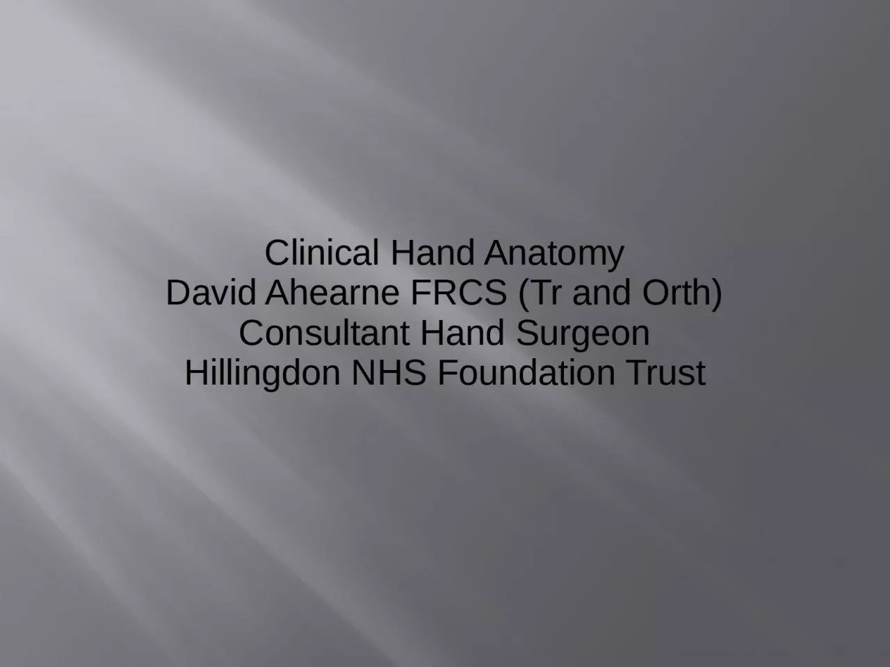
/
Clinical Hand Anatomy David Ahearne FRCS (Tr and Orth) Clinical Hand Anatomy David Ahearne FRCS (Tr and Orth)

Clinical Hand Anatomy David Ahearne FRCS (Tr and Orth) - PowerPoint Presentation

jalin
jalin . @jalin
Follow
0 views
Uploaded On 2024-03-15

Clinical Hand Anatomy David Ahearne FRCS (Tr and Orth) - PPT Presentation

Consultant Hand Surgeon Hillingdon NHS Foundation Trust Acknowledgements Mr H Belcher Hand Surgeon QVH East Grinstead Aids to the examination of the PNS Advanced Examination Techniques in Orthopaedics ID: 1048545

injury hand ulnar extensor hand injury extensor ulnar tendon flexor avulsion repair nervesensation tendons median digitorum examination trauma nervemotorabductor

Share:

Link:

Embed:

Download Presentation from below link

Download Presentation The PPT/PDF document "Clinical Hand Anatomy David Ahearne FRCS..." is the property of its rightful owner. Permission is granted to download and print the materials on this web site for personal, non-commercial use only, and to display it on your personal computer provided you do not modify the materials and that you retain all copyright notices contained in the materials. By downloading content from our website, you accept the terms of this agreement.


Presentation Transcript

1. Clinical Hand AnatomyDavid Ahearne FRCS (Tr and Orth)Consultant Hand SurgeonHillingdon NHS Foundation Trust

2. AcknowledgementsMr H Belcher Hand Surgeon, QVH, East Grinstead.Aids to the examination of the PNSAdvanced Examination Techniques in OrthopaedicsNetters Concise Atlas of Orthopaedic Anatomy.

3. Referrals to Hand SurgeonsTraumaInfectionDegenerativeInflammatoryIdiopathicTumour

4. HistoryAgeDominanceOccupationHistory of ComplaintLength of timePrimary symptom eg Pain, stiffness, instability.Level of Dysfunction

5. Trauma HistorySharpKnifeBluntHammerBlastIndustrial accidents.Indirect

6. Types of Trauma

7. Flexor Tendon InjuriesFlexor ZonesHelp determine Tendons Injured.

8. Trauma AssesmentMechanism of InjuryPosition of fingers at time of injuryTime of injuryDysfunctionExaminationVascular assessmentNeurological assessmentIndividual Tendons.

9. Aim of treatmentRestoration of functionDepends on needs of PatientsConcert Pianist (Dexterity)Labourer (Pain Free and Stable)

10. Examination of the HandSystematic approachDetermine cause of reduced function.NeurologicalMuscularTendonJointFunctional Position

11. Hand functional positions

12. Abnormal Functional Position

13. Neurological ExaminationNerve supplySensationMotorStrength

14. Median NerveSensation

15. Median NerveMotorAbductor Pollicis BrevisOpponens Pollicis

16. Median NerveFlexor Digitorum Superficialis

17. Ulnar NerveSensation

18. Ulnar nerveMotorAbductor Digiti MinimiFirst Dorsal Interosseous Muscle

19. Radial NerveSensation

20. Radial NerveMotorNo Muscle Supplied in handPosterior Interosseous Nerve main branch

21. JointRange of movementMCPJ0-90 degreesPIPJ0-110 degreesDIPJ0-90 degreesActivePassivePain on movementStabilityStatic Bony ContourLigamentsDynamicMuscular Contractions

22. TendonsAre all workingAre they StiffStiffness intrinsic extrinsic

23. Flexor TendonsFlexor Digitorum Profundus

24. Flexor TendonsFlexor Digitorum SuperficialisImmobilise other fingers to isolate FDSBottom picture shows decrease PIPJ movement due to Hypoplastic FDS

25. Tenodesis Effect,

26. Extensor Communis (Left)Extensor Proprius (Right)

27. MuscleAre they contractingLevel of PowerMRC Grading

28.

29. Hand incisions

30. Friday Night Boxers Fracture

31. Thenar WastingConsequence of Chronic Carpal Tunnel Syndrome

32. Ulnar nerve Lesion

33. Boutonniere Deformity (Central Slip Rupture)

34. Paronychia

35. Extensor tendon laceration

36. Repair of extensor tendom

37. Spiral fracture Metacarpals

38. FDP Tendon Injury

39. Rheumatoid Hand (Ulnar deviation)

40. Osteoarthritis of Base of Thumb

41. PIN Injury

42. Flexor Tendon Laceration

43. Multiple Extensor Tendons

44. PIPJ Dislocation

45. OA Base of Thumb

46.

47. Swan Neck Deformity

48. AIN Injury

49. Tip Avulsion Injury

50. Ulnar Collateral Ligament Rupture

51. Bony Avulsion of Above

52. Fixation of above

53. Ring Avulsion Injury

54. Operative repair of above

55. Severe Tip avulsion

56. Local Skin Flap to Repair

57. Volar Plate Injury

58. Peri-lunate Dislocation

59. Degloving Injury