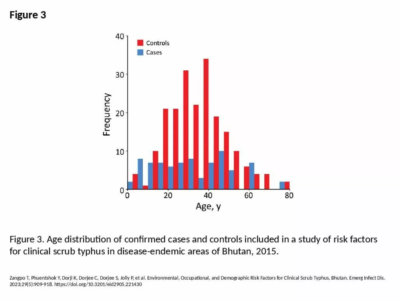
PPT-Figure 3 Figure 3. Age distribution of confirmed cases and controls included in a study

Author : jayden | Published Date : 2024-09-23

Zangpo T Phuentshok Y Dorji K Dorjee C Dorjee S Jolly P et al Environmental Occupational and Demographic Risk Factors for Clinical Scrub Typhus Bhutan Emerg Infect

Presentation Embed Code

Download Presentation

Download Presentation The PPT/PDF document "Figure 3 Figure 3. Age distribution of c..." is the property of its rightful owner. Permission is granted to download and print the materials on this website for personal, non-commercial use only, and to display it on your personal computer provided you do not modify the materials and that you retain all copyright notices contained in the materials. By downloading content from our website, you accept the terms of this agreement.

Figure 3 Figure 3. Age distribution of confirmed cases and controls included in a study: Transcript


Zangpo T Phuentshok Y Dorji K Dorjee C Dorjee S Jolly P et al Environmental Occupational and Demographic Risk Factors for Clinical Scrub Typhus Bhutan Emerg Infect Dis 2023295909918 httpsdoiorg103201eid2905221430. -1- Figure 1 -2- Figure 3 Figure 4 Figure 5loss resistance is zero, and that the radiation resistance is the textbook figure of 35 ohms. The feedpointimpedance would then be 15+0+35 = 50 ohms, and th John . Dement. 1. , Laura . Welch. 2. , Knut . Ringen. 2. , 3. , Patricia Quinn. 2. , Anna Chen. 4. , Scott . Haas. 4.  . 1. Division of . Occupational & Environmental . Medicine, Duke . University Medical . encouraged to reuse our maps and graphs . for your own . purposes and free to translate, . provided the . content is not altered and the source . is acknowledged. . Either copy the images from the following slides or download directly from: . for your own . purposes and free to translate, . provided the . content is not altered and the source . is acknowledged. . Either copy the images from the following slides or download directly from: . Yoo D, Kim Y, Lee E, Lim J, Hong S, Lee I, et al. Transmission Dynamics of African Swine Fever Virus, South Korea, 2019. Emerg Infect Dis. 2021;27(7):1909-1918. https://doi.org/10.3201/eid2707.204230. Quan C, Wang Q, Zhang J, Zhao M, Dai Q, Huang T, et al. Avian Influenza A Viruses among Occupationally Exposed Populations, China, 2014–2016. Emerg Infect Dis. 2019;25(12):2215-2225. https://doi.org/10.3201/eid2512.190261. Minetti A, Kagoli M, Katsulukuta A, Huerga H, Featherstone A, Chiotcha H, et al. Lessons and Challenges for Measles Control from Unexpected Large Outbreak, Malawi. Emerg Infect Dis. 2013;19(2):202-209. https://doi.org/10.3201/eid1902.120301. Messenger SL, Smith JS, Orciari LA, Yager PA, Rupprecht CE. Emerging Pattern of Rabies Deaths and Increased Viral Infectivity. Emerg Infect Dis. 2003;9(2):151-154. https://doi.org/10.3201/eid0902.020083. Luk E, Lo J, Li A, Lau M, Cheung T, Wong A, et al. Scarlet Fever Epidemic, Hong Kong, 2011. Emerg Infect Dis. 2012;18(10):1658-1661. https://doi.org/10.3201/eid1810.111900. Sola C, Devallois A, Horgen L, Maïsetti J, Filliol I, Legrand E, et al. Tuberculosis in the Caribbean: Using Spacer Oligonucleotide Typing to Understand Strain Origin and Transmission. Emerg Infect Dis. 1999;5(3):404-411. https://doi.org/10.3201/eid0503.990311. Boisier P, Rahalison L, Rasolomaharo M, Ratsitorahina M, Mahafaly M, Razafimahefa M, et al. Epidemiologic Features of Four Successive Annual Outbreaks of Bubonic Plague in Mahajanga, Madagascar. Emerg Infect Dis. 2002;8(3):311-316. https://doi.org/10.3201/eid0803.010250. Guo Z, Wang Z, Zhang S, Li X, Li L, Li C, et al. Aerosol and Surface Distribution of Severe Acute Respiratory Syndrome Coronavirus 2 in Hospital Wards, Wuhan, China, 2020. Emerg Infect Dis. 2020;26(7):1583-1591. https://doi.org/10.3201/eid2607.200885. Brown EM, McTaggart LR, Dunn D, Pszczolko E, Tsui K, Morris SK, et al. Epidemiology and Geographic Distribution of Blastomycosis, Histoplasmosis, and Coccidioidomycosis, Ontario, Canada, 1990–2015. Emerg Infect Dis. 2018;24(7):1257-1266. https://doi.org/10.3201/eid2407.172063. *Includes case reports indicating the presence of at least one of the following risks 6 weeks to 6 months prior to onset of acute, symptomatic hepatitis C: 1) using injection drugs; 2) having sexual contact with suspected/confirmed hepatitis C patient; 3) being a man who has sex with men; 4) having multiple sex partners concurrently; 5) having household contact with suspected/confirmed hepatitis C patient; 6) having had occupational exposure to blood; 7) being a hemodialysis patient; 8) having received a blood transfusion; 9) having sustained a percutaneous injury; and 10) having undergone surgery..

Download Document

Here is the link to download the presentation.
"Figure 3 Figure 3. Age distribution of confirmed cases and controls included in a study"The content belongs to its owner. You may download and print it for personal use, without modification, and keep all copyright notices. By downloading, you agree to these terms.

Related Documents