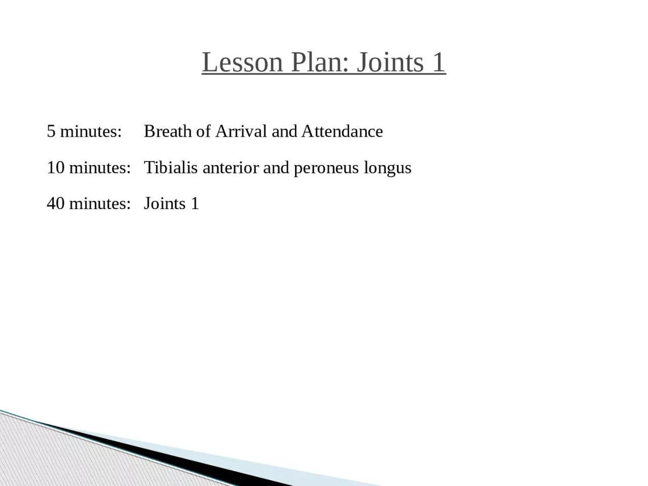
/
Lesson Plan:  Joints  1 5 minutes:	Breath Lesson Plan:  Joints  1 5 minutes:	Breath

Lesson Plan: Joints 1 5 minutes: Breath - PowerPoint Presentation

joanne
joanne . @joanne
Follow
342 views
Uploaded On 2022-06-08

Lesson Plan: Joints 1 5 minutes: Breath - PPT Presentation

of Arrival and Attendance 10 minutes Tibialis anterior and peroneus longus 40 minutes Joints 1 Classroom Rules Punctuality everybodys time is precious Be ready to learn by 900 well have you out of here by 130 ID: 914942

synovial joints types joint joints synovial joint types ynovial aka lateral ankle medial rotation membrane movement foot metatarsal insertion

Share:

Link:

Embed:

Download Presentation from below link

Download Presentation The PPT/PDF document "Lesson Plan: Joints 1 5 minutes: Breat..." is the property of its rightful owner. Permission is granted to download and print the materials on this web site for personal, non-commercial use only, and to display it on your personal computer provided you do not modify the materials and that you retain all copyright notices contained in the materials. By downloading content from our website, you accept the terms of this agreement.


Presentation Transcript

Slide1

Lesson Plan: Joints 1

5 minutes: Breath

of

Arrival and Attendance

10 minutes:

Tibialis

anterior and peroneus

longus

40 minutes:

Joints

1

Slide2

Classroom Rules

Punctuality- everybody's time is precious:

Be ready to learn by 9:00, we'll have you out of here by 1:30

Tardiness: arriving late, late return after breaks, leaving early

The following are not allowed:

Bare feet

Side talking

Lying down

Inappropriate clothing

Food or drink except water

Phones in classrooms, clinic or bathrooms

You will receive one verbal warning, then you'll have to leave the room.

Slide3

Tibialis Anterior and Peroneus Longus

Slide4

Tibialis

Anterior

Origin

:

Upper

2/3 of lateral tibia

Interosseous membrane Insertion: Base of 1st metatarsal Medial cuneiform (plantar) Actions: Ankle dorsiflexion Foot inversion

Slide5

Tibialis Anterior

Origin

:

Upper

2/3 of lateral tibia Interosseous membrane

Insertion

:

Base of 1st metatarsal Medial cuneiform (plantar) Actions: Ankle dorsiflexion Foot inversion

Slide6

Tibialis Anterior

Origin

:

Upper

2/3 of lateral tibia Interosseous membrane

Insertion

:

Base of 1st metatarsal Medial cuneiform (plantar) Actions: Ankle dorsiflexion Foot inversion

Slide7

Tibialis Anterior

Origin

:

Upper

2/3 of lateral tibia Interosseous membrane

Insertion

:

Base of 1st metatarsal Medial cuneiform (plantar) Actions: Ankle dorsiflexion Foot inversion

Slide8

Peroneus Longus

Origin

:

Proximal

2/3 of lateral fibula Insertion:

Base

of 1

st metatarsal Medial cuneiform (plantar) Actions: Ankle plantarflexion Foot eversion

Slide9

Peroneus Longus

Origin

:

Proximal

2/3 of lateral fibula Insertion:

Base

of 1

st metatarsal Medial cuneiform (plantar) Actions: Ankle plantarflexion Foot eversion

Slide10

Peroneus Longus

Origin

:

Proximal

2/3 of lateral fibula Insertion:

Base

of 1

st metatarsal Medial cuneiform (plantar) Actions: Ankle plantarflexion Foot eversion

Slide11

Peroneus Longus

Origin

:

Proximal

2/3 of lateral fibula Insertion:

Base

of 1

st metatarsal Medial cuneiform (plantar) Actions: Ankle plantarflexion Foot eversion

Slide12

Tibialis Anterior and Peroneus Longus

Slide13

Joints 1

“Opportunity is missed by most people because it is dressed in overalls and looks like work.”

–Thomas Edison

Slide14

Joint (AKA: articulation or arthrosis) Where bones come together or

join.

Joints

Slide15

PhysiologyEnable the body to move.Bear the weight of the body.Provide stability.

Joints

Slide16

Fibrous / SynarthroticCartilaginous /

Amphiarthrotic

Synovial /

Diarthrotic

Structural and Functional Classification

Slide17

Fibrous / Synarthrotic Connected by dense

fibrous

connective tissue, consisting mainly of collagen.

Extremely

limited movement. Examples: cranial sutures, facial sutures, teeth, and tibiofibular joints

Structural and Functional Classification

Slide18

Cartilaginous / Amphiarthrotic Connected by

cartilage

.

Slightly

movable

. Examples: costochondral joints, pubic symphysis, and intervertebral

disk joints

Structural and Functional Classification

Slide19

Synovial / Diathrotic Contains a joint capsule that contains synovial fluid to nourish and lubricate the articulating surfaces.

Freely

movable.

Examples: glenohumeral, iliofemoral . . . see Synovial Joints section for more examplesStructural and Functional Classification

Slide20

Articular cartilageJoint capsule Joint cavity

Synovial membrane

Synovial fluid

Synovial sheath

Bursa

Meniscus Synovial Joints

Slide21

Articular cartilage Hyaline cartilage covering an epiphysis.

Synovial Joints

Slide22

Joint capsule

Double-layered structure around a synovial

joint. The

outer layer is

fibrous

and forms ligaments, and the inner layer is the synovial membrane.

Synovial Joints

Slide23

Joint cavity Space within a joint capsule. Lined with a

synovial

membrane.

Synovial Joints

Slide24

Synovial membrane Membrane that lines cavities of freely moving joints,

synovial

sheaths, and

bursae

.

Synovial Joints

Slide25

Synovial fluid Viscous fluid secreted by synovial

membranes. Provides nutrition

and lubrication.

Synovial Joints

Slide26

Synovial sheath Tube-like structure lined with synovial membrane that surrounds long

tendons

.

Synovial Joints

Slide27

Bursa Collapsed sac-like structure with an interior lining of synovial , membrane. Contains

synovial

fluid. Plural

is

bursae

.Synovial Joints

Slide28

Meniscus Fibrocartilage pads

found in select joints such as the knee and

jaw. Helps

the joint move smoothly and serves as a shock

absorber.

Plural is menisci.Synovial Joints

Slide29

Hinge Pivot Ellipsoidal /

condyloid

Saddle

Ball and socket

Gliding / planar Types of Synovial Joints

Slide30

Hinge Limited to flexion and extension .

Types of Synovial Joints

Slide31

Pivot Limited to rotation .

Types of Synovial Joints

Slide32

Ellipsoidal / condyloid Limited to flexion, extension, abduction, and adduction.

Types of Synovial Joints

Slide33

Saddle Allowing flexion, extension, abduction, adduction, opposition, reposition, and circumduction, but not rotation.

Types of Synovial Joints

Slide34

Ball and socket Allowing all movements except gliding . Offers

the

greatest

range of motion.

Types of Synovial Joints

Slide35

Gliding / planar Limited to planar movements but movement may be permitted in all

planes

.

Types of Synovial Joints

Slide36

Flexion Bending or decreasing the

angle

of a joint.

Extension

Straightening or

increasing the

angle of a joint .Types of Synovial Joints

Slide37

Types of Synovial Joints

Flexion

Bending or decreasing the

angle of a joint.

Extension

Straightening or

increasing the angle of a joint .

Slide38

Types of Synovial Joints

Flexion

Bending or decreasing the

angle

of a joint.

Extension Straightening or increasing the angle of a joint .

Slide39

Hyperextension A continuation of extension beyond

anatomic position.

Types of Synovial Joints

Slide40

Abduction Movement away ,

from

the median plane.

Adduction

Movement

toward ,

the

median plane.

Types of Synovial Joints

Slide41

Abduction Movement away ,

from

the median plane.

Adduction

Movement

toward ,

the

median plane.

Types of Synovial Joints

Slide42

Supination Lateral (outward) rotation of the forearm so that the palm

is

turned

up

.

Pronation

Medial (inward)

rotation

of the forearm so that the palm is turned down .Types of Synovial Joints

Slide43

Plantarflexion Extension of the ankle

such that the toes are

pointing

downward

, increasing

the ankle angle anteriorly.

Dorsiflexion

Flexing the ankle

dorsally so that the toes are moving toward the shin .Types of Synovial Joints

Slide44

Inversion Elevation of the medial , edge

of the foot so that the sole is

turned

inward (or medially).

Eversion

Elevation of the

lateral

, edge of the foot so that the sole is turned outward (or laterally).Types of Synovial Joints

Slide45

Circumduction Cone -shaped

range of motion that occurs when the distal

end

moves in a circle and the proximal end is fixed.

Types of Synovial Joints

Slide46

Rotation Circular movement when a bone moves around its own central axis .

Types of Synovial Joints

Slide47

Right and left rotation Rotation

for joints that lie

within

the median axis.

Types of Synovial Joints

Slide48

Lateral and medial rotation Rotation for joints that lie

outside

of the

median

axis.

Types of Synovial Joints

Slide49

Upward and downward rotation Rotation of the

scapula

so that the

glenoid fossa faces either upward or downward.

Types of Synovial Joints

Slide50

Elevation Raising or lifting a body part. Moving superiorly

.

Depression

Lowering or dropping a

body part. Moving

inferiorly

.

Types of Synovial Joints

Slide51

Protraction Movement forward or anteriorly

.

Retraction

Movement backward or

posteriorly

.

Types of Synovial Joints

Slide52

Opposition Movement in which the tip of the thumb

comes into contact

with

the tip of any other digit on the same hand.

Types of Synovial Joints

Slide53

Lateral deviation Side to side movement in the transverse plane.

Types of Synovial Joints

Slide54

Anterior pelvic tilt Entire pelvis tilted forward

in the

frontal

plane.

Posterior pelvic tilt

Entire pelvis

tilted

backward in the frontal plane.Types of Synovial Joints

Slide55

Glenohumeral Glenoid fossa of the scapula –

Humerus

; AKA: shoulder joint.

S

ynovial Joints

Slide56

Iliofemoral Ilium of the pelvic bone – Femur; AKA: hip joint.

S

ynovial Joints

Slide57

Humeroulnar Humerus – Ulna; AKA: elbow joint.

S

ynovial Joints

Slide58

Humeroradial Humerus – Radius; AKA: elbow joint.

S

ynovial Joints

Slide59

Tibiofemoral Tibia – Femur; AKA: knee joint.

S

ynovial Joints

Slide60

Talocrural Talus – Tibia; AKA: ankle joint.

S

ynovial Joints

Slide61

Interphalangeal Phalanx – Phalanx; AKA: IP joint.

S

ynovial Joints

Slide62

Temporomandibular Temporal bone – Mandible; AKA: TMJ, jaw joint.

S

ynovial Joints

Slide63

Atlantoaxial Atlas (C1) – Axis (C2); “No-no” joint.

S

ynovial Joints

Slide64

Radioulnar (proximal) Radius – Ulna.

S

ynovial Joints

Slide65

Carpometacarpal of the thumb Carpal – Metacarpal; AKA: CM joint.

S

ynovial Joints

Slide66

Radiocarpal Radius – Carpal; AKA: wrist joint.

S

ynovial Joints

Slide67

Metacarpophalangeal Metacarpal – Phalanx; AKA: MCP joint.

S

ynovial Joints

Slide68

Metatarsophalangeal Metatarsal – Phalanx; AKA: MTP joint.

S

ynovial Joints

Slide69

Atlantooccipital Atlas (C1) – Occiput; “Yes-yes” joint.

S

ynovial Joints

Slide70

Intervertebral facet Between each vertebrae from C2-L5; AKA: facet joints.

S

ynovial Joints

Slide71

Acromioclavicular Acromion process of the scapula – Clavicle; AKA: AC joint.

S

ynovial Joints

Slide72

Sternoclavicular

Sternum – Clavicle; AKA: SC joint.

S

ynovial Joints

Slide73

Intercarpal Carpal – Carpal.

S

ynovial Joints

Slide74

Carpometacarpal Carpal –

Metacarpal

; AKA: CM

joint

.

Synovial Joints

Slide75

Lumbosacral facet L5 – S1.

S

ynovial Joints

Slide76

Patellofemoral Patella – Femur.

S

ynovial Joints

Slide77

Tarsometatarsal Tarsal – Metatarsal; AKA: TM joint.

S

ynovial Joints

Slide78

Intertarsal Tarsal – Tarsal.

S

ynovial Joints