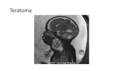
/
REPORT Systemic to pulmonary vascular malformation HMM Pouwels B REPORT Systemic to pulmonary vascular malformation HMM Pouwels B

REPORT Systemic to pulmonary vascular malformation HMM Pouwels B - PDF document

joyce
joyce . @joyce
Follow
343 views
Uploaded On 2022-08-26

REPORT Systemic to pulmonary vascular malformation HMM Pouwels B - PPT Presentation

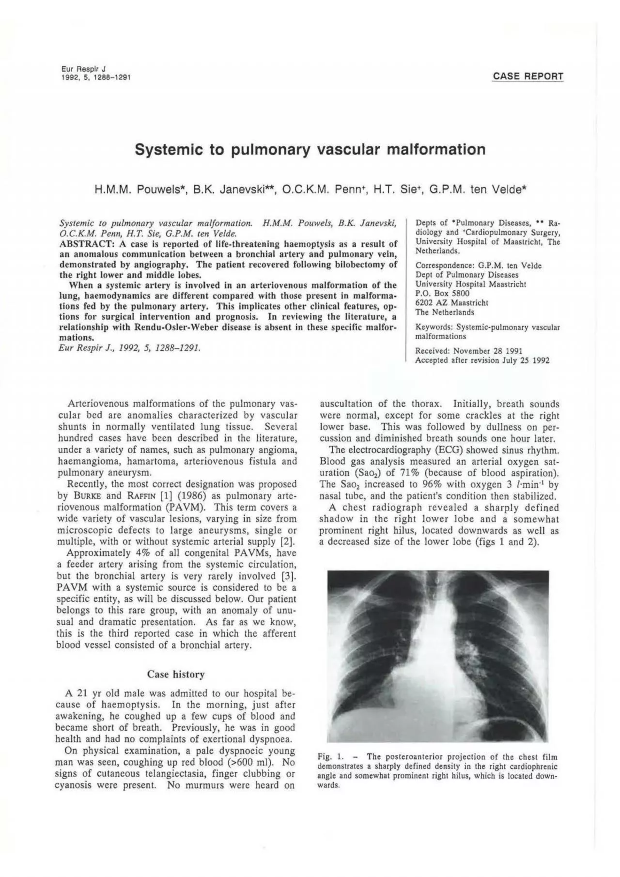
Eur Resplr J 1992 5 12881291 CASE OCKM Penn HT Sie GPM ten Velde Systemic to pulmonary vascular malformation HMM Pouwels BK Janevski OCKM Penn HT Sie GPM ID: 942354

systemic pulmonary left artery pulmonary systemic artery left bronchial arteriovenous blood malformation pavm vascular arteries lobe chest malformations supply

Share:

Link:

Embed:

Download Presentation from below link

Download Pdf The PPT/PDF document "REPORT Systemic to pulmonary vascular ma..." is the property of its rightful owner. Permission is granted to download and print the materials on this web site for personal, non-commercial use only, and to display it on your personal computer provided you do not modify the materials and that you retain all copyright notices contained in the materials. By downloading content from our website, you accept the terms of this agreement.


Presentation Transcript

Eur Resplr J 1992, 5, 1288-1291 CASE REPORT Systemic to pulmonary vascular malformation H.M.M. Pouwels*, B.K. Janevski**, O.C.K.M. Penn+, H.T. Sie+, G.P.M. ten Velde* Systemic to pulmonary vascular malformation. H.M.M. Pouwels, B.K. Janevski, O.C.K.M. Penn, H.T. Sie, G.P.M. ten Velde. Depts of •Pulmonary Diseases, • • Ra· diology and University Hospital of Maastricht, The Netherlands. ABSTRACT: A case is reported of life-threatening haemoptysis as a result of an anomalous communication between a bronchial artery and pulmonary vein, demonstrated by angiography. The patient recovered following bilobectomy of the right lower and middle lobes. Correspondence: G.P.M. ten Velde Dept of Pulmonary Diseases University Hospital Maastricht P.O. Box 5800 When a systemic artery is involved in an arteriovenous malformation of pt·esent in malforma­tions fed by the pulmonary artery. This implicates other clinical features, op­ tions for surgical intervention and prognosis. In reviewing the literature, a relationship with Rendu-Osler-Weber disease is absent in these specific malfor­mations. 6202 AZ Maastricht The Netherlands Keywords: Systemic-pulmonary vascular malformations Eur Respir J., 1992, 5, 1288-1291. Arteriovenous malformations of the pulmonary vas­ cular bed are anomalies characterized by vascular shunts in normally ventilated lung tissue. Several hundred cases have been described in the literature, under a variety of names, such as pulmonary angioma, haemangioma, hamartoma, arteriovenous fistula and pulmonary aneurysm. Recently, the most correct designation was proposed by BURKE and RAFFIN [1] (1986) as pulmonary arte­ riovenous malformation (PA VM). This term covers a wide variety of vascular lesions, varying in size from microscopic defects to large aneurysms, single or multiple, with or without systemic Our patient belongs to this rare group, with an anomaly of unu­ sual and dramatic presentation. As far as we know, this is the third reported case in which the afferent blood vessel consisted be­ cause of haemoptysis. In the morning, just after awakening, he coughed up a few cups of blood and became short of breath. Previously, he was in good health and had no complaints of exertional dyspnoea. On physical examination, a pale dyspnoeic young man was seen, coughing up red blood �(600 ml). No signs of cutaneous telangiectasia, finger per­ cussion and diminished breath sounds one hour later. The electrocardiography (ECG) showed sinus rhythm. Blood gas analysis measured an arterial oxygen sat­ blood aspiration). increased to 96% with oxygen 3 l·min·' by nasal tube, and the patient's condition then stabilized. A chest radiograph revealed a sharply defined shadow in the right lower lobe and a somewhat prominent right hilus, located downwards as well as a decreased size of the lower lobe (figs 1 and 2). Fig. 1. -The posteroanterior projection of the chest film demonstrates a sharply defined density in the right cardiophrenic angle and somewhat prominent right hilus, which is located down­wards. SYSTEMIC TO PULMONARY VASCULAR MALFO

RMATION 1289 Fig. 2. -The lateral projection of the chest film demonstrates marked vascular signs in the middle lobe and a decreased size of the lower lobe. On rigid bronchoscopy blood clots obstructing the right lower lobe bronchus were seen. Digital subtrac­ tion angiography of the pulmonary arteries revealed no abnormalities. Subsequently, a selective arteriogram of the bron­ chial arteries was performed (fig. 3). The arterial, capillary and venous phase of the left bronchial arte­ riogram were normal. However, the right selective bronchial angiogram revealed a hypertrophic and tor­ tuous right bronchial artery, with hypertrophic branches running towards a large malformation in the right lower and middle lobe. The venous drainage of this malformation occurred through the pulmonary veins from the middle and lower lobes toward the left atrium. With this procedure a left-to-left shunt was estab­ lished, forming a communication between the right bronchial artery and pulmonary vein, through a vascular mass. One day later the patient coughed up 400 ml bright red blood and his situation became precarious. After haemodynamic parameters had been stabilized, a sur­ gical intervention was performed. At thoracotomy, large convolutions of expanded bronchial arteries were seen at the dorsal side of the lower branch of the right bronchus. Side bran­ ches to the upper lobe were carefully dissected, and bilobectomy of the right lower and middle lobe was performed. Histopathology showed almost complete consolidation of the lobes because of extensive haemorrhage. In the bronchial wall, bron­ chial arteries were enlarged with focal media atrophy. No specific site of haemorrhage was found, as is often the case in pulmonary malformations. Fig. 3. -Selective arteriogram of the right bronchial artery performed by intra-arterial digital subtraction angiography (DSA). The opacified bronchial artery is enlarged with hypertrophic and tortuous branches to the lower and the middle lobe. The is an increase of blood supply to this region and an irregular accumu­ lation of contrast in the lower lobe. Note also, early venous fill­ ing inferiorly. Discussion Several theories have been proposed for the origin of PA VM; however, its embryological genesis and natural course remain unclear [2, 4, 5). Most malfor­ mations are believed to be congenital anomalies. PAVM with systemic blood supply is a rare lesion. In large series from the Mayo Clinic, only 3 out of 101 patients wilh PAVM derived their blood supply from a systemic feeder artery [6]. BosHER et al. f3) de­ scribed an "interesting variation of PA VM", in a large survey. In this series, in only 12 cases out of 350, the arterial blood supply derived from a systemic source (systemic-pulmonary arteriovenous malformation (S­PAVM)). No relationship with Rendu-Osler- Weber disease has been mentioned in the literature when the afferent supply is from the systemic circulation. Dyspnoea and the classical triad of cyanosis, clubbing of the digits and polycythaemia, as a result of under­ saturation in "conventional PA VM", are not present in

systemic arterial to pulmonary venous mal­ formation (7, 8), because it is a left-to-left shunt. This was already observed by MAtER et al. [9] in 1948. 1290 H.M.M. POUWELS ET AL. Shunts from systemic arteries to pulmonary vessels may originate from anomalous branches of the aorta and from bronchial [1 0, 11], intercostal, epicardial pericardiophrenic, lateral thoracic, oesophageal and, more frequently, internal mammary arteries [5, 12]. Accessory bronchial arteries may originate from these vessels and may contribute to the blood supply of S-PAVM. On physical examination the only sign of a S-PAVM is often a continuous systolic extra­ cardiac murmur, which should not be misinterpreted for persistent ductus arteriosus. Because of lack of symptoms, the discovery of these malformations is often an accidental finding. S-PAVM may be pre­ sented at any age, but most patients are young asymptomatic adults, predominantly male [13]. The right lung is involved more often than the left and, most importantly, they tend to be solitary, but may involve several lobes. S-PA VMs are subject to sys­ temic pressure, and so tend to grow and are more apt to rupture. Due to the rarity of these lesions, there is no experience of their evolution. Theoretically, complications such as haemoptysis are more likely to occur [9]. In the clinical work-up, chest radiographs are usu­ally abnormal, but lesions may be minimal and aspecific. A clear distinction between those malfor­mations with a systemic artery supply and those without is possible with angiography of pulmonary arteries, as well as of selective systemic arteries [14]. Hence, because of different haemodynamic and clinical findings, PAVM, with and without systemic blood vessel supply, can be distinguished according to the criteria summarized in table 1. DINES and eo-workers [6, 10] and other authors proposed conservative surgical intervention in sys­temic artery to pulmonary malformations because of future enlargement of the shunt, risk of rupture and infection. The majority of reported cases underwent a minimal surgical resection, including the entire removal of the malformation. Silastic spheres, gel-foam particles and autologuous tissue are dangerous in these malforma­tions, because of possible passage of the material into the pulmonary veins, with resultant systemic embolism [15]. Occlusion with balloon therapy is still an issue and depends on patient characteristics, local facilities and, most importantly, the necessary experience [16]. Spi­ nal cord injury, oesophageal necrosis, bronchial is­ chaemia and left main-stem bronchial stenosis are reported as serious complications [16, 17]. Our patient represents the first case with life­threatening haemoptysis occurring in congenital systemic to pulmonary arteriovenous malformations (S-PAVMs), namely of a bronchial artery to pul­ . - S-PAVM PAVM Female � male Relationship with ROW (40-65%) Classical trial of symptoms Right-to-left shunt Single or multiple Local or diffuse Hereditarity Angiography of a. pulmonalis is mandatory S-PAVM Female male No relationship with R

OW Absent Left-to-right of left-to-left shunt Single Local Absent Angiography of a. pulmonalis and selective systematic arteries is mandatory PAVM: pulmonary arteriovenous malformation; S-PAVM: systemic to pulmonary arteriovenous malformation; ROW: Rendu-Osler-Weber. Pulmonary angiograms sometimes demonstrate an early wash-out of contrast, when the vascular shunt is towards the pulmonary artery. They fail to show a defect, when the vascular communication has its drainage to the pulmonary vein, as in our case. Obviously, for the same reason, right heart catheteri­zation reveals no abnormality in these left-to-left shunts. Selective angiography of systemic arteries is mandatory before any surgical or other medical intervention and should be performed as soon as possible. References 1. Burke CM, Raffin TA. -Pulmonary arteriovenous malformations, aneurysms and reflections. Chest, 1986; 89(6): 771-772. 2. Burke CM, Safai C, Nelson DP, Raffin TA. - Pul­ monary arteriovenous malformations: a critical update. Am Rev Respir Dis, 1986; 134: 334-339. 3. Bosher LH, Blake DA, Byrd BR. -An analysis of the pathologic anatomy of pulmonary arteriovenous aneu­rysms with particular reference to the applicability of local excision. Surgery, 1959; 45: 91-104. 4. Anabtawi IN, Ellison RG, Ellison LT. -Pulmonary SYSTEMIC TO PULMONARY VASCULAR MALFORMATION 1291 arteriovenous aneurysms and fistulas: anatomical variations, embryology and classification. Ann Thorac Surg, 1965; 1: 277-285. 5. Brundage BH, Gomez AC, Cheitlin MD, Gmelich JT. -Systemic artery to pulmonary vessel fistulas. Chest, 1972; 62(1): 19-23. 6. Dines DE, Seward JB, Bernatz PE. -Pulmonary ar· teriovenous fistulas. Mayo Clin Proc, 1983; 58: 176-181. 7. Hearne SF, Burbank MK. -Internal mammary artery­ to pulmonary artery fistulas. Circulation, 1980; 62: 1131-1135. 8. Voll A, Marstrander F, Wexels P. - Systemic­ pulmonary shunt. Dis Chest, 1964; 45: 396-401. 9. Maier HC, Himmelstein A, Riley RL, Bunin JJ. -Arteriovenous fistula of the lung. J Thorac Surg, 1948; 17: 13-26. 10. Dines DE, Arms RA, Bernatz PE, Gomes MR. -Pulmonary arteriovenous fistulas. Mayo Clin Proc, 1974; 49: 460-465. 11. Lawrence EA, Rumel WR. -Arteriovenous fistula of the lung. J Thorac Surg, 1950; 20: 142-150. 12. Pillay R, Mitchell A, Jackson JW. -Arterial fistula between the left internal mammary artery and left pulmo­ nary artery. Thorax, 1982; 37: 386-387. 13. Kleiner JP, . - non­ resolving pulmonary infiltrate. Chest, 1980; 78: 327-328. 14. Gomes MR, Bernatz PhE, Dines DE. -Pulmonary arteriovenous fistulas. Ann Thorac Surg, 1969; 7: 582-593. 15. Stoll JF, Bettman MA. -Bronchial artery emboliza­ tion to control hemoptysis: a review. Cardiovasc lntervent Radiology, 1988; 11: 263-269. 16. Soo Hoo GW, Julien PJ, Brown HV, Belman MJ. -Improvement in exercise performance after pulmonary arteriovenous malformation embolization. Chest, 1990; 97(4): 1016-1018. 17. Girard Ph, Baldeyron P, Lemoinze G, Grunewald D. -Left main-stem bronchial stenosis complicating bronchial artery embolization. Chest, 1990; 97(5): 1246-1248.