/
Courtesy of CRC Press/Taylor & Francis Group Courtesy of CRC Press/Taylor & Francis Group

Courtesy of CRC Press/Taylor & Francis Group - PowerPoint Presentation

julia
julia . @julia
Follow
342 views
Uploaded On 2022-06-08

Courtesy of CRC Press/Taylor & Francis Group - PPT Presentation

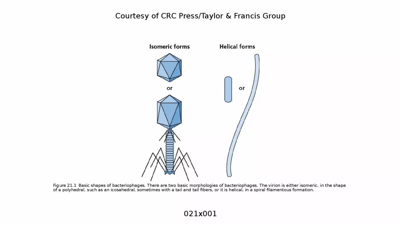
Figure 211 Basic shapes of bacteriophages There are two basic morphologies of bacteriophages The virion is either isomeric in the shape of a polyhedral such as an icosahedral sometimes with a tail and tail fibers or it is helical in a spiral filamentous formation ID: 915658

bacterial bacteriophage courtesy figure bacteriophage bacterial figure courtesy crc press taylor amp francis group genome bacteriophages membrane dna cell

Share:

Link:

Embed:

Download Presentation from below link

Download Presentation The PPT/PDF document "Courtesy of CRC Press/Taylor & Franc..." is the property of its rightful owner. Permission is granted to download and print the materials on this web site for personal, non-commercial use only, and to display it on your personal computer provided you do not modify the materials and that you retain all copyright notices contained in the materials. By downloading content from our website, you accept the terms of this agreement.


Presentation Transcript

Slide1

Courtesy of CRC Press/Taylor & Francis Group

Figure 21.1 Basic shapes of bacteriophages. There are two basic morphologies of bacteriophages. The virion is either isomeric, in the shape of a polyhedral, such as an icosahedral, sometimes with a tail and tail fibers, or it is helical, in a spiral filamentous formation.

021x001

Slide2

Courtesy of CRC Press/Taylor & Francis Group

Figure 21.2 Bacteriophages with membrane envelopes. Some bacteriophages, such as ϕ6 shown here, are encapsulated by membrane envelopes, derived from the bacterial cell membrane. During formation (bottom panel), bacteriophage proteins (brown) are integrated into the bacterial membrane, excluding the bacterial surface proteins (red).

021x002

Slide3

Courtesy of CRC Press/Taylor & Francis Group

Figure 21.3 Replication of bacteriophages. Bacteriophages replicate within bacterial cells following attachment and injection of the bacteriophage genome into the cell. Once new bacteriophage particles are made, they are released from the bacterial cell via lysis.

021x003

Slide4

Courtesy of CRC Press/Taylor & Francis Group

Figure 21.4 Lysogenic bacteriophages. Lysogenic bacteriophages integrate their nucleic acids into the bacterial chromosome.

021x004

Slide5

Courtesy of CRC Press/Taylor & Francis Group

Figure 21.5 Chronic and pseudolysogenic bacteriophage infections. Chronic bacteriophage infections are similar to lytic infections, however, the bacterial cells do not lyse. Instead, the bacteriophages bud from the bacterial membranes and leave the bacteria intact. Pseudolysogenic infections do not integrate into the chromosome, therefore, the bacteriophage nucleic acids do not replicate with the bacterial DNA and do not segregate into daughter cells upon division, as they would in lysogens.

021x005

Slide6

Courtesy of CRC Press/Taylor & Francis Group

Figure 21.6 Integration and excision of the bacteriophage genome. To generate the prophage, the bacteriophage genome enters the bacterial cell cytoplasm as linear DNA, which then circularizes. The circular bacteriophage genome recombines with the bacterial genome at the attP and attB sites, respectively. Integration requires the bacteriophage Int and bacterial IHF proteins. Excision needs the additional Xis bacteriophage protein and reforms attP and attB from the attL and attR sequences that flank the prophage.

021x006

Slide7

Courtesy of CRC Press/Taylor & Francis Group

Figure 21.7 Bacteriophage MS2. The structure, morphology, and size of bacteriophage MS2. The capsid is made of the maturation protein and 180 copies of the coat protein. The genome is 3,569 bases of RNA, encoding four proteins.

021x007

Slide8

Courtesy of CRC Press/Taylor & Francis Group

Figure 21.8 Bacteriophage λ. The structure, morphology, and size of bacteriophage λ. The capsid is made of many copies of proteins that form the head, tail, and tail fibers. The genome is 48,490 bases of double-stranded DNA, encoding 73 proteins.

021x008

Slide9

Courtesy of CRC Press/Taylor & Francis Group

Figure 21.9 Bacteriophage λ genome entry via LamB. The receptor for bacteriophage

λ on E. coli is the maltose porin protein LamB. Binding to this porin enables the genome to pass through the outer membrane. It then passes through the inner membrane via the mannose permease complex to reach the bacterial cytoplasm.

021x009

Slide10

Courtesy of CRC Press/Taylor & Francis Group

Figure 21.10 Bacteriophage T4. The structure and mechanism of nucleic acid injection into E. coli by bacteriophage T4. The tail of bacteriophage T4 has a contractile outer sheath and an inner core that is a rigid tube. When the tail fibers bind to the outer membrane, the sheath contracts and the tube pierces the outer membrane, peptidoglycan, and inner membrane, depositing the double-stranded DNA genome into the cytoplasm.

021x010

Slide11

Courtesy of CRC Press/Taylor & Francis Group

Figure 21.11 Comparison of bacteriophage T4 and Type 6 Secretion Systems. The structure of the T4 bacteriophage is similar to λ and many other bacteriophage with a head and tail capsid structure. The tail of T4 is also similar to the bacterial Type 6 Secretion Systems, which evolved from bacteriophage structures.

021x011

Slide12

Courtesy of CRC Press/Taylor & Francis Group

Figure 21.12 Bacteriophage 

X174 genome. This is a representation of the genes encoded by the bacteriophage 

X174

genome, which was the first DNA sequence to be determined using the Sanger sequencing method. This identified the coding regions of the bacteriophage.

021x012

Slide13

Courtesy of CRC Press/Taylor & Francis Group

Figure 21.13 The temporary tail of bacteriophage 

X174. To deliver its single-stranded circular DNA genome into the E. coli cytoplasm, bacteriophage 

X174

targets LPS on the surface of the bacterial cell, then polymerizes copies of the H protein into a tube through the outer membrane, peptidoglycan, and inner membrane. The circular genome of the bacteriophage is then able to pass through to the bacterial cytoplasm, at which point the H proteins dissociate.

021x013

Slide14

Courtesy of CRC Press/Taylor & Francis Group

Figure 21.14 Generalized and specialized transduction. Bacterial DNA can end up packaged within bacteriophages. In generalized transduction (left), random DNA is packaged into the capsid, which can include the bacterial DNA (blue), as well as the prophage genome (red). In specialized transduction (right), some bacterial DNA (blue) may be excised with the prophage (red) and end up packaged into the bacteriophages. In both cases, bacterial DNA is carried by the bacteriophage virion to the next bacterial cell.

021x014

Slide15

Courtesy of CRC Press/Taylor & Francis Group

Figure 21.15 Bacteriophage-mediated chromosomal rearrangements. The presence of prophages within a bacterial chromosome or plasmid can result in rearrangements. This can occur due to homologous recombination between two prophage genomes, indicated here with Φ. This can also occur when the prophage excises and reintegrates elsewhere, sometimes taking some of the bacterial sequence with it in the process.

021x015

Slide16

Courtesy of CRC Press/Taylor & Francis Group

Figure 21.16 Outer membrane vesicles to survive bacteriophages. Blebs of the outer membrane have the same surface proteins and structures as the bacteria that they came from, therefore, bacteriophage adsorption occurs on outer membrane vesicles (OMVs) as readily as it does on bacterial cells. This can help protect the bacterial cell, by diverting the bacteriophages to the OMVs.

021x016

Slide17

Courtesy of CRC Press/Taylor & Francis Group

Figure 21.17 Restriction digestion of bacteriophage DNA. When the bacteriophage DNA enters the cell, it is subject to the restriction-modification system. If the bacteriophage genome contains any recognition sequences for restriction enzymes expressed by the bacteria, the bacteriophage DNA will be digested. It is therefore neither replicated nor integrated into the chromosome.

021x017

Slide18

Courtesy of CRC Press/Taylor & Francis Group

Figure 21.18 Abortive infection system. Bacterial population level resistance to bacteriophages means that the infected cell sacrifices itself, but the surrounding cells survive. The bacteriophage replication is interrupted, resulting in premature lysis of the bacterial cell. Because no intact bacteriophage particles are made, the neighboring bacterial cells in the population survive. Compare to Figure 21.3.

021x018

Slide19

Courtesy of CRC Press/Taylor & Francis Group

Figure 21.19 Phage-inducible chromosomal islands. Some pathogenicity islands use infection by bacteriophages as a means to spread to new bacterial cells. Rather than succumbing to infection, the bacterial cell shuts down the replication of the bacteriophage genome, excises the pathogenicity island, replicates it, and packages it into the bacteriophage capsid. The pathogenicity island is then released with the lysis of the bacterial cell, spreading to other bacteria.

021x019