/
Determination of the Blood Pressure Determination of the Blood Pressure

Determination of the Blood Pressure - PowerPoint Presentation

julia
julia . @julia
Follow
343 views
Uploaded On 2022-06-11

Determination of the Blood Pressure - PPT Presentation

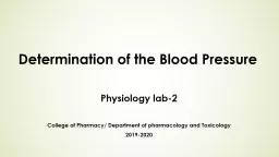
Physiology lab7 College of Pharmacy Department of pharmacology and Toxicology 20202021 Dr Shahad S Aldeen DEFINITION Blood pressure BP is the lateral pressure exerted by the column of blood on the wall of the artery ID: 916210

blood pressure method systolic pressure blood systolic method diastolic pulse artery cuff heart increased arteries exercise sphygmomanometer arterial manometer

Share:

Link:

Embed:

Download Presentation from below link

Download Presentation The PPT/PDF document "Determination of the Blood Pressure" is the property of its rightful owner. Permission is granted to download and print the materials on this web site for personal, non-commercial use only, and to display it on your personal computer provided you do not modify the materials and that you retain all copyright notices contained in the materials. By downloading content from our website, you accept the terms of this agreement.


Presentation Transcript

Slide1

Determination of the Blood Pressure

Physiology lab-7

College of Pharmacy/ Department of pharmacology and Toxicology

2020-2021

Dr. Shahad S.

Aldeen

Slide2

DEFINITION

Blood pressure (BP) is the lateral pressure exerted by the column of blood on the wall of the artery.

Slide3

Systolic pressure is the maximum pressure in the arteries during systole. It indicates:a. The extent of work done by the heartb. The force with which the heart is working

c. The degree of pressure which the arterial walls have to withstand.

Diastolic pressure

is the minimum pressure at the end of ventricular diastole. It is the measure of constant stretch to which walls of the arteries are subjected. It is more important than systolic pressure because:

a. It is less fluctuating

b. It is the constant load against which the heart has to work

c. It is the pressure of peripheral resistance and depends mainly on the tone of the arteries.

Slide4

Pulse pressure is the difference between the systolic and diastolic pressures. It is the rise in pressure caused by the ejection of blood into the aorta by ventricular contraction. It is a measure of stroke volume and compliance of arteries.

Mean arterial pressure

is the average pressure present throughout the cardiac cycle. It is responsible for pushing the blood through the systemic circulatory system. It is equal to diastolic pressure + 1/3 pulse pressure

Slide5

Blood Pressure Determinations

AIM

To determine the blood pressure of the given subject at rest and after moderate exercise.

APPARATUS

Sphygmomanometer and stethoscope.

PRINCIPLE

The pressure of blood in the artery (brachial artery) is balanced against the

pressure of air in a rubber cuff surrounding the artery. The pressure of air

in the cuff is then measured by means of a mercury manometer

.

Slide6

METHODS:1. Palpatory method

2. Auscultatory method

3. Oscillatory method

.

Slide7

ProcedurePalpatory method: The subject is asked to sit on a stool. The cuff is tied around the upper arm with the lower border of the cuff not less than 2.5 cm above the cubital fossa. The outlet valve of the bulb is closed. The radial pulse is palpated while the cuff is being inflated to a pressure slightly above the level at which the radial pulsation is no longer felt. The pressure at which the pulsation was obliterated is read in the mercury manometer. The outlet valve is opened. The manometric reading is noted at the point where the pulsation reappears. The average of the two readings gives the systolic pressure. The diastolic pressure cannot be determined by this method.

Slide8

procedureAuscultatory method: By palpatory method, only the systolic blood pressure could be measured. By auscultatory method, both the systolic and diastolic blood pressure can be measured. The chest piece of the stethoscope is placed over the brachial

artery.The

pressure in the cuff is raised above the systolic pressure (by about 30 mm Hg) previously determined by the palpatory method. The pressure is then lowered gradually (2–3 mm per second). The sounds that are heard are the Korotkoff’s sounds. The first sound (phase one) that occurs is a sharp tapping sound, indicates the peak systolic pressure, the second and third phases, initially murmurs in quality and then louder and more banging, are due to turbulent flow of blood through a partially occluded vessel. In the fourth phase, the sound becomes muffled and dull and the fifth phase accurately gives true diastolic pressure, which is disappearance of the sound (Figure 1).

Slide9

Figs 1: (A) Mercury sphygmomanometer; (B) Aneroid sphygmomanometer; (C) Riva-Rocci cuff; (D) Korotokoff’s sounds

A

B

C

D

Systolic

Bp

Diastolic

Bp

Slide10

procedureOscillatory method:

This is another method of determining blood pressure. By this method, both the systolic and diastolic blood pressure are determined. The pressure at which oscillations appear in the mercury manometer gives the systolic pressure and the pressure at which it disappears give the diastolic blood pressure. However, this method is not accurate.

Slide11

Important precautions in the use of sphygmomanometer: 1. The manometer should be placed at the level of the heart.

2. The lower border of the cuff should be 2.5 cm above the cubital

fossa. For children, a narrow cuff should be used.

3. Blood pressure should be preferably taken in the left arm

Slide12

NORMAL VALUESThe average systolic pressure in healthy adults is 100–140 mm Hg. The average diastolic pressure is 60–90 mm Hg. In children it is closer to the lower end of the scale and in the elderly, it reaches or even exceeds the higher figure. The difference between the systolic and diastolic pressure is the pulse pressure 40–60 mm Hg.

Slide13

Observing the Effect of Various Factorson Blood Pressure and Heart Rate

Arterial blood pressure is directly proportional to cardiac output (CO, amount of blood pumped out of the left ventricle per unit time) and peripheral resistance (PR) to blood flow, that is,

BP = CO * PR

Peripheral resistance is increased by blood vessel constriction (most importantly the arterioles), by an increase in blood viscosity, and by a loss of elasticity of the arteries (seen in arteriosclerosis). Any factor that increases either the cardiac output or the peripheral resistance causes an almost immediate reflex rise in blood pressure.

Slide14

Physiological Variations

Blood pressure is slightly lower in women than men.

Persons with slender build have got a lower blood pressure than those of heavy build.

During sleep, systolic pressure is less.

Emotional excitement and muscular exercise cause an increase in the blood pressure. It is also increased after meals.

The blood pressure especially the diastolic is highest in the standing position, lower in the sitting and lowest while the subject is lying down.

Slide15

Effect of moderate exercise on blood pressureDuring exercise, there is a moderate increase in systolic blood pressure. This is due to an increase in cardiac output caused by an increased heart rate and myocardial contractility (stroke volume increases) due to increased sympathetic activity, and increased venous return.

The effects of vasoconstriction in inactive regions are overcome by vasodilatation in active muscles. Hence, the diastolic pressure at the pre-exercise level is slightly reduced.

Pulse pressure is increased.

Slide16

The Pulse

The term pulse refers to the alternating flows of pressure (expansion and then recoil) in an artery that occur with each contraction and relaxation of the left ventricle.

Normally the pulse rate (or the count of arterial pulse per minute) equals the heart rate (beats per minute), and the pulse averages 70 to 76 beats per minute in the resting state.

The pulse may be felt easily on any superficial artery when the artery is compressed over a bone or firm tissue such as at the neck (carotid artery), on the inside of the elbow (brachial artery), at the wrist (radial artery).

Slide17

Thank you