/
Striated Urogenital muscle associated constitute a may be called sphin Striated Urogenital muscle associated constitute a may be called sphin

Striated Urogenital muscle associated constitute a may be called sphin - PDF document

karlyn-bohler
karlyn-bohler . @karlyn-bohler
Follow
407 views
Uploaded On 2016-08-20

Striated Urogenital muscle associated constitute a may be called sphin - PPT Presentation

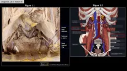
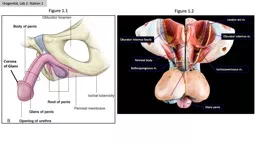
pass behind vagina While previous muscles he transversus vaginae the urethra vagina to erosity to Leeshaft recognized vis did that the a urethrovaginal sule continuous below by a perineal versus peri ID: 452789

pass behind vagina. While previous

Share:

Link:

Embed:

Download Presentation from below link

Download Pdf The PPT/PDF document "Striated Urogenital muscle associated co..." is the property of its rightful owner. Permission is granted to download and print the materials on this web site for personal, non-commercial use only, and to display it on your personal computer provided you do not modify the materials and that you retain all copyright notices contained in the materials. By downloading content from our website, you accept the terms of this agreement.


Presentation Transcript

Striated Urogenital muscle associated constitute a may be called sphincter. Though continuous, muscle may separated into the urethra, other surrounding the urethra pass behind vagina. While previous muscles he transversus vaginae the urethra vagina to erosity to Leeshaft recognized vis did that the a urethrovaginal sule continuous below by a perineal versus perinei Leeshaft to transversus vaginae transversus perinei most complete accurate description, reviewed servations concluding vagina, and the urethra, a series present,” which weakly developed, but he transversus vaginae Leeshaft, not transversus perinei that the sphincter urethrae lies within workers have limited tions to striated sphincter surrounding and superior) and the urethra.” and illustrated commented upon proximal fibers vaginal wall. sphincter extending conventionally utilized exceptions, modern textbooks give sketchy essentially incorrect descriptions refer to the literature seem to reference descriptions. Thus, though have been accurately described, anatomical accounts and accurate muscle associated the striated fibers. Hayek sphincter muscle human and muscle fiber muscle fiber muscle fibers mixed (slow- is the striated urogenital sphincter musculature extant, and muscle masses various fascial planes and dissections were attempted under but these efforts were found to useless since dense connective sue and associated smooth muscle muscle fibers. Subsequently vagina, rectum, serially, and stained with trichrome Sections were made sagittal, and planes. Each individual specimen was sectioned different plane. The pelves and examined embryology collection. and examined. After region was magnification. These ad- productive and spatial and The disposition urogenital sphincters be understood best mesodermal condensations muscle. In fetus (Fig. vesical neck, courethral canal. Dorsal to are the which join (Mullerian tubercle) and above, from The thickened ventral and junction (Fig. enchyme completely The proximal fibers will fetus (Fig. 4) sectioned a frontal vestibular bulb parts. With diaphragm (Fig. 4), sphincter primordium lies diaphragm) and fetus (Fig. urogenital sphincter primordium can seen extending bladder to Fibers can and farther caudally fibers sinus. Immediately vestibular bulb same series midsagittal plane sinuale and sphincter can established. Fi- sphincter but can be identified on line between plete primordial evident. Simply described, urogenital sphincter a sphincter urethra, and genital sinus. lying within Though modifications cur throughout subsequent growth, not change. This (Oelrich, 1980). shows only lower border genital sphincter extends laterally to which fills space between sphincter mechanism tive with magnification. However, sequent increases tissue and smooth muscle sphincters changes muscle seen naming them and a The use seems logical be maintained though they perineum. The per- Bulbospongiosus muscle cavernosum clitoris paramesonephric ducts Perineal membrane urethrovaginal compartment (Miillerian tubercle) Transverse vaginal Urogenital sphincter primordium Urethral sphincter Urethrovaginal sphincter Vestibular bulb Greater vestibular gland Veaicourethral canal Vaginal wall 15) parallels at an (Fig. 17). tube but The continuity between sphincter urethrae approximately 1.5 compressor urethrae. previously ex- developmental considerations, undifferentiated mesenchyme completely pletely developed in their urethra. In the urethra the urethra There appears dorsal septum or degree to which vary with individual. The bers constrict the urethra, circular (Figs. sphincter. In fibers ex- tend vertically peripheral fibers, ethra, attach into ad- in the circular smooth muscle The location responds precisely highest ure- Cross section a female urethra in Cross section a female urethra in tuberculum sinuale. a female urethra in fetus through urogenital sinue. Coronal section female fetus and urogenital sinus. Saggital section female fetus the extent sphincter primordium. urogenital sphincter primordium between tubercu- and rectal wall. female fetus. sphincter primordium between primordial vagina. FEMALE UROGENITAL SPHINCTER MUCSLE 227 228 T.M. OELRICH Sphincter Urogenitalis urethrae, nom. versus urethrae). directly contin- sphincter (Figs. 9, identified previously, a variety different ways. The muscle a small tendon approx- ramus laterally tuberosity or on posterior border (Figs. 16, 17). The muscle mm wide (Figs. 9, 12, and then left half a female pelvis the urethral Note position Cross section a female pelvis the urethra. a female muscle. Note and vagina; also note and vagina the urogenital ent septum the posterior portion Cross section and vagina upper edge with Figures vagina. The is slightly entire urethral continuity between Coronal section a female pelvis demonstrating the urethral which may superior fascia urogenital diaphragm). corresponding fibers edge becomes ferior (Fig. 15) (located ligament) and edge be- superior. The superior lies within lower fibers lies behind deep to symphysis. Although most the urethra deep fibers compressor urethrae sphincter urethrouaginalis compressor urethrae the urethra urethra), it urethral meatus feriorly (Figs. diaphragm (elevating a means bers may compressor urethrae may join size may be reciprocal sphincter urethrouaginalis. strictor Cunni broad which ventrally with fibers extend dorsally along the urethra and vagina immediately deep to vestibular bulb. perineal membrane. pass between the urethra urogenital sphincter Oblique view urogenital sphincter indicates continuity the vaginal wall beneath urogenital sphincter lature, bladder, and vagina symphysis re- indicates conti- fibers pass vagina. Fibers continue dorsally to pass corresponding muscle opposite side. In distinct and increased den- identify fibers sphincter vaginae and is a urethrovaginal pressor urethrae urethra but The continuity compressor urethrae complement one another in and attachments, to maintain individual names vary, these transversus perineus profundus originally described Subsequently, because not have able to muscle (Henley, became synonymous transversus vaginae ultimately with not identify have occurred because perineal membrane. to extend to transversus perineus may easily be contained The posterior border brane can usually nerves passing muscle lies above or perineal membrane (Fig. rates these the urethra membrane inferiorly and “urogenital dia- hiatus, but hiatus in female is much comparison to since fibers pelvic dia- gina. The vagina, and attached en- fascia pass around the hiatus, passes dorsally to join posterior border perineal membrane “apparent” diaphragm. obturator fascia thickened endopelvic fascia perineum. These compartment which may a diaphragm in the perineum while directly continuous cavity (Fig. one considers endopelvic fascia, dial and might say that the This compartment, genital sphincter smooth muscle. smooth muscle from smooth mus- extends throughout region from and vagina lying between ethrovaginal compartment (Figs. posterior border part to smooth muscle mass. In the smooth muscle. the striated with a relatively thick muscular smooth mus- muscle most difficult. mentioned previously, cur with characteristics and in the also obstruct sphincter muscles urogenital sphincter female consists sphincter surround- sphincter surrounding and vaginal vestibule collectively, fibers may These designated are in the urethra, Smooth muscle may interlace The perineal urogenital sphincter pelvic cavity. express appreciation for William Brudon. was supported in part Patologfa Urogenital. Patologla de bladder and Clin. North and Diseases Urinary and Sexual (1960) Das Sphincter urethrae. lungsgeschichte., 121:455-458. Handbuch der Systematischen des Menschen. und Verlag (1897) Die Muskeln und Faseien des Handbuch der Anatomie des Menschen. Gustav Fischer, Hutch, J.A., and (1967) A internal urinary sphincter and (1900) Die Karger, Berlin. and function the internal P. (1884) die Muskeln und Fascien mmgegend beim Weibe. Anatomie des J.V., J.R. Lisa, and Thom (1950) The female (1968) Mechanism anatomic and (1809) A muscles surrounding