/
www.biocare.netison of the 3.    de Vries TJ,  Expression of gp100, MA www.biocare.netison of the 3.    de Vries TJ,  Expression of gp100, MA

www.biocare.netison of the 3. de Vries TJ, Expression of gp100, MA - PDF document

karlyn-bohler
karlyn-bohler . @karlyn-bohler
Follow
392 views
Uploaded On 2015-09-03

www.biocare.netison of the 3. de Vries TJ, Expression of gp100, MA - PPT Presentation

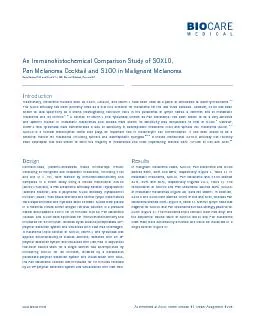
However S100 often reacts with a host Conversely S100 has been shown to lack sensitivity in epithelioid melanomas when compared to MART1 showed MART1 and Tyrosinase to be positive in 917 33 ID: 120948

However S100 often reacts

Share:

Link:

Embed:

Download Presentation from below link

Download Pdf The PPT/PDF document "www.biocare.netison of the 3. de Vrie..." is the property of its rightful owner. Permission is granted to download and print the materials on this web site for personal, non-commercial use only, and to display it on your personal computer provided you do not modify the materials and that you retain all copyright notices contained in the materials. By downloading content from our website, you accept the terms of this agreement.


Presentation Transcript

www.biocare.netison of the 3. de Vries TJ, Expression of gp100, MART-1, tyrosinase, and S100 in paraffin-embedded primary melanomas and locoregional, lymph node, and visceral metastases: implications for diagnosis and immunotherapy. A study conducted by the EORTC Melanoma Cooperative HMB-45, S-100, NK1/C3, and MART-1 in metastatic melanoma. Hum Pathol. 2004 Feb; 35(2):217-23.ns: a comparison 6. Nakajima T, Immunohistochemical demonstration of S100 protein in malignant melanoma and pigmented nevus, and its diagnostic application. Cancer. 1982 Sep 1; 50(5):912-8.8. Nonaka D, Chiriboga L, Rubin BP. Sox10: a pan-schwannian and melanocytic marker. Am J Surg Pathol. 2008 Sep; 32(9):1291-8.9. Mohamed A, et al. SOX10 expression in malignant melanoma, carcinoma, and normal tissues. Appl Immunohistochem Mol Morphol. 2013 10. Tacha D, A Newly Developed Mouse Monoclonal SOX10 Antibody is a Highly Sensitive and Specific Marker for Malignant Melanoma, Comparison of five antibodies as markers in the diagnosis of melanoma in cytologic preparations. Am J Clin Pathol. 2002 Immunohistochemical distinction of epithelioid histiocytic proliferations from epithelioid melanocytic nevi. Am J 13. Fernandes BF, 14. de Vries TJ, High expression of immunotherapy candidate proteins gp100, MART-1, tyrosinase and TRP-1 in uveal melanoma. Br J Cancer. 1998 Nov; 78(9): 1156–61. However, S100 often reacts with a host Conversely, S100 has been shown to lack sensitivity in epithelioid melanomas when compared to MART-1 showed MART-1 and Tyrosinase to be positive in 91.7% (33/36) and 97.2% (35/36) of uveal melanomas, respectively. Moreover, MART-1 and Tyrosinase when used together as Pan Melanoma Cocktail stained 100% of uveal melanomas.Uveal melanomas differ from cutaneous melanomas in the respect that the expression of these immunotherapy antigens was much more In another study, 100% (50/50), 98% (49/50) and 92% (46/50) of primary cutaneous malignant melanomas were shown to be positive with S100, Pan Melanoma Cocktail and HMB45 respectively, thus demonstrating the high sensitivity of Pan Melanoma when compared to S100. Nonaka et al showed SOX10 nuclear expression in 97% (76/78) of melanomas, whereas S100 was expressed in 91% than SOX10 and S100 in malignant and metastatic melanomas. However, it is well known that MART-1 and Tyrosinase stain a much lower Overall, S100 stained 88% of malignant melanomas (Table 1). When examining the literature, some publications have shown higher staining percentages in melanomas. Our results could be due to TMA tissue sampling. However, there were clear cases of melanoma that were strongly Melanoma stained 93% (67/72) of all melanomas vs. S100 which stained 88% (63/72). We only observed one case of melanoma that was e only observed one case of melanoma that was and Pan better choice for screening of metastatic and malignant melanoma due to the specificity and sensitivity demonstrated in our study. A potential double stain that may include SOX10, MART-1 and Tyrosinase may be suitable for tumors of unknown origin or in differential diagnosis of melanoma and its mimics. 5A Melanoma stained with SOX10, MART-1 and Tyrosinase (FR)Table 1: Comparison of SOX10, Pan Melanoma and S100 Expression in Melanoma Melanoma Type 1A 1B 1CFigure 1: MelanomaMelanoma stained with SOX10 Melanoma stained with Pan Melanoma Cocktail Melanoma stained with S100 3A 3B 3CFigure 3: Desmoplastic MelanomaDesmoplastic melanoma stained with SOX10Desmoplastic melanoma stained with Pan Melanoma CocktailDesmoplastic melanoma stained with S100 2A 2C 2B 2DFigure 2: Metastatic MelanomaMetastatic melanoma stained with SOX10Metastatic melanoma stained with S100 (Arrows: melanin pigment)Metastatic melanoma stained with Pan Melanoma CocktailMetastatic melanoma stained with SOX10 and Pan Melanoma Cocktail 4A Figure 4: Normal Lymph Node An Immunohistochemical Comparison Study of SOX10,Pan Melanoma Cocktail and S100 in Malignant Melanoma www.biocare.netDavid Tacha, PhD and Charlie Yu, MD; Biocare Medical, Concord CA [BC34] (nuclear), a Pan Melanoma antibody cocktail (cytoplasmic) were deparaffinized and hydrated down to water. Slides were placed A melanoma triple cocktail of SOX10, MART-1 and Tyrosinase was polymer detection system and visualized with Fast Red. A sequential two-color double stain for a single section was accomplished by the Pan Melanoma Cocktail was incubated for 30 minutes followed stained 88%, 90% and 88%, respectively (Figure 1, Table 1). In 81%, 89% and 81%, respectively (Figures 2A-C, Table 1). The combination of SOX10 and Pan Melanoma stained 92% (24/26) Melanoma stained 50% (Figure 3, Table 1). Normal lymph node was Traditionally, melanoma markers such as S100, HMB45, and MART-1 have been used as a panel of antibodies to identify melanoma.The S100 antibody has been primarily used as a first-line screener for melanoma for the last three decades. However, S100 has been A cocktail of MART-1 and Tyrosinase (known as Pan Melanoma) has been shown to be a very sensitive However, MART-1 and Tyrosinase have demonstrated a lack of sensitivity in desmoplastic melanoma (DM) and spindle cell melanoma (SCM).been developed that was shown to stain the majority of melanomas and most importantly, stained 98% (47/48) of DM and SCM.As Presented at ASDP, Poster Session #1, Poster Assignment #278