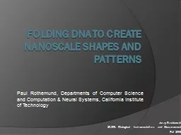
/
Folding DNA to create  nanoscale Folding DNA to create  nanoscale

Folding DNA to create nanoscale - PowerPoint Presentation

kittie-lecroy
kittie-lecroy . @kittie-lecroy
Follow
347 views
Uploaded On 2018-10-08

Folding DNA to create nanoscale - PPT Presentation

shapes and patterns Paul Rothemund Departments of Computer Science and Computation amp Neural Systems California Institute of Technology Jerzy Szablowski 20309 Biological Instrumentation and ID: 686692

molecule dna assembled structures dna molecule structures assembled staples molecules assembly properly structure afm 2006 folding 2003 patterns rothemund shapes pixels methods

Share:

Link:

Embed:

Download Presentation from below link

Download Presentation The PPT/PDF document "Folding DNA to create nanoscale" is the property of its rightful owner. Permission is granted to download and print the materials on this web site for personal, non-commercial use only, and to display it on your personal computer provided you do not modify the materials and that you retain all copyright notices contained in the materials. By downloading content from our website, you accept the terms of this agreement.


Presentation Transcript

Slide1

Folding DNA to create nanoscale shapes and patterns

Paul Rothemund, Departments of Computer Science and Computation & Neural Systems, California Institute of Technology

Jerzy

Szablowski

20.309

: Biological Instrumentation and

Measurement

Fall 2008Slide2

NanopatterningAvailable methods: Atomic force microscopy Scanning tunneling microscopy micro-contact printing

Lithography Self-assembly

Fabrication of sub-micron structures allowing for arbitrary positioning of molecules and, in some cases, single atoms.

Source: IBM, 1990

Zhang, 2003

Rothemund

, 2006

(“Top-bottom” method)

(“Bottom-up” method)Slide3

Self assembly using DNACan assembly multiple structures at a time.Can occur in less stringent conditions than “top-down” methods (AFM, STM…).Does not require expensive equipmentComplexity of patterns formed is not as high as in the other cases.Some exciting studies emerged:

Nanowire

mesh, by Yan et al,2003

Chen and

Seeman

, 1991

 These approaches very sensitive to DNA concentrations, and require multiple reactions and purification steps. Yet patterns complexity doesn’t match other methods of nanofabrication. Is it worth it?

Rothemund

, 2006Slide4

How to build an arbitrary 2-D pattern with 6-nm resolution out of DNA.Draw out a shape you would like to obtain, determine the desired size.

Sketch a single molecule of DNA

through the shape, as in the picture below.

Proceed with a custom written software.

(

Rothemund

, 2006)Slide5

Computer aided design of DNA assembliesDesign primers binding to two distant sites of the DNA molecules: this will fold DNA into a desired structure.

It might be necessary to introduce additional bridges that will stabilize the structure.

Synthesize linkers and scaffold, add

togather

in a tube and cool down from 95C to 20C at 1C per minute.

6nm pixelSlide6

Results

All shapes based on a single M13mp18 DNA molecule. Containing up to 273 linkers. Many of the structures are assembled properly. Regardless of stoichiometry.

70% assembled properly.

1% assembled properly,

rest slanted.

88% assembled properly,

with additional DNA staples.

Based on probability

Stronger structures – with lower energy are more likely to assemble.

Mechanic disruption: inserting the AFM tip can destroy the DNA structures. Sometimes also: AFM imaging artifact.

100 nm

100 nm

100 nmSlide7

Assembling higher order structuresStructures can be assembled by adding“staples” on the side of an assembled molecule. Staples are put in on the rim which previously held its structure:

Scheme.Real result

100nm

Many other structures can be made! Both contained and of unlimited size.Slide8

Implanting the “pixels”Brighter specks on the image represent labeled DNA. Up to 200 pixels can be placed. Labels are pieces of DNA and anything that is attached to it, such as: - biotin, fluorophores… proteins?Slide9

ApplicationsUnlimited: each pixel can be a specific aptamer binding a protein or a small molecule. Biotin also can serve as a very efficient adapter for binding. We could place molecules arbitrarily in space.Some ideas include:Nanobreadbord: with

nanowires, gold nanoparticles, maybe enzymes, fluorophores…Small “catalyzing centers”, where molecules are patterned in a way favoring certain chemical reactions.

n

ano

-Microarrays?

Future: 3D structures, greater number of pixels.Slide10

Summary“DNA origami”, allows for folding a DNA molecule into a highly predictable two-dimensional shape with 6nm resolution.It requires “stitching” various parts of the molecule by short complementary primers (staples).All these molecules can be implanted with DNA adapters (pixels) that can bind range of moleculesBut sometimes only a fraction of molecules fold properly.Slide11

References and questionsReferencesRothermund, P. W. K. Folding DNA to create nanoscale shapes and patterns. Nature 440, 298–302 (2006).

Yan, H., Park, S. H., Finkelstein, G., Reif, J. H. & LaBean, T. H. DNA-templated self-assembly of protein arrays and highly conductive nanowires

. Science 301,1882–-1884 (2003).

Zhang

, S. Fabrication of novel biomaterials through molecular self-assembly.

Nat.

Biotechnol.

21, 1171–1178 (2003).

Chen J, Seemen, N.C. The synthesis from DNA of a molecule with the connectivity of a cube. Nature, 350, 631-633 (1991)

Questions

?Slide12

What about twisting DNA? Other steric hindrances?It does twist. To avoid deleterious strain, staples are placed either facing upwards, or downwards, which in the longer run reduces the strain.It also bends because of the DNA stiffness and curving on the edges. That’s why AFM images show Slide13

Do you have more cool images? Slide14

Would not M13 genome dimerize, leaving no space for staples? Yes it would, but apparently thanks to very high concentration of staples and conditions of folding – it rarely does.

Secondary structure of M13 genome, from supplementary material.