/
THE CIRCULATORY SYSTEM THE CIRCULATORY SYSTEM

THE CIRCULATORY SYSTEM - PowerPoint Presentation

kittie-lecroy
kittie-lecroy . @kittie-lecroy
Follow
342 views
Uploaded On 2019-11-20

THE CIRCULATORY SYSTEM - PPT Presentation

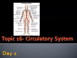
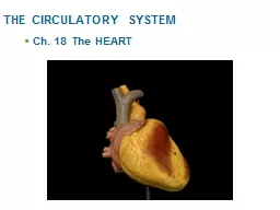
THE CIRCULATORY SYSTEM Ch 18 The HEART ASSIGNMENT Most Of The Anatomy We Have Already Done In The Lab So Make Sure To Do The of slide 1 through 29 As I Will Likely Not Take The Time To Review This Material ID: 766034

heart cardiac ventricles valves cardiac heart valves ventricles blood depolarization cells figure muscle node valve ventricular coronary repolarization atria

Share:

Link:

Embed:

Download Presentation from below link

Download Presentation The PPT/PDF document "THE CIRCULATORY SYSTEM" is the property of its rightful owner. Permission is granted to download and print the materials on this web site for personal, non-commercial use only, and to display it on your personal computer provided you do not modify the materials and that you retain all copyright notices contained in the materials. By downloading content from our website, you accept the terms of this agreement.


Presentation Transcript

THE CIRCULATORY SYSTEM Ch. 18 The HEART

ASSIGNMENT : Most Of The Anatomy We Have Already Done In The Lab, So Make Sure To Do The * of slide 1 through 29. As I Will Likely Not Take The Time To Review This Material.

* PARTS OF THE CARDIOVASCULAR SYSTEM * 1. * 2. * 3.

I. Heart Anatomy * A. Size, Location, and Orientation * Size: * Location: * Orientation of A pex & Base Point of maximal intensity (PMI) Diaphragm Sternum 2nd rib Midsternal line base apex STUDY QUESTION EXAMPLES : 1. Describe the Heart’s Size, Location, Orientation, Coverings, and Wall Layers

B. Coverings of the Heart 2A Review See text p. 18 * Serous Membranes - *P_________ - *V_________ - *for Heart named: *Cavity - * and that cavity is in the M____________________ (page 665)

B. Coverings of Heart … Figure 11.1a–b 1. * Pericardium: Description = Fibrous pericardium – Function: Parietal pericardium Visceral Pericardium = epicardium = is the same as the Visceral Pericardium * Serous fluid : Outer Sac Inner Sac

Figure 18.1c (c) Superior vena cava Left lung Aorta Parietal pleura (cut) Pericardium (cut) Pulmonary trunk Diaphragm Apex of heart The Heart = pump of the cardiovascular system

C. * Layers of the Heart Wall Figure 11.2b 1. * Epicardium 2. * Myocardium What type of tissue: 3. * Endocardium – endothelium: is what type of tissue

D. Fibrous Cardiac Skeleton (page 666 ) Cardiac muscle bundles Connective Tissue * Thickest in what areas and why? Electrical Characteristics: * Can action potentials pass through it? Importance: limits the spread of action potentials through the cardiac muscle to certain areas, or pathways (… in “III-A. Electrical Events”) 2. What is the Fibrous Skeleton and what is its purpose?

* Atria: * Auricles * Ventricles : * Interatrial & Interventricular Septa: location & description? *Sulci = describe?Coronary Sulcus*Anterior Interventricular Sulcus : location? What is within it? * Posterior Interventricular Sulcus : same questions as above? E. Chambers and Associated Great Vessels Overview

E. Chambers and Associated Great Vessels … 1. * Atria * Pectinate Muscles Fossa Ovalis Foramen OvaleRight *Superior Vena Cava: Blood from where to where?*Inferior Vena Cava: same?Coronary Sinus (later )Left * Pulmonary Veins: from where to where?

2. *Ventricles& Blood & Vessels– 2 pumps Figure 11.4 Right vs. Left * Shapes : * Thickness, why? * Trabeculae carneae * Papillary Muscles * Right: Pulmonary Trunk  blood goes where? * Left: Aorta– Blood goes where? * Coronary Arteries: Branch of ? Blood goes where?

F . Pathway of Blood Through the Heart 1. 2 PUMP SYSTEM with two circulations Blood Flow Direction: Pulmonary Circulation : Systemic Circulation : 2. Blood Vessels in Pathway 5. Describe the path a blood cell would take starting with the Right Atria and ending back at the RA. 6. Explain how the heart is like 2 pumps.

G. *Coronary Circulation =? Figure 11.2a * Coronary Arteries: a) * Supply the heart muscle w what? b) * Branch from: 2. * Branches of the Left Coronary Artery = 3. * Branches of the Right Coronary Artery =

Figure 18.7a Right coronary artery Right marginal artery Posterior interventricular artery Anterior interventricular artery Circumflex artery Left coronary artery Aorta

Figure 18.7b Superior vena cava Anterior cardiac vein s Small cardiac vein Middle cardiac vein Great cardiac vein Coronary sinus (b) The major cardiac veins Pulmonary Trunk Aorta G. Coronary Circulation … 5. Cardiac Veins - * Function? - Coronary Sinus =

6. Homeostatic ImbalancesSTUDENTS DO* Angina pectoris * Myocardial Infarction (heart attack )

H. FETAL CIRCULATION * Lungs are functional? 1. Alteration of Atrial Blood Flow Foramen Ovale After Birth = Fossa Ovalis 2 . Alteration of Blood Flow from Pulmonary Trunk Ductus Arteriosis Ligamentum Arteriosum 3 . Umbilical Cord with Umbilical Artery and Veins

I . Heart Valves 1. Function: prevent backflow of blood 2. * Atrioventricular (AV) valves— between what cavities? *Bicuspid (mitral) valve __________ side* Tricuspid valve ___________sideParts: Cusps, Chordae Tendinae, Papillary muscles *When ventricles contract: these valves do what?

H. Heart Valves … *Parts: Describe and give functionCusps Chordae Tendinae Papillary muscles * What happens to these valves when the atria and then the ventricles contract?

Figure 11.5a, step 1 Returning blood fills relaxed atria and ventricles (open AV valves) Ventricles 3. Operation of the AV valves AV valves open (a) H. Heart Valves …

Figure 11.5a, step 2 Returning blood fills relaxed atria and ventricles (open AV valves) Atria contract – more blood forced into ventricles Relaxed Ventricles Operation of the AV valves … AV valves open (a)

Figure 11.5a, step 4 Ventricles contract, blood forced against AV flaps / into arteries AV valves close preventing backflow to atria (LUB of LUB-dup sound) (a) Operation of the AV valves … Chordae tendineae prevent valve flaps from everting

H. Heart Valves … 4. Semilunar valves Function: Figure 11.2c Pulmonary semilunar valve – ____side Aortic semilunar valve – ______side * When Ventricles contract, what happens to valve? 3 cusps each

Figure 11.5b, step 1 As ventricles contract semilunar valves are forced open Aorta Pulmonary trunk Semilunar valve open 5. Operation of the semilunar valves (b)

Figure 11.5b, step 2 Aorta Pulmonary trunk Semilunar valve open Semilunar valve closed As ventricles relax, blood flows back from arteries, forcing semilunar valves to close Operation of the semilunar valves … (b) All this to ensure that… ‘dup’ of LUB-dup sound

* Review Skeletal Muscles (Text pages 282-288) * Number of nuclei? * Sarcolemma = * Myofibrils = * Sarcoplasmic Reticulum = * Sarcomeres = * T-tubules = * Triads = II. Cardiac Muscle Fibers—Histology A. Microscopic Anatomy of Cardiac Cells Thin Myofilaments Thick Myofilaments Skeletal Muscle CellScaroplasmic ReticulumT Tubules MyofibrilsSarcomere

A. Microscopic Anatomy of Cardiac Cells… 1. * Cardiac muscle cell characteristics: see list under skeletal muscles, previous slide (text pp. 676-677) 2. Endomysium anchored to: 3. T tubules: . Triads? . 4. SR: .  Source of Ca+2: . 5. Mitochondria: Nucleus Cardiac muscle cell Sarcolemma Z disc Mitochondrion Mitochondrion T tubule Sarcoplasmic reticulum Intercalated disc

A. Microscopic Anatomy of Cardiac Cells … 6. * Intercalated discs = 7. Functional Syncytium: Autorhythmic Cells = Function: 8. Long Absolute Refractory Period Function: Desmosomes Gap junctions Intercalated discs

III. Heart Physiology A . Mechanism and Events of Contraction 1. * Review of Skeletal Muscle Cells * Depolarization -via AP Proceeds down T tubulesCausing SR to release Ca+2 *Repolarization occurs while * Excitation-Contraction Coupling occurs Ca+2 attaches to Troponin Myosin Cross-bridges form Thin Myofilament slides inward Sarcomeres shorten Cells shorten  Muscle shortens *Na+-K+ pump redistributes ions & SR reabsorbs Ca+2

Skeletal Fibers Cardiac Cells Muscle Tension Have gap junctions between cells (intercalated discs) Source of Ca+2: mostly extracellular through Ca+2 channels Supply of ATP: Aerobic Respiration only No Tetany Have Pace-Maker cells which initiate the contraction cycle Cells contract together as a unit 2. Differences between skeletal and cardiac muscle contraction physiology– Cardiac cells have:

3. Mechanism and Events of Cardiac Cell Contraction a . Stimulus : comes from A utorhythmic Cells  Pacemaker Potentialb. Action Potential– initiated by Pacemaker Potentialc . Depolarization in Cardiac Muscle Cells 1 in figurei) Na+ channels open ii) slow Ca 2+ channels open  iii) Influx of extracellular Ca 2+ causes Depolarization Na + Slow Ca 2+ Ca2+

d. Excitation- Contraction coupling 3 Figure 18.12 AP in Cardiac Muscle Cells Cardiac Muscle / Contraction 2 i n figure i v) Contraction begins v) Plateau Phase Ca+ channels slowly begin to close Contraction K+ channels closed e . Repolarization Ca channels close K+ channels open Ca+ pumped back to SR and extracellular Muscle Contraction ends f. Hyperpolarization– NO g. Na-K+ Pump redistributes ions and SR reuptake Ca+2

III. Heart Physiology … B. Electrical Pathway: 1. Setting the Basic Rhythum a. Not Dependent on: b. Intrinsic Ability Due to: i ) ___________Junctions ii) Intrinsic Conduction System with Autorhythmic Cellsc. Intrinsic Conduction System = Autorhythmic Cells: Non-Contractile; all linked togetherFunctions: i ) ii) SA Node AV Node

2. Sequence of Excitation i ) Sinoatrial (SA) node – Function: Normal75 beats/min = Sinus Rhythm: Needs help of: Intrinsic Rate: Moves to: Atrioventricular (AV) node – functions: Location: Interatrial septum, inferiorly and just above Tricuspid Valve

2. Sequence of Excitation … iii) Atrioventricular Bundle = AV bundle (bundle of His)  Location: Very short in: iv) Bundle branches: carry impulses what direction: Purkinje fibers: Location: Function:

(a) Anatomy of the intrinsic conduction system showing the sequence of electrical excitation Internodal pathway Superior vena cava Right atrium Left atrium Purkinje fibers Inter- ventricular septum 1 sinoatrial (SA) node (pacemaker) generates impulses. 2 Impulses pause (0.1 s) at atrioventricular (AV) node. atrioventricular (AV) bundle connects atria to ventricles. 4 bundle branches conduct impulses thru interventricular septum. 3 Purkinje fibers depolarize contractile cells of both ventricles. 5 Action potential succession during one heartbeat.

A. Electrical Events … 5 . Electrocardiography = Picks up: i ) Electrodes, Leads: pick up electrical activity Clinically: 12 leads ii) Electrocardiogram, ECG or EKG = Vertical axis = Horizontal axis = Figure 18.6 Electrocardiogram

An electrocardiogram (ECG a.k.a. EKG) tracing Sinoatrial node Atrioventricular node Atrial depolarization QRS complex Ventricular depolarization Ventricular repolarization P-Q Interval S-T Segment Q-T Interval 0.1

1 Atrial depolarization, initiated by the SA node, causes the P wave. P R T Q S SA node AV node 2 3 With atrial depolarization complete, the impulse is delayed at the AV node. Ventricular depolarization begins at apex, causing the QRS complex. Atrial repolarization occurs. P R T Q S P R T Q S Depolarization Repolarization Electrocardiography … Waves & Intervals– sequence of = P-Q interval (or P-R interval), begin atrial excitation to begin ventricle excitation; includes Atrial depolarization and depolarization through rest of conduction pathway 0.1 s just after it begins, Atria Contract P-Q interval

4 5 6 Ventricular depolarization is complete. Ventricular repolarization begins at apex, causing the T wave. Ventricular repolarization is complete. P R T Q S P R T Q S P R T Q S Depolarization Repolarization Sequence of deflection waves of an ECG tracing = S-T segment, ventricles completely depolarized, all cells in plateau phase Q-T Segment Q-T Segment = Ventricular Depolarization through Ventricular Repolarization

An electrocardiogram (ECG a.k.a. EKG) tracing Sinoatrial node Atrioventricular node Atrial depolarization QRS complex Ventricular depolarization Ventricular repolarization P-Q Interval S-T Segment Q-T Interval Begin V. D epolarization to end V. R epolarization

Abnormalities in EKG Q-T interval: if elongated or shortened  R : enlarged  Enlarged Ventricles S-T segment : if elevated or depressed  Cardiac IschemiaNormal Junction Rhythum: Nonfunctional SA node 2nd Block: missing P waves Ventricular Fibrillation Heart Attack Electrical Shock Repolarization Abnormal

C . Heart Sounds: Valves Students do Tricuspid valve sounds typically heard in right sternal margin of 5th intercostal space Aortic valve sounds heard in 2nd intercostal space at right sternal margin Pulmonary valve sounds heard in 2nd intercostal space at left sternal margin Mitral valve sounds heard over heart apex (in 5th intercostal space) in line with middle of clavicle K now: Heart M urmur Incompetent Stenotic