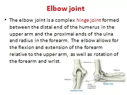
Additional Professor Department of Orthopaedics Objectives Understand the spectrum of Disease Common Paediatric fractures Common Adult Injuries Develop evaluation of elbow injuries Diagnose and choose treatment ID: 911958
Download Presentation The PPT/PDF document "Elbow Injuries DR R B Kalia," is the property of its rightful owner. Permission is granted to download and print the materials on this web site for personal, non-commercial use only, and to display it on your personal computer provided you do not modify the materials and that you retain all copyright notices contained in the materials. By downloading content from our website, you accept the terms of this agreement.
Slide1
Elbow Injuries
DR R B Kalia,
Additional Professor
Department of Orthopaedics.
Slide2Objectives
Understand the spectrum of Disease
Common Paediatric fractures
Common Adult Injuries
Develop evaluation of elbow injuries
Diagnose and choose treatment
Slide3Introduction
“
Pity the young surgeon whose first case is a fracture around the elbow
”
Slide4Question 1- Identify this injury in a 3 years old boy
Dislocation elbow
Supracondylar fracture Humerus
Separation distal humeral
physis
Lateral condyle Humerus fracture
Slide5Introduction- Elbow fractures
5% to 10% of all fractures in children are fractures of the elbow.
High potential for complications-difficult to manage.
Supracondylar fractures- 50% to 70% of all elbow fractures
F
requently in children between the ages of 3- 10 years.
Slide6CRITOE
C
apitellum
(2years)
R
adius(4years)
I
nternal (or medial) epicondyle(6years)
T
rochlea(8 years)
O
lecranon(10 years)
E
xternal (or lateral) epicondyle(12 years)
Slide7Common Fractures
The supracondylar Humerus
The
transphyseal
distal Humerus
The lateral humeral condyle
The medial humeral epicondyle (often associated with elbow dislocation)
Slide8Uncommon FRACTURES
The capitellum
Coronoid
Medial condyle
Lateral epicondyle
Intracondylar
or T-condylar fractures
Slide9Supracondylar Fractures of the Humerus
Devastating long-term complications.
Anteriorly- the brachial artery and median nerve
Laterally, the radial nerve crosses
The ulnar nerve passes behind the medial epicondyle
Slide10ANATOMY
Coronoid Fossa
Olecranon Fossa
Slide11Extension type 97.7%
Flexion type 2.3%
In extension type fracture line runs upwards and backwards
And in flexion type it runs downwards and backwards
Classification -Supracondylar fracture
Slide12Extension Type Fr
When forced into hyperextension, the olecranon can act as a fulcrum through which an extension force can propagate a fracture across the medial and lateral columns
Slide13Flexion
Supraconylar
Fr
A posteriorly applied force with the elbow in flexion creates a flexion-type supracondylar humeral fracture (
arrow
).
Slide14CLASSIFICATION-
Gartland
Classification
After initial classification as either extension or flexion injuries.
Classified according to the amount of radiographic displacement.
Slide15Q 2- How Do You Classify?
Flexion/Extension?
Gartland
I,II,III?
Slide16How Do You Classify?
Flexion/
Extension
?
Gartland
I,II,
III
?
Slide17RADIOGRAPHIC FINDINGS
The elbow is painful and difficult to move
True AP and lateral radiographs of fractures are required
“ Bad x-rays lead to bad decisions.”
Slide18TREATMENT -
Goal
“
Avoid catastrophes”
Vascular compromise
Compartment syndrome
“minimize embarrassments”
cubitus
varus
,
iatrogenic nerve palsies
Slide19Emergency Treatment
Immobilized - simple splint(radiolucent splint).
Contraindication- Ischemic hand or tented skin,
Radiographs should be obtained before splinting, or should be used.
Slide20If distal extremity is initially ischemic?
Align the fracture fragments
Re
evaluaie
vascularity
Avoid Flexion >90 degrees
Slide21Treatment of Nondisplaced Fractures ?
Long-arm cast immobilization for 3 weeks .
Radiographs are repeated at one week –Check
Slide22Extension
Gartland
III- Treatment?
Slide23Treatment of Displaced Fractures (types II and III).
Require reduction.
Reduction can be accomplished in closed fashion.
Maintaining the reduction?
Cast immobilization, traction, and percutaneous pin fixation.
Adequate closed reduction cannot be achieved- open reduction pinning
Slide24CR and K-wire fixation
Slide25Per op Images
Slide26Healed fracture at 4 weeks
Slide27Collateral circulation
Slide28Viable hand with abnormal pulses ?
Close observation.
Unidentified vascular pathology-thrombus formation-
an ischemic limb.
Pulse oximetry- valuable tool after closed reduction and pinning.
Pulseless, viable limb-ischemic-
arteriography and thrombolytic therapy
Slide29COMPLICATIONS
Early complications
Vascular injury
Peripheral nerve palsies
Volkmann's ischemia (compartment syndrome).
Late complications
Malunion
Stiffness
Myositis
ossificans
.
Slide30Vascular Injury-Spectrum?
A diminished pulse/Without a pulse/With an ischemic limb.
Complete transection of the brachial artery
An intimal tear
Compression either between the fracture fragments or over the anteriorly displaced fragment.
Indirect injury is usually the result of compression due to the swelling.
Slide31Management
Thorough assessment of the skin and neurologic status
If ischemic- manipulated into an extended position.
If fails to provide distal circulation
Closed reduction and pinning
Reduction of the fracture frequently restores the circulation
Slide32Peripheral Nerve Injury
10% to 15% of supracondylar humeral fractures.
The anterior interosseous nerve is the most commonly injured nerve with extension-type supracondylar fractures
Usually recover spontaneously
If within 8 to 12 weeks function is not returning-NCV/EMG nerve has not been
transected
.
Transected
-
reanastomosis
with grafting or tendon transfers
Slide33Compartment Syndrome?
Best managed by closed reduction and pinning.
A
fasciotomy
is essential to decompress the increased pressure
Splinting and active and passive range-of-motion exercises -essential to maintain joint mobility until function returns.
Slide34Volkmann's Ischemic Contracture (Chr
Compartment Syndrome)
Ischemic paralysis and contracture of the muscles of the forearm and hand
Primarily resulted from obstruction of arterial blood flow, resulting in death of the muscles which get replaced by fibrous tissue
Slide35Malunion
:
Cubitus
Varus
and
Cubitus
Valgus
Posteromedially
displaced fractures tend to develop
Cubitus
varus
angulation-more common
Posterolaterally
displaced fractures tend to develop valgus deviation.
Slide36T/T -Osteotomy and k-wire fixation
Slide37Lateral Condyle Fractures
These fractures are the second most common children’s
elbow fracture to need operative treatment.
(1) The fracture heals slowly.
(2) Late deformity can occur.
(3) Non-union is a recognized complication
Slide38Treatment
Undisplaced lateral condyle-
Long arm cast.
Displaced fractures –
stabilized by Open reduction K-wire fixation.
Slide39Complete Articular Fractures of the Distal
Humerus—T-Fracture
Slide40Radial Neck
30◦ of angulation can be treated conservatively provided there is no displacement- Long arm cast
Slide41Displaced fractures ?
Need to be reduced but closed reduction can be difficult
Slide42Post Op
Slide43Injuries of Adult Elbow
Slide44Spectrum
Olecranon & Proximal Ulnar Fractures
Radial Head Fractures
Elbow Dislocations
Slide45Anatomy-Olecranon Fractures-
The triceps attaches to the olecranon
Principle force which displaces the fracture.
Slide46Symptoms
H/O Trauma: Direct/Indirect
Pain
Swelling
Inability to extend against gravity
Tenderness
Slide47Signs
Swelling
Contusion
Gap at fracture site
Extension lag
Slide48Mayo Classification
Type 1: Minimally displaced-
Nonoperative
Type 2: Displaced without
ulnohumeral
instability-SurgeryType 3: Displaced with
ulnohumeral
instability-
Surgery
Slide49OR&IF- Tension band wiring
Slide50Post Op
Slide51Complex fractures or fracture-dislocations
Tension band wire constructs can fail- Plating is the choice
Slide52RADIAL HEAD FRACTURES-Mechanism
Fractures when it collides with the capitellum
Fall onto the outstretched hand
Slide53Classification- Mason
Type 1- Nondisplaced fractures- nonoperative treatment
Type 2- Displaced fractures involving part of the radial head- Screws
Type 3- comminuted fractures -excision
Slide54Type 2
Slide55SIMPLE ELBOW DISLOCATIONS
Stable after manipulative reduction.
Acute
redislocations
and chronic recurrent dislocations are uncommon.
Mobilization of the elbow within 2 weeks results in less stiffness and pain
Slide56Question 1- Identify this injury in a 3 years old boy?
Dislocation elbow
Supracondylar fracture Humerus
Separation distal humeral
physis
Lateral condyle Humerus fracture
Slide57Questions?