/
MYOCARDIAL DISEASE Myocardial disease MYOCARDIAL DISEASE Myocardial disease

MYOCARDIAL DISEASE Myocardial disease - PowerPoint Presentation

layla
layla . @layla
Follow
66 views
Uploaded On 2023-05-21

MYOCARDIAL DISEASE Myocardial disease - PPT Presentation

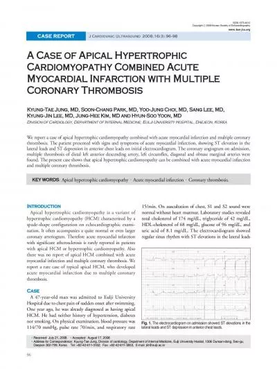
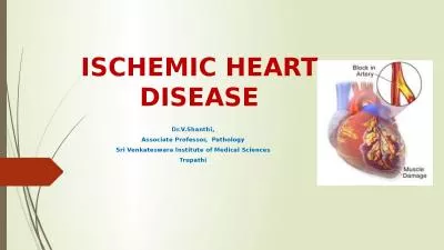
is a disaese of the myocardium that is not due to an ischaemic valvular or hypertensive heart disease It may be caused by an acute or chronic inflammatory pathology myocarditis ID: 998736

ventricular cardiac left disease cardiac ventricular disease left sudden cardiomyopathy death heart hypertrophy patients myocardial due myocarditis failure clinical

Share:

Link:

Embed:

Download Presentation from below link

Download Presentation The PPT/PDF document "MYOCARDIAL DISEASE Myocardial disease" is the property of its rightful owner. Permission is granted to download and print the materials on this web site for personal, non-commercial use only, and to display it on your personal computer provided you do not modify the materials and that you retain all copyright notices contained in the materials. By downloading content from our website, you accept the terms of this agreement.


Presentation Transcript

1. MYOCARDIAL DISEASE

2. Myocardial disease , is a disaese of the myocardium that is not due to an ischaemic, valvular or hypertensive heart disease, It may be caused by:an acute or chronic inflammatory pathology (myocarditis) idiopathic myocardial disease (cardiomyopathy).

3. MyocarditisThis is an acute inflammatory condition that can have an infectious, toxic or autoimmune aetiology. Myocarditis can complicate many infections in which inflammation may be due directly to infection of the myocardium or the effects of circulating toxins. Viral infections are the most common causes, such as Coxsackie and influenza A and B viruses. Myocarditis may occur several weeks after the initial viral symptoms and susceptibility is increased by corticosteroid treatment, immunosuppression, radiation, previous myocardial damage and exercise.

4. Some bacterial and protozoal infections may be complicated by myocarditis; for example, approximately 5% of patients with Lyme disease (Borrelia burgdorferi) develop myopericarditis, which is often associated with AV block. Toxic aetiologies include drugs, which may directly injure the myocardium (e.g. cocaine, lithium and anticancer drugs such as doxorubicin) or which may cause a hypersensitivity reaction and associated myocarditis (e.g. penicillins and sulphonamides), lead and carbon monoxide. Occasionally, autoimmune conditions such as systemic lupus erythematosus and rheumatoid arthritis are associated with myocarditis.

5. PathologyIn the acute phase myocarditic hearts are flabby with focal haemorrhages; in chronic cases they are enlarged and hypertrophied.Histologically an inflammatory infiltrate is present– lymphocytes predominating in viral causes; - Polymorphonuclear cells in bacterial causes; - eosinophils in allergic and hypersensitivity causes

6. Clinical featuresMyocarditis may be an acute or chronic process; its clinical presentations range from an asymptomatic state associated with limited and focal inflammation to fatigue, palpitations, chest pain, dyspnoea and fulminant congestive cardiac failure due to diffuse myocardial involvement.Physical examination includes soft heart sounds, a prominent third sound and often a tachycardia. A pericardial friction rub may be heard.

7. InvestigationsChest X-ray may show some cardiac enlargement, depending on the stage and virulence of the disease. ECG demonstrates ST- and T wave abnormalities and arrhythmias. Heart block may be seen with diphtheritic myocarditis, Lyme disease and Chagas’ disease .Cardiac enzymes are elevated. Viral antibody titres may be increased. However, since enteroviral infection is common in the general population, the diagnosis depends on the demonstration of acutely rising titres.Echocardiography may reveal left ventricular dysfunction that is sometimes regional (due to focal myocarditis)Endomyocardial biopsy may show acute inflammation Viral RNA can be measured from biopsy material using polymerase chain reaction (PCR).

8. TreatmentThe underlying cause must be identified, treated, eliminated or avoided. Bed rest is recommended in the acute phase of the illness and athletic activities should be avoided for 6 months. Heart failure should be treated conventionally with the use of diuretics, ACE inhibitors/ARB, beta-blockers, spironolactone ± digoxin. Antibiotics should be administered immediately where appropriate. NSAIDs are contraindicated in the acute phase of the illness but may be used in the late phase. The use of corticosteroids is controversial and no studies have demonstrated an improvement in left ventricular ejection fraction or survival following their use. The administration of high-dose intravenous immunoglobulin on the other hand appears to be associated with a more rapid resolution of the left ventricular dysfunction and improved survival

9. CARDIOMYOPATHYcardiomyopathies are defined as "a heterogeneous group of diseases of the myocardium associated with mechanical and/or electrical dysfunction that usually (but not invariably) exhibit inappropriate ventricular hypertrophy or dilatation and are due to a variety of causes that frequently are genetic."

10. Dilated cardiomyopathy (DCM)DCM has a prevalence of 1 in 2500 and is characterized by dilatation of the ventricular chambers and systolic dysfunction with preserved wall thickness.

11. Familial DCM is predominantly autosomal dominant and can be associated with over 20 abnormal loci and genes . Many of these are genes encoding cytoskeletal or associated myocyte proteins (dystrophin in X-linked cardiomyopathy; actin, desmin, troponin T, beta myosin heavy chain, sarcoglycans, vinculin and lamin a/c in autosomal dominant DCM) .

12. Sporadic DCM can be caused by multiple conditions:myocarditis – Coxsackie, adenoviruses, erythroviruses, HIV, bacteria, fungae, mycobacteria, parasitic (Chagas’ disease)toxins – alcohol, chemotherapy, metals (cobalt, lead, mercury, arsenic)autoimmuneendocrineneuromuscular.

13. Clinical featuresDCM can present with heart failure, cardiac arrhythmias, conduction defects, thromboembolism or sudden death.Increasingly, evaluation of relatives of DCM patients is allowing identification of early asymptomatic disease, prior to the onset of these complications.

14. InvestigationsChest X-ray demonstrates generalized cardiac enlargement.ECG may demonstrate diffuse non-specific ST segment and T wave changes. Sinus tachycardia, conduction abnormalities and arrhythmias (i.e. atrial fibrillation, ventricular premature contractions or ventricular tachycardia) are also seen.Echocardiogram reveals dilatation of the left and/or right ventricle with poor global contraction function .Cardiac MR may demonstrate other aetiologies of left ventricular dysfunction (e.g. previous myocardial infarction) or demonstrate abnormal myocardial fibrosis . Coronary angiography whenever there is evidence of ischemiaBiopsy is generally not indicated outside specialist care.

15. TreatmentTreatment consists of the conventional management of heart failure with the option of cardiac resynchronization therapy and ICDs in patients with NYHA III/IV grading.Cardiac transplantation is appropriate for certain patients.

16. Hypertrophic cardiomyopathy (HCM)HCM includes a group of inherited conditions that produce hypertrophy of the myocardium in the absence of an alternate cause (e.g. aortic stenosis or hypertension). It is the most common cause of sudden cardiac death in young people and affects 1 in 500 of the population. The majority of cases are familial autosomal dominant, due to mutations in the genes encoding sarcomeric proteins . The most common causes of HCM are mutations of the β-myosin heavy chain and myosin-binding protein C.

17. Clinical featuresHCM is characterized by: variable myocardial hypertrophy frequently involving the interventricular septum and disorganization (‘disarray’) of cardiac myocytes and myofibrils.Twenty-five per cent of patients have dynamic left ventricular outflow tract obstruction due to the combined effects of hypertrophy, systolic anterior motion (SAM) of the anterior mitral valve leaflet and rapid ventricular ejection. The salient clinical and morphological features of the disease vary according to the underlying genetic mutation. For example, marked hypertrophy is common with β myosin heavy chain mutations whereas mutations in troponin T may be associated with mild hypertrophy but a high risk of sudden death.The hypertrophy may not manifest before completion of the adolescent growth spurt, making the diagnosis in children difficult.

18.

19. Symptomsmany are asymptomatic and are detected through family screening of an affected individual or following a routine ECG examinationchest pain, dyspnoea, syncope or pre-syncope (typically with exertion), cardiac arrhythmias and sudden death are seen sudden death occurs at any age but the highest rates (up to 6% per annum) occur in adolescents or young adults. dyspnoea occurs due to impaired relaxation of the heart muscle or the left ventricular outflow tract obstruction that occurs in some patients.

20. Signsdouble apical pulsation (forceful atrial contraction producing a fourth heart sound)jerky carotid pulse because of rapid ejection and sudden obstruction to left ventricular outflow during systole ejection systolic murmur due to left ventricular outflow obstruction late in systole – it can be increased by manoeuvres that decrease after-load, e.g. standing or Valsalva, and decreased by manoeuvres that increase after-load and venous return, e.g. squattingpan-systolic murmur due to mitral regurgitation (secondary to SAM)fourth heart sound (if not in AF).

21. InvestigationsECG abnormalities of HCM include left ventricular hypertrophy, ST and T wave changes, and abnormal Q waves especially in the infero-lateral leads.Echocardiography is usually diagnostic and in classical HCM there is asymmetric left ventricular hypertrophy (involving the septum more than the posterior wall), systolic anterior motion of the mitral valve, and a vigorously contracting ventricle . Cardiac MR can detect both the hypertrophy but also abnormal myocardial fibrosis .Genetic analysis, where available, may confirm the diagnosis and provide prognostic information for the patient and relatives.

22. Treatment

23. HCMHCM with obstructionHCM without obstructionBBBB +/- disopyramide if obstruction persistDiuretics for congestive symptomVerapamide in stead of disopyramideSurgical myectomy or alcohol septal ablationDual chamber cardiac pacingBB &/or verapamilDiuretics for congestionTransplantation

24. The management of HCM includes treatment of symptoms and the prevention of sudden cardiac death in the patient and relatives.Risk factors for sudden death:massive left ventricular hypertrophy (> 30 mm on echocardiography)family history of sudden cardiac death (< 50 years old) non-sustained ventricular tachycardia on 24-hour Holter monitoringprior unexplained syncopeabnormal blood pressure response on exercise (flat or hypotensive response).

25. The presence of these cardiac risk factors is associated with an increased risk of sudden death, and patients with one or more should be assessed for implantable cardioverter– defibrillator (ICD).

26. Restrictive cardiomyopathyThis is a rare condition in which there is normal or decreased volume of both ventricles with bi-atrial enlargement, normal wall thickness, normal cardiac valves and impaired ventricular filling with restrictive physiology but near normal systolic function. The restrictive physiology produces symptoms and signs of heart failure. Conditions associated with this form of cardiomyopathy include amyloidosis (commonest), sarcoidosis,…. The idiopathic form of restrictive cardiomyopathy may be familial.

27. Clinical featuresPatients with restrictive cardiomyopathy may present with dyspnoea, fatigue and embolic symptoms. On clinical examination there will be elevated jugular venous pressure with diastolic collapse (Friedreich’s sign) and elevation of venous pressure with inspiration (Kussmaul’s sign), hepatic enlargement, ascites and dependent oedema. Third and fourth heart sounds may be present.Irrgular pulse (AF)

28. InvestigationsChest X-ray may show pulmonary venous congestion. The cardiac silhouette can be normal or show biatrial enlargement.ECG may demonstrate low-voltage QRS and ST segment and T wave abnormalities.Echocardiography shows a normal systolic ejection fraction but there is diastolic dysfunction.Cardiac MR may demonstrate abnormal myocardial fibrosis in amyloidosis or sarcoidosis .Cardiac catheterization and haemodynamic studies may help distinguish between restrictive cardiomyopathy and constrictive pericarditis, although volume loading may be required. Endomyocardial biopsy in contrast with other cardiomyopathies is often useful in this condition and may permit a specific diagnosis, such as amyloidosis, to be made.

29. TreatmentThere is no specific treatment. Cardiac failure and embolic manifestations should be treated. Cardiac transplantation is necessary in some severe cases, especially the idiopathic variety. In primary amyloidosis combination therapy with melphalan plus prednisolone with or without colchicine may improve survival. However, patients with cardiac amyloidosis have a worse prognosis than those with other forms of the disease, and the disease often recurs after transplantation.

30. Arrhythmogenic right ventricular cardiomyopathy In this condition, patches of the right ventricular myocardium are replaced with fibrous and fatty tissue. It is inherited as an autosomal dominant trait and has a prevalence of approximately 10 per 100 000. The dominant clinical problems are ventricular arrhythmias, sudden death and right-sided cardiac failure. The ECG typically shows a slightly broadened QRS complex and inverted T waves in the right precordial leads. MRI is a useful diagnostic tool and is often used to screen the first-degree relatives of affected individuals. Patients at high risk of sudden death can be offered an ICD.

31.

32. Acquired cardiomyopathies

33. Stress (Tako-tsubo/octopus pot) cardiomyopathyThis is a recently described acute and reversible cardiomyopathy that occurs in the absence of coronary artery disease and is usually triggered by profound psychological stress. It is more common in middle–old aged women and classically affects the left ventricular apex causing apical ballooning

34. Peri-partum cardiomyopathyThis rare condition affects women in the last trimester of pregnancy or within 5 months of delivery. It presents as a dilated cardiomyopathy, is more common in obese, multiparous women over 30 years old and is associated with preeclampsia.Nearly half of patients will recover to normal function within 6 months but in some patients it can causes progressive heart failure and sudden death.

35. Alcoholic CMPLong-term exposure to alcohol is an important preventable cause of DCM. The risk is seen when more than 90 g of alcohol (about seven standard alcoholic drinks) is consumed daily for more than 5 years; lower exposure may lead to cardiomyopathy in women. Abstinence from alcohol may lead to sustained improvements in ventricular function.

36. Tachycardia cardiomyopathyProlonged periods of supraventricular or ventricular tachycardia will lead to dilated cardiomyopathy. Cardioversion and ablation may be necessary to restore sinus rhythm and allow for recovery of cardiac function.

37.

38.

39.

40.