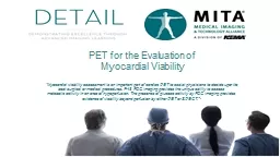
hypoperfusion The presence of glucose activity by FDG imaging provides evidence of viability beyond perfusion by either PET or SPECT 7 PET for the Evaluation of Myocardial Viability Add presenter info here ID: 776149
Download Presentation The PPT/PDF document " “Myocardial viability assessment is a..." is the property of its rightful owner. Permission is granted to download and print the materials on this web site for personal, non-commercial use only, and to display it on your personal computer provided you do not modify the materials and that you retain all copyright notices contained in the materials. By downloading content from our website, you accept the terms of this agreement.
Slide1
“Myocardial viability assessment is an important part of cardiac PET to assist physicians to decide upon the best surgical or medical procedures. F-18 FDG imaging provides the unique ability to assess metabolic activity in an area of hypoperfusion. The presence of glucose activity by FDG imaging provides evidence of viability beyond perfusion by either PET or SPECT.” 7
PET for the Evaluation of Myocardial Viability
Add presenter info here
Slide2These materials were prepared in good faith by MITA as a service to the profession and are believed to be reliable based on current scientific literature. The materials are for educational purposes only and do not replace either the need for individualized patient diagnosis and treatment planning by qualified physicians based on existing good practices or the need for implementation by qualified radiologists or other qualified healthcare practitioners. Neither MITA nor its members are responsible for any diagnostic or treatment outcomes. MITA, its members, and contributors do not assume any responsibility for the user’s compliance with applicable laws and regulations. MITA does not endorse the proprietary products or processes of any one company.
Legal Disclaimers
Slide3Overview
Information about myocardial viability is necessary in the management of patients with ischemic cardiomyopathy in that only viable myocardial segments benefit from revascularization
Viable myocardium exhibits an affinity for glucose compared to irreversible damaged heart muscle
FDG PET has been showed to be the gold standard when assessing myocardial viability
Slide4Objectives
Review the ischemic cascade in acute and chronic CAD
Review various states of myocardial viability
Review predictors of survival in patients with heart failure
Evaluate how glucose metabolism may identify high risk patients
Slide518.
Dilsizian and Narula Atlas of Nuclear Cardiology, 3rd Ed. 2009; Figure 9-15, p212
Ischemia: Supply and Demand
Slide6Myocardial Hibernation
18.
Dilsizian
and
Narula
Atlas of Nuclear Cardiology, 3
rd
Ed. 2009; Figure 9-21B, p215
Slide7N-13
FDG
Blood Flow vs. Metabolism: Mismatch
18.
Dilsizian
and
Narula
Atlas of Nuclear Cardiology, 3
rd
Ed. 2009; (L) Figures 8-23, 8-24, p194 and (R) Figure 8-26B, p195
Slide819. Di Carli, et al. J Thorac Cardiovasc Surg 1998; 116(6):997-1004
Survival by PET Viability Pattern and Treatment
With PET Mismatch
Time (months)
P
= 0.007
CABG
Medicine
Survival Probability
1.0
0.8
0.6
0.4
0.2
0.0
0
12
60
48
24
36
Without PET Mismatch
Time (months)
P
= 0.12
CABG
Medicine
Survival Probability
1.0
0.8
0.6
0.4
0.2
0.0
0
12
60
48
24
36
Viability determined by presence of mismatch more accurately predicted the success of the intervention
Prognosis of Patients with Defects and LV Dysfunction
Slide920.
D’Egidio, et al. JACC Cardiovascular Imaging 2009; 2(9):1960-68
PARR-2 = PET and Recovery after Revascularization-2
Mismatch and Clinical Benefit: The PARR-2 Trial
Quantitative Scoring of Mismatch Size
Slide10Mismatch and Clinical Benefit: The PARR-2 Trial
Progressive revascularization benefit with increasing mismatch (>7%)
PARR-2 = PET and Recovery after Revascularization-2
20. D’Egidio, et al. JACC Cardiovascular Imaging 2009; 2(9):1960-68
Hazard ratio decreases
with increasing mismatch score
Figure 2.
Interaction hazard ratios and 95% confidence interval at various levels of mismatch as a continuous variable
Slide11Clinical Scenario: Evaluation for Ischemic EtiologySPECTPETRest OnlyStress/RestRest OnlyStress/RestWith Angina/ischemia equivalentRAMAWithout Angina/ischemic equivalentRAMA
21. Adapted from: 2013 ACCF/ACR/ASE/ASNC/SCCT/SCMR Appropriate Utilization of Cardiovascular Imaging in Heart Failure. JACC 2013; 61(21)
Compared to SPECT: PET may increase accuracy for detection of multi-vessel disease, provide myocardial perfusion reserve for detection of patients with CAD and allow assessment of glucose metabolism that may then identify high-risk patients
Appropriateness ratings: R=Rarely; M=May Be; A=Always
Appropriate Use Criteria in Heart Failure
Slide12Clinical Scenario: Viability evaluation amenable to revascularizationSPECTPETRest/RedistStress/RestRest OnlyStress/RestSeverely reduced ventricular function(EF <30)AAAAModerately reduced ventricular function (EF 30-39%)MAAMMild ventricular function abnormality(EF 40-49%)MAMA
PET validated by PARR-1, PARR-2: Higher sensitivity for viable myocardium vs. SPECT
Appropriateness ratings: R=Rarely; M=May Be; A=Always
Appropriate Use Criteria in Heart Failure
21. Adapted from: 2013 ACCF/ACR/ASE/ASNC/SCCT/SCMR
Appropriate Utilization of Cardiovascular Imaging in Heart Failure. JACC 2013; 61(21)
Slide13Summary
The physics of PET and pharmacokinetics of the tracers are more optimal for MPI
1-5, 9-10
Cardiac PET addresses the need for improved interpretive certainty and greater efficiency
1-4
Cardiac PET performs well even with challenging patient types (e.g. pharm stress, obese, female) and more accurately identifies multi-vessel disease
1,3-4,6,7,17
PET can help improve the management of patients with known or suspected CAD, heart failure and cardiac sarcoidosis
1-3,6,7,18-24
Slide14Summary
Quantification of myocardial blood flow adds incremental prognostic value
18,22,23
PET can help to implement a strategy for the reduction of radiation exposure from cardiac imaging procedures
25-26
Slide15References
Bateman TM, Heller GV,
McGhie
IA, et al. Diagnostic accuracy of
r
est/stress ECG-gated Rb-82 myocardial perfusion PET: Comparison with ECG-gated Tc-99m
sestamibi
SPECT. J
Nucl
Cardiol
2006; 13:24-33
Merhige ME, Breen WJ, Shelton V, et al. Impact of myocardial perfusion imaging with PET and (82)Rb on downstream invasive procedure utilization, costs, and outcomes in coronary disease management. J
Nucl
Med 2007; 48:1069-1076
Yoshinaga
K, Chow BW, Williams K, et al. What is the prognostic
v
alue of myocardial perfusion imaging using rubidium-82 positron emission tomography?
J Am
Coll
Cardiol
2006; 48:1029-39
Bateman TM. Cardiac positron emission
t
omography and the role of adenosine pharmacologic
s
tress.
Amer
J
Cardiol
2004; 94:19-24
Gould KL. Reversal of coronary atherosclerosis: Clinical promise as the basis for non-invasive management of coronary
a
rtery disease. Circulation 1994; 90:1558-1571
Chow BJ, Wong JW,
Yoshinaga
K, et al. Prognostic significance of dipyridamole-induced ST depression in patients with normal 82Rb PET myocardial perfusion imaging. J
Nucl
Med 2005; 46:1095-1101
Slide16ASNC Model Coverage Policy: Cardiac positron emission tomographic imaging. J Nucl Cardiol 2013; 20:916-47Botvinik EH, Ed: Nuclear medicine self-study program III: Nuclear medicine cardiology. Society of Nuclear Medicine, Reston, VA; 1998 Mullani NM, Goldstein RA, Gould KL, et al. Myocardial perfusion with rubidium-82. Measurement of extraction fraction and flow with external detectors. J Nucl Med 1983; 24:898-906Dilsizian V, Narula J, Braunwald E, Eds: Atlas of Nuclear Cardiology 2003; Current Medicine Group LLC. DOI 11007/978-1-4615-6496-6Machac J, Bacharach S, Bateman T, et al. PET myocardial perfusion and glucose metabolism imaging. J Nucl Cardiol 2006; 13(6):e121-51Dorbala S, Vangala D, Sampson U, et al. Value of vasodilator left ventricular ejection fraction reserve in evaluating the magnitude of myocardium at risk and the extent of angiographic coronary artery disease: A 82Rb PET/CT study. J Nucl Med 2007; 48:349-358
References
Slide17Iskander S and Iskandrian A. A risk assessment using single-photon emission computed tomographic technetium-99m sestamibi imaging. J Am Coll Cardiol 1998; 32:57-62McArdle BA, Dowsley TF, deKemp RA, et al. Does rubidium-82 have superior accuracy to SPECT perfusion imaging for the diagnosis of obstructive coronary disease? J Amer Coll Cardiol 2012; 60(8):1828-37Dorbala S, Di Carli MF, Beanlands RS, et al. Prognostic value of stress myocardial perfusion positron emission tomography: Results from a multicenter observational registry. J Amer Coll Cardiol 2013; 61(2):176-184Heller GV, Hendel RC, Eds: Handbook of nuclear cardiology: Cardiac SPECT and Cardiac PET. Springer-Verlag London ©2013Chow BJ, Dorbala S, Di Carli MF, et al. Prognostic value of PET myocardial perfusion imaging in obese patients. JACC Cardiovascular Imaging 2014; 7(3):278-87Dilsizian V and Narula J, Eds: Atlas of Nuclear Cardiology 3rd Edition 2009. Current Medicine Group LLC; ISBN 1573403105
References
Slide18Di Carli M, Maddahi J, Rokhsar S, et al. Long term survival of patients with coronary artery disease and left ventricular dysfunction: Implications for the role of myocardial viability assessment in management decisions. J Thorac Cardiovasc Surg 1998; 116(6):997-1004D’Egidio G, Nichol G, Williams KA, et al. Increasing benefit from revascularization is associated with increasing amounts of myocardial hibernation: A substudy of the PARR-2 trial. JACC Cardiovasc Imag 2009; 2(9):1060-68Patel MR, White RD, Abbara S, et al. 2013 ACCF/ACR/ASE/ASNC/SCCT/SCMR. Appropriate utilization of cardiovascular imaging in heart failure. J Amer Coll Cardiol May 2013; 61(21)Ziadi MC, Dekemp RA, Williams KA, et al. Impaired myocardial flow reserve on rubidium-82 positron emission tomography imaging predicts adverse outcomes in patients assessed for myocardial ischemia. J Amer Coll Cardiol 2011; 58(7):740-48Murthy VL, Naya M, Foster CR, et al. Improved cardiac risk assessment with non-invasive measures of coronary flow reserve. Circulation 2011; 124(20):2215-2224
References
Slide19Skali H, Schulman A, Dorbala S. 18-F FDG PET/CT for the assessment of myocardial sarcoidosis. Curr Cardiol Reports 2013; 15(4):352Einstein EJ. Effects of radiation exposure from cardiac imaging: How good are the data? J Am Coll Cardiol 2012; 59(6):553-565Cerqueira MD, Allman KC, Ficaro EC, et al. ASNC information statement: Recommendations for reducing radiation exposure in myocardial perfusion imaging. J Nucl Cardiol; published online 26 May 2010
References
Slide20Important Safety Information
Image interpretation errors can occur with PET imaging. A negative image does not rule out recurrent prostate cancer and a positive image does not confirm its presence. Clinical correlation, which may include histopathological evaluation, is recommended. Hypersensitivity reactions, including anaphylaxis, may occur in patients who receive PET radiopharmaceuticals. Emergency resuscitation equipment and personnel should be immediately available.PET/CT imaging contributes to a patient’s overall long-term cumulative radiation exposure, which is associated with an increased risk of cancer. Safe handling practices should be used to minimize radiation exposure to the patient and healthcare providers. Adverse reactions, although uncommon, may occur when using PET radiopharmaceuticals. Always refer to the package insert prior to use.