PPT-Neoplasms of the esophagus
Author : lindy-dunigan | Published Date : 2017-08-20
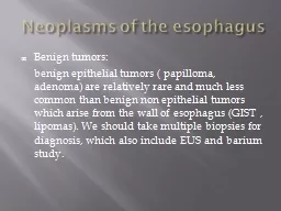
Benign tumors benign epithelial tumors papilloma adenoma are relatively rare and much less common than benign non epithelial tumors which arise from the wall of
Presentation Embed Code
Download Presentation
Download Presentation The PPT/PDF document "Neoplasms of the esophagus" is the property of its rightful owner. Permission is granted to download and print the materials on this website for personal, non-commercial use only, and to display it on your personal computer provided you do not modify the materials and that you retain all copyright notices contained in the materials. By downloading content from our website, you accept the terms of this agreement.
Neoplasms of the esophagus: Transcript
Download Rules Of Document
"Neoplasms of the esophagus"The content belongs to its owner. You may download and print it for personal use, without modification, and keep all copyright notices. By downloading, you agree to these terms.
Related Documents