/
The impact of the use of new X-Ray technology to ensure the The impact of the use of new X-Ray technology to ensure the

The impact of the use of new X-Ray technology to ensure the - PowerPoint Presentation

lindy-dunigan
lindy-dunigan . @lindy-dunigan
Follow
373 views
Uploaded On 2016-06-15

The impact of the use of new X-Ray technology to ensure the - PPT Presentation

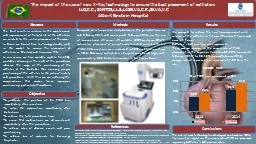
catheters LUZCC SANTOSJLSLASELVACRSILVAVC Albert Einstein Hospital Abstract The best results in relation to the maintenance and improvement of helpful life of the catheters are related to the initial placement of the same ID: 363652

2013 placement positioning catheters placement 2013 catheters positioning central procedure february january reduce catheter picc rate 2014 study results

Share:

Link:

Embed:

Download Presentation from below link

Download Presentation The PPT/PDF document "The impact of the use of new X-Ray techn..." is the property of its rightful owner. Permission is granted to download and print the materials on this web site for personal, non-commercial use only, and to display it on your personal computer provided you do not modify the materials and that you retain all copyright notices contained in the materials. By downloading content from our website, you accept the terms of this agreement.


Presentation Transcript

Slide1

The impact of the use of new X-Ray technology to ensure the best placement of

catheters

LUZ,C.C.; SANTOS,J.L.S.;LASELVA,C.R.;SILVA,V.C

Albert Einstein Hospital

Abstract

The best results in relation to the maintenance and improvement of helpful life of the catheters, are related to the initial placement of the same.

In Brazil, we do not have technology already sold in the market to ensure the placement of catheters, as Sherlock and Vasonova TPS. In our service we have available mobile RX (XRD), portable, allowing us the end of the procedure, observe the image of the positioning of the catheter at the bedside, thus ensuring proper positioning of the catheter to the patient's safety and procedure . With this, we are reducing the complications that comes from improper positioning, particularly thrombosis.

Objective

To evaluate the position of the PICC line, immediately after insertion.To solve the malposition immediately after detection.To reduce the total procedure time.To esure that medications are administered immediately after the procedure.To reduce rates of adverse events and poor positioning.To reduce loss of materials for dressings duplicate.

Methods

Comparative retrospective study between the period of January and February 2013 and January and February 2014, on the use of conventional XR versus XR portable XRD-Carestream DRX-1 system, for placement of peripherally inserted central catheter (PICC). 324 insertions were evaluated in 2013, and 332 insertions in 2014. Data were collected in a hospital considered large, with approximately 800 beds, tertiary care, in Sao Paulo, Brasil.

Results

Conclusions

References

The use of new technology for radiological confirmation - DRX has a positive impact on the success rate of PICC line placement, improving Catheter´s bad position rates.

We managed to reduce the time from assessment until release of the catheter in 1 hour. (There was 2 hours and now is 1 hour).

Catheter´s bad position rates improved.

There was an increase in the success rate of PICC placement in the Superior Vena Cava 82.20% (January and February 2013) to 90.10% (January-February 2014) average, which represents an increase of 7.90 % in this rate.

-

Pittiruti

M, The intracavitary ECG method for positioning the tip of central venous catheters: results of an Italian multicenter study. J Vasc Access. 2012 Jul-Sep;13(3):357-65. -Katheria AC, A randomized controlled trial of ultrasound-guided peripherally inserted central catheters compared with standard radiograph in neonates. J Perinatol. 2013 Oct;33(10):791-4.-Roberto P. Central venous line placement is not compromised by the choice between different insertion sites. Study performed in cardiovascular surgery patient. Rev Port Cir Cardiotorac Vasc. 2013 Jan-Mar;20(1):13-7