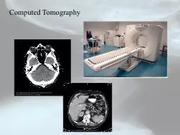
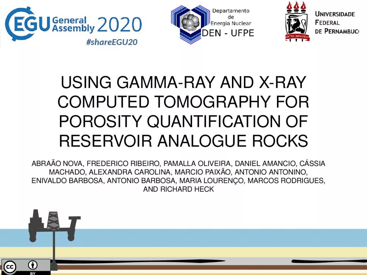
/
USING GAMMA-RAY AND X-RAY COMPUTED TOMOGRAPHY FOR POROSITY QUANTIFICATION OF RESERVOIR USING GAMMA-RAY AND X-RAY COMPUTED TOMOGRAPHY FOR POROSITY QUANTIFICATION OF RESERVOIR

USING GAMMA-RAY AND X-RAY COMPUTED TOMOGRAPHY FOR POROSITY QUANTIFICATION OF RESERVOIR - PowerPoint Presentation

scarlett
scarlett . @scarlett
Follow
43 views
Uploaded On 2024-02-03

USING GAMMA-RAY AND X-RAY COMPUTED TOMOGRAPHY FOR POROSITY QUANTIFICATION OF RESERVOIR - PPT Presentation

ABRAÃO NOVA FREDERICO RIBEIRO PAMALLA OLIVEIRA DANIEL AMANCIO CÁSSIA MACHADO ALEXANDRA CAROLINA MARCIO PAIXÃO ANTONIO ANTONINO ENIVALDO BARBOSA ANTONIO BARBOSA MARIA LOURENÇO MARCOS RODRIGUES AND RICHARD HECK ID: 1044551

porosity ray samples shareegu20 ray porosity shareegu20 samples µct gamma quantification results image data tomography method barbosa methods3 time

Share:

Link:

Embed:

Download Presentation from below link

Download Presentation The PPT/PDF document "USING GAMMA-RAY AND X-RAY COMPUTED TOMOG..." is the property of its rightful owner. Permission is granted to download and print the materials on this web site for personal, non-commercial use only, and to display it on your personal computer provided you do not modify the materials and that you retain all copyright notices contained in the materials. By downloading content from our website, you accept the terms of this agreement.


Presentation Transcript

1. USING GAMMA-RAY AND X-RAY COMPUTED TOMOGRAPHY FOR POROSITY QUANTIFICATION OF RESERVOIR ANALOGUE ROCKSABRAÃO NOVA, FREDERICO RIBEIRO, PAMALLA OLIVEIRA, DANIEL AMANCIO, CÁSSIA MACHADO, ALEXANDRA CAROLINA, MARCIO PAIXÃO, ANTONIO ANTONINO, ENIVALDO BARBOSA, ANTONIO BARBOSA, MARIA LOURENÇO, MARCOS RODRIGUES, AND RICHARD HECK#shareEGU20

2. X-ray micro-computed tomography (µCT)Characterization of rock properties High-resolution 3D digital images.Porous systems in reservoir rocks. 1Introduction#shareEGU20

3. #shareEGU20#shareEGU20a) Imagem em HUb) Huangc) IsoDatad) Lie) Momentse) Otsue) PVEThe challenge:Reliable quantification of porosity in rocks which present pore sizes ranging from centimeter to nanometer scale;Intrinsic limits of the x-ray imaging method. 1Introduction

4. Problems that are related to the physical aspects of the µCT imagem Method:Image artifacts;Bam hardening effect. #shareEGU20#shareEGU20Ring ArtifactBeam hardening effect1Introduction

5. The computational costs of processing for large image volumes, necessary for the reliable definition of some petrophysical characteristics of rock samples, like porosity and permeability.#shareEGU201Introduction

6. A method is presented for quantification of porosity, based in data from gamma ray tomography. The objective was to create a protocol for core samples scanning in a gamma tomograph, in order to perform porosity calculation with reliable results within the minimum operational time. The study was carried out in in sandstones and limestones samples (bioclastic mudstones and wackstones)The results obtained was compared with the porosity quantification results produced with the application of traditional segmentation methods in µCT images of the same samples. Also, the results of gamma tomography and µCT images analyses were compared with porosity data obtained with a porosimeter for the same samples. The traditional hard-data obtained with the porosimeter was used as a reference to compare with the other two sets of results. 2Objective#shareEGU20

7. These analyses were performed in core samples of limestones and sandstones analogous of Brazilian oil reservoirs.#shareEGU20Sandstone cores from Botucatu FmBioclastic limestones (wacstones and packstones) from Jandaira FmSampleDiametre (cm)Length (cm)B12.544.44B22.544.19C12.543.68C22.544.04Materials and Methods3Samples dimension

8. #shareEGU20CoreXR-µCTImagingRγ-CTscaningRadiodensity/Histogram evaluationAttenuation CoefficientPorosityBasic Workflow for Evaluation of Samples Image SegmentationMaterials and Methods3

9. Gamma Ray attenuation method#shareEGU20(a) Radioactive source;(b) Reference sample without pores;(c) Detector; (d) Porous rock sample.Experimental procedure for determining porosity with the Gamma Ray attenuation methodMaterials and Methods3

10. #shareEGU20Porosity    NIST XCOMDataMineral DensityDataLinear Attenuation Coefficient of Mineralsγ-Ray CTLinear Attenuation coefficient of the SamplesSample Thickness2Materials and Methods

11. #shareEGU20RockSampleDetectorGamma radiation beamRadiation SourceScan directionShielding and collimatorShielding and collimatorRadiation Source: 137Cs (662 keV);Half-life: 30,1 years;Radioactive activity: 3.70x10⁹ Bq; NaI(Tl) Scintillation Crystal DetectorLead collimator with circular hole 5.5 mm in diameter and length 73 mm.2Materials and MethodsGamma-Ray CT

12. 135º0º45º90º16 points in each planeAxial view#shareEGU20Distant by 1 mmAxial viewMaterials and Methods3

13. #shareEGU20Scan ParameterValueElectric current (µA)70 µAMaximal tension (kV)150 kVAluminum Filter0.25 mmVoxel Resolution30 µmIntegration time500 msX-RAY µCT Scan ParameterMaterials and Methods3X-Ray CTGrayscale image

14. #shareEGU20X-RAY µCT imageCroped ROIBinary imageSampleThresholdB11325B21325C12630C22630PVE segmentationMaterials and Methods3

15. 4Results and Discussion#shareEGU20CB SamplePorosity (%)γR-CTXR-µCTHeB128.5120.3626.52B227.8320.1324.15C140.7933.7033.73C240.7932.1535.80

16. Scan time around 25 minVolume reconstruction around 15 minImage segmentation and porosity arond 60-120 min#shareEGU20γR-CTXR-µCTHe30 min2-3 hour5 minIt Is an experimental approachWhat is important in this new approach is to test the possibilityof obtaining activated pore measurements, in less time and cost, than in the extreme two methodsTime required in each methodology4Results and Discussion

17. The results suggest that Industrial application of γ-ray CT for precise evaluation of large number of core samples can be highly effective. The method allows the porosity quantification of a larger number of cores, with less operational and computational costs.Furthermore, the γ-ray data can be integrated with data provided by conventional X-ray µCT image processing to complement information regarding morphological aspects. This can be done for a fewer number of cores, due the higher complexity of this process and costs in terms of time and computation.4Conclusion#shareEGU20

18. 6Acknowledgment#shareEGU20

19. #shareEGU20Thank you!abe.alves2@gmail.comAbraão Alves Vila NovaDepartment of Nuclear Energy - Federal University of Pernambuco – Brazil

20. Using Gamma-Ray and X-Ray Computed Tomography for Porosity Quantification of Reservoir Analogue RocksAbraão Nova, Frederico Ribeiro, Pamalla Oliveira, Daniel Amancio, Cássia Machado, Alexandra Carolina, Marcio Paixão, Antonio Antonino, Enivaldo Barbosa, Antônio Barbosa, Maria Lourenço, Marcos Rodrigues, and Richard Heck#shareEGU20