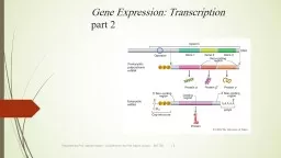
/
Dr. Sabah Mohamed Alharazy, Nephrology Unit, Department of Medicine, P Dr. Sabah Mohamed Alharazy, Nephrology Unit, Department of Medicine, P

Dr. Sabah Mohamed Alharazy, Nephrology Unit, Department of Medicine, P - PDF document

lois-ondreau
lois-ondreau . @lois-ondreau
Follow
429 views
Uploaded On 2016-07-01

Dr. Sabah Mohamed Alharazy, Nephrology Unit, Department of Medicine, P - PPT Presentation

December 03 2013 Alharazy S Kong Mohd B ID: 384759

December 2013; Alharazy

Share:

Link:

Embed:

Download Presentation from below link

Download Pdf The PPT/PDF document "Dr. Sabah Mohamed Alharazy, Nephrology U..." is the property of its rightful owner. Permission is granted to download and print the materials on this web site for personal, non-commercial use only, and to display it on your personal computer provided you do not modify the materials and that you retain all copyright notices contained in the materials. By downloading content from our website, you accept the terms of this agreement.


Presentation Transcript

Research Article J Clin Cell Immunol 2014, 5:1 Research Article Alharazy S (2014) Urinary Monocyte Chemoattractant Protein and Lupus Nephritis Activity. J 10.4172/2155-9899.1000187 Denition of LN activityA. Active LN was dened by the presence of one or more of the Proteinuria with or without any of the following features Presence of haematuria and/or red cell castsb) Proteinuria was measured as spot morning urine protein creatinine �index (uPCI) and was positive if the value was 1000 mg /mmol (NR Renal SLEDAI score  4 [13]. B. Relapse/ are of LN was dened as recurrence of renal disease activity aer a period of remission  3 months for the purpose of this C. Remission was dened as absence or reduction of renal disease activity and no change in immunosuppressive therapy for at least 3 D. Inactive LN was dened by the presence of one or more of the following criteria: I. Proteinuria (uPCI)mg/ mmol with/ without any of the Each patient was evaluated using clinical and laboratory parameters. erythrocyte sedimentation rate (ESR), serum complement levels C3 directions. Standard or test samples were added to each well and le the correction wavelength set at 540 nm. All samples were assayed in duplicate. uMCP-1 levels were expressed as concentrations normalized (SN (1-SN))TP+FN=Z×() 0.90 1 –0.901.96× 3.842×() TP FN NSN 34.578 distributed data were presented as median ± interquartile range (IQR). variables. Correlation between uMCP-1 levels with relevant laboratory Alharazy S (2014) Urinary Monocyte Chemoattractant Protein and Lupus Nephritis Activity. J 10.4172/2155-9899.1000187 regression to evaluate the independent predictors of LN activity. uMCP-1 and all relevant standard markers of LN activity with a pwere included in the regression model. e SPSS soware version 18.0 was used for statistical analysis. Probability (p) values ofconsidered signicant.ResultsDemographic and clinical characteristics of the study patients One hundred patients with SLE and biopsy proven LN were recruited. eir baseline clinical characteristics are listed in Table 1. Patients with active LN had lower serum albumin, higher angiotensin system (RAS) blockers (ACE-inhibitors and/or ARBs and/ use of immunosuppressive medications between the two groups viz Urinary MCP-1 levels and correlation with clinical and 1 [9,317.5 pg/ mg creatinine, IQR (548.3-40,170)] than those with 92 (92)43 (91.5)49 (92.5)0.578 (8)4 (8.5)4 (7.5)41(41)24 (51.1)17 (32.1)0.1455 (55)21 (44.7)34 (64.2)4 (4)2 (4.3)2 (3.8)Age, mean ± SD years36.90 ± 10.6236.40 ± 9.97 37.33 ± 11.240.74LN duration (years) 7 (1-24)7 (1-24)7 (1-17)0.561 (1)1 (2.1)0 (0) - WHO class II ± V 6 (6)3 (6.4)3 (5.7) - WHO class III ± V34 (34)15 (31.9)19 (35.8) - WHO class IV ± V52 (52)26 (55.3)26 (49.1)5 (5)1 (2.1)4 (7.5)0.712 (2)1 (2.1)1 (1.9)8 (0-19) 9 (0-16)8 (0-19) 0.933 (0-15)3.58 (0-9)3 (1-15)0..55� -Stage 1 (eGFR 90)61 (61)25 (53.2)36 (67.9)22 (22)10 (21.3)12 (22.6)0.06 -Stage 3 (eGFR30-59)14 (14) 9 (19.1) 5 (9.4) -Stage 4 (eGFR15-29) 3 (3) 3 (6.4) 0 (0%)Continuous variables were calculated by Mann-Whitney U test or Kruskal-Wallis test, and categorical variables by Pearson’s chi-square test. SD: Standard Active LN n=47Inactive LN n=53p value12.2 (8.6-16.6)12.3 (8.5-15.6)0.57/L)7.7 ± 3.67.1 ± 2.650.45234 ± 112256 ± 83.50.19Serum albumin (NR 35-50 g/L)37.78 ± 5.5441.88 ± 3.5969 (33-252)63 (41-158)0.2993.61 ± 46.0199.75 ± 31.540.43uPCI (NR)1100 (100-5100)200 (100-500)Serum C3 (NR 79-152 mg/dL)100.5 ± 36.39109.62 ± 39.940.24Serum C4 (NR 16-38 mg/dL)21.46 ±12.8222.94 ± 110.548 (0-18)2 (0-10)4 (0-16)0 (0-3.5)52 (98.1)0.1210 (18.9)0.4219 (40.4)11 (20.8)12 (25.5)24 (45.3)20 (42.6)22 (41.5)0.9129 (61.7)39 (73.6)0.11Values are in median (interquartile range) or mean (standard deviation)deviation)values were calculated by Mann-Whitney U test or Independent sample T test]LN: Lupus Nephritis; WBC: White Blood Cell count; eGFR: Estimated Glomerular Filtration Rate; uPCI: Urine Protein Creatinine Index; SLEDAI-2K: Systemic Lupus Erythematosus Disease Activity Index-2K; anti-dsDNA: Anti Double Stranded DNA; Alharazy S (2014) Urinary Monocyte Chemoattractant Protein and Lupus Nephritis Activity. J 10.4172/2155-9899.1000187 creatinine with a maximum Youden index of 0.48, the sensitivity of uMCP-1 for early diagnosis of active LN was 0.87 with a specicity (95% CI: 0.37-0.63: p=0.95) and those for serum C3 and serum C4 werp=0.95: p)and that for SLEDAI-2K renal score were 0.96 Figure 1:uMCP-1 levels in patients with and without active lupus nephritis (LN). The horizontal line across the boxes represent the median value of uMCP-1 levels among patient groups: the areas between the upper and lower limits of boxes represent the interquartile range; the vertical lines protruding from the box represent the maximum and minimum values of uMCP-1 levels respectively. uMCP-1 levels were signi�cantly higher in patients with active LN compared to those with inactive Figure 2:The correlation between uMCP-1 levels and SLEDAI-2K global and renal scores. A. Positive correlation between uMCP-1 and SLEDAI-2K (global) (r=0.28, Alharazy S (2014) Urinary Monocyte Chemoattractant Protein and Lupus Nephritis Activity. J 10.4172/2155-9899.1000187 leucocyturia were 0.72 (95% CI: 0.60-0.84: p=0.001) and 0.65 (95% CI: that for serum creatinine was 0.58 (95% CI: 0.44-0.71: p=0.95: p=0.21) us, uMCP-1 was superior to the serological and usual biochemical markers but was not as good as proteinuria (uPCI) and SLEDAI-2K r: Spearman’s rho correlation coef�cient; eGFR: Estimated Glomerular Filtration Rate; C3: Complement 3; C4: Complement 4; anti dsDNA: Anti Double Stranded DNA; Figure 3:Receiver operating characteristic curves (ROC) of uMCP-1 and serological markers for the diagnosis of LN activity in SLE patients. The AUC was 0.82 (95% CI: 0.73-0.:1: p=0.001), the symbol (+) represents the best cut-off value (4,247 pg/mL) with a sensitivity and a speci�city of 0.87 and 0.61 respectively. The AUC for anti dsDNA Ab titres was 0.50 (p=0.95) and that for serum C3 and serum C4 were 0.37 (p=0.50) and 0.43 (p=0.26). These later were lower than that for uMCP-1. Alharazy S (2014) Urinary Monocyte Chemoattractant Protein and Lupus Nephritis Activity. J 10.4172/2155-9899.1000187 biomarker that could be used for the monitoring of LN disease activity activity 100 patients with LN, we evaluated the role of uMCP-1 levels as a non-invasive biomarker for LN activity and investigated its correlations with current standard laboratory markers and disease activity indices.MCP-1 is a chemotactic cytokine that is expressed by various renal cells in response to stimulation with proinammatory cytokines and immune complexes (IC) [21]. is in turn leads to mononuclear pathogenesis and progression of glomerular and tubulointerstitial and tubulointerstitial reported that MRL/lpr mice with MCP-1-deciency exhibit prolonged survival when compared to MCP-1+/+MRL/lpr mice as they lack renal renal injury and are thus protected from renal damage. Furthermore, renal damage. Furthermore, In patients with LN, the presence of MCP-1 in the urine indicates its intrarenal expression [10,27,28] and this correlated signicantly with the degree of leukocyte inltration in the kidneys [28]. In follow up studies, uMCP-1 levels were also found to be increased in patients Figure 4:Receiver operating characteristic curve (ROC) of uMCP-1 compared with standard parameters for LN activity. A. uMCP-1 compared with urinary parameters and SLEDAI-2K (renal). B) uMCP-1 compared with blood parameters and eGFR. The black solid curve in Figure A and B represents the uMCP-1; the area under the curve (AUC) was 0.82 (p=0.001). The AUC for proteinuria was 0.94 (p)and those for haematuria and leucocyturia were 0.72 (p=0.001) and 0.65 (p=0.23) respectively. The AUC for SLEDAI-2K was 0.96 (p)The AUC for serum albumin was 0.24 (p=0.001) and that for serum creatinine was 0.58 (p=0.21). The AUC for eGFR was 0.41 (p=0.21). Thus, uMCP-1 was better than haematuria, leucocyturia serum albumin, serum creatinine and eGFR but was not as good as proteinuria (uPCI) and SLEDAI-2K renal score for detection of LN activity. 0.81 (Hosmer & Lemeshow’s), 0.66 (Cox & Snell), 0.89 (Nagelkerke). Model x=103.:3, p<0.001. Hosmer-Lemeshow χ Alharazy S (2014) Urinary Monocyte Chemoattractant Protein and Lupus Nephritis Activity. J 10.4172/2155-9899.1000187 with active LN and reduced with treatment-induced disease remission remission It appears that uMCP 1 may be a useful clinical marker for predicting and monitoring LN activity. Singh et al. [33] in a longitudinal study (20 patients) reported that uMCP-1 can distinguish between those patients with active LN from those with inactive renal disease or stable SLE.Our study (n=100) also demonstrated that uMCP-1 levels were signicantly elevated in patients with active LN compared to those with inactive renal disease. uMCP-1 levels also correlated directly with proteinuria and inversely with serum albumin. ese ndings ndings and Alzawawy et al. [35]. e two latter authors also observed the same signicant correlation between uMCP-1 levels and proteinuria. proteinuria. In a recent report of LN patients (n=83) with juvenile onset of SLE, Watson et al. [36] also found that uMCP-1 levels were higher in those patients with active renal disease. Like us, they found no association between uMCP-1 levels and serum creatinine or eGFR. On the other hand, other studies [27,34,37,38] have reported that uMCP-1 levels levels with SLE.Our ndings of a high correlation between uMCP-1 with global SLEDAI-2K and renal SLEDAI-2K scores had previously been reported by many authors. ese include Rovin et al. [11], Chan et al. [34], El-shehaby et al. [37] and Rosa et al. [38]. Due to the lag time between the urine samples obtained for uMCP-1 assays and renal biopsies, we were not able to correlate this biomarker with LN classes as the histological features of LN would have changed with time and treatment.ere were no associations between the third and fourth components of serum complement (C3, C4) and anti dsDNA antibody (C3, C4) and anti dsDNA antibody that uMCP-1 levels were associated with serum complements C3 and C4. Whereas Chan et al. [34] demonstrated a signicant correlation between urinary mRNA expression of MCP-1 with SLE disease activity indices and anti dsDNA antibody titres in patients with active LN during treatment with immunosuppressive medications. Similary, Kiani et al. [39] in a longitudinal study of SLE patients (n=87) found a signicant correlation between uMCP-1 levels with anti dsDNA positivity.Contrary to all the above, Tucci et al. [27] found the uMCP-1 levels to be decreased in his cohort of LN patients during treatment with IV Cyclophosphamide. In our study, uMCP-1 levels were higher in those inhibitor and azathioprine with LN disease activity, uMCP-1 levels did not dier with the use of these immunosuppressive medications. On the other other found that uMCP-1 levels were not related to the cumulative dose of steroids or immunosuppressive medications neither received within 30 days preceding a renal are nor were the uMCP-1 levels related to the use of RAS blockers. Whereas Wada et al. [10] reported that uMCP-1 levels decreased aer commencing high dose corticosteroid therapy in in serum complement levels were not associated with LN activity. ese were tested at 2-monthly intervals which may be too short an interval ndings are consistent with those reported by others [41,42]. Moroni et al. demonstrated that only anti-C1q antibodies associated signicantly signicantly Esdaile et al. evaluated anti dsDNA Ab, C3, C4, and Clq as predictors predictors raised anti dsDNA Ab levels was associated with active renal disease [43,44] and that decline in C3 or C4 levels coincided with an increase in LN activity [45].In a recent report, Abujam et al. demonstrated uMCP-1 to be superior to serum C4 and urinary CXCL-10/IP-10 in this regard but of equivalent ecacy to anti ds DNA Ab titres and serum C3 in dierentiating active LN from non renal SLE [46]. In this current study, the ROC curve for uMCP-1 showed that it had a good diagnostic prole for early detection of LN activity and outperformed anti ds DNA Ab range from histopathologically severe disease to varying levels of Early treatment of LN may lead to earlier and more complete to immunosuppressive medications and their toxicities and results in Alharazy S (2014) Urinary Monocyte Chemoattractant Protein and Lupus Nephritis Activity. J 10.4172/2155-9899.1000187 the majority of such patients. We are following this cohort of 100 LN patients longitudinally and hope to report these results in the near This paper was presented as a poster [SU183] at the International Society of Nephrology (ISN), World Congress of Nephrology (WCN) 2013 Hong Kong, May 31-4 June 2013. We thank the Dean and Medical Director of the UKMMC for his kind permission to publish these data. We are grateful to Rahimah Ismail and Ra�dah Mamat for helping with preparation of urine samples for the MCP-1 assays. This study was supported by a grant from the Faculty of Medicine, UKM [FF-446- Korbet SM, Lewis EJ, Schwartz MM, Reichlin M, Evans J, et al. (2000) Factors predictive of outcome in severe lupus nephritis. Lupus Nephritis Collaborative Reyes-Thomas J, Blanco I, Putterman C (2011) Urinary biomarkers Hewitt SM, Dear J, Star RA (2004) Discovery of protein biomarkers for renal Brunner HI, Mueller M, Rutherford C, Passo MH, Witte D, et al. (2006) Urinary neutrophil gelatinase-associated lipocalin as a biomarker of nephritis in childhood-onset systemic lupus erythematosus. Arthritis Rheum 54: 2577- Fiehn C, Hajjar Y, Mueller K, Waldherr R, Ho AD, et al. (2003) Improved clinical outcome of lupus nephritis during the past decade: Importance of early diagnosis and treatment. Ann Rheum Dis 62: 435-439. Houssiau FA, Vasconcelos C, D’Cruz D, Sebastiani GD, de Ramon Garrido E, et al. (2004) Early response to immunosuppressive therapy predicts good renal outcome in lupus nephritis. Arthritis Rheum 50: 3934-3940. Rovin BH, Phan LT (1::8) Chemotactic factors and renal in�ammation. Am J Kelley VR, Rovin BH (2003) Chemokines: therapeutic targets for autoimmune Kuroiwa T, Lee EG (1998) Cellular interactions in the pathogenesis of lupus nephritis: the role of T cells and macrophages in the ampli�cation of the Wada T, Yokoyama H, Su SB, Mukaida N, Iwano M, et al. (1996) Monitoring urinary levels of monocyte chemotactic and activating factor re�ects disease 11. Rovin BH, Song H, Birmingham DJ, Hebert LA, Yu CY, et al. (2005) Urine chemokines as biomarkers of human systemic lupus erythematosus activity. J Hochberg MC (1997) Updating the American College of Rheumatology revised criteria for the classi�cation of systemic lupus erythematosus. Arthritis Rheum Gladman DD, Ibañez D, Urowitz MB (2002) Systemic lupus erythematosus 14. Bombardier C, Gladman DD, Urowitz MB, Caron D, Chang CH (1992) of the SLEDAI. A disease activity index for lupus patients. The Committee on Kong N et al (2011) Pulse Cyclophosphamide versus added dual �ltration plasmapheresis (DFPP) therapy for severe lupus nephritis. UKM research Jones S, Carley S, Harrison H (2003) An introduction to power and sample size estimation. Emerg Med J 20: 453-458. Sidiropoulos PI, Kritikos HD, Boumpas DT (2005) Lupus nephritis �ares. Lupus Youden WJ (1950) Index for rating diagnostic tests. Cancer 3: 32-35. Cameron JS (1999) Lupus nephritis. J Am Soc Nephrol 10: 413-424.20. Schwartz N, Su L, Burkly LC, Mackay M, Aranow C, et Segerer S, Nelson PJ, Schlöndorff D (2000) Chemokines, chemokine receptors, and renal disease: from basic science to pathophysiologic and therapeutic Stahl RA, Thaiss F, Disser M, Helmchen U, Hora K, et al. (1993) Increased expression of monocyte chemoattractant protein-1 in anti-thymocyte antibody- Rovin BH, Doe N, Tan LC (1996) Monocyte chemoattractant protein-1 levels in Saitoh A, Suzuki Y, Takeda M, Kubota K, Itoh K, et al. (1998) Urinary levels of monocyte chemoattractant protein (MCP)-1 and disease activity in patients with Tesch GH, Maifert S, Schwarting A, Rollins BJ, Kelley VR (1999) Monocyte chemoattractant protein 1-dependent leukocytic in�ltrates are responsible for Hasegawa H, Kohno M, Sasaki M, Inoue A, Ito MR, et al. (2003) Antagonist of monocyte chemoattractant protein 1 ameliorates the initiation and progression of lupus nephritis and renal vasculitis in MRL/lpr mice. Arthritis Rheum 48: Tucci M, Barnes EV, Sobel ES, Croker BP, Segal MS, et al. (2004) Strong a functional polymorphism in the monocyte chemoattractant Noris M, Bernasconi S, Casiraghi F, Sozzani S, Gotti E, et al. (1995) Monocyte Wada T, Yokoyama H, Tomosugi N, Hisada Y, Ohta S, Wada T, Furuichi K, Sakai N, Iwata Y, Yoshimoto K, et al. (2000) Up-regulation of monocyte chemoattractant protein-1 in tubulointerstitial lesions of human Tesar V, Masek Z, Rychlík I, Merta M, Bartůnková J, et al. (1::8) Cytokines and adhesion molecules in renal vasculitis and lupus nephritis. Nephrol Dial Tsai CY, Wu TH, Yu CL, Lu JY, Tsai YY (2000) Increased excretions beta2-microglobulin, IL-6, and IL-8 and decreased excretion of Tamm-Horsfall glycoprotein in urine of patients with active lupus nephritis. Nephron 85: 207- diagnostic and prognostic marker in patients with lupus nephritis �are. Lupus Chan RW, Lai FM, Li EK, Tam LS, Wong TY, et al. (2004) Expression of chemokine and �brosing factor messenger RNA in the urinary sediment of Alzawawy A, Zohary M, Ablordiny M, chemoattractantprotein-1 (Mcp-1) level in patients with lupus nephritis. Int J Watson L, Midgley A, Pilkington C, Tullus K, Marks S, monocyte chemoattractant protein 1 and alpha 1 acid glycoprotein as biomarkers of renal disease activity in juvenile-onset systemic lupus erythematosus. Lupus El-Shehaby A, Darweesh H, El-Khatib M, Momtaz M, Marzouk S, et al. (2011) of urinary biomarkers, TNF-like weak inducer of apoptosis (TWEAK), osteoprotegerin (OPG), monocyte chemoattractant protein-1 (MCP- Rosa RF, Takei K, Araújo NC, Loduca SM, Szajubok JC, et al. (2012) Monocyte chemoattractant-1 as a urinary biomarker for the diagnosis of activity of lupus Kiani AN, Johnson K, Chen C, Diehl E, Hu H, et al. (2009) Urine osteoprotegerin and monocyte chemoattractant protein-1 in lupus nephritis. J Rheumatol 36: Alharazy S (2014) Urinary Monocyte Chemoattractant Protein and Lupus Nephritis Activity. J 10.4172/2155-9899.1000187 40. Illei GG, Tackey E, Lapteva L, Lipsky PE (2004) Biomarkers in systemic lupus Moroni G, Trendelenburg M, Del Papa N, Quaglini S, Raschi E, et al. (2001)help in diagnosing a renal �are in lupus nephritis. Am Esdaile JM, Joseph L, Abrahamowicz M, Li Y, Danoff D, et al. (1996) Routinetests in systemic lupus erythematosus: is there a need for more Spronk PE, Bootsma H, Kallenberg CG (1998) Anti-DNA antibodies as earlydisease exacerbations in SLE. Guideline for treatment? Clin Rev Bootsma H, Spronk P, Derksen R, de Boer G, Wolters-Dicke H, et al. (1995)Prevention of relapses in systemic lupus erythematosus. Lancet 345: 1595- Ho A, Barr SG, Magder LS (2001) A decrease in complement is associatedwith increased renal and hematologic activity in patients with systemic lupuserythematosus. Arthritis Rheum 44: 2350-2367. Abujam B, Cheekatla S, Aggarwal A (2013) Urinary CXCL-10/IP-10 and MCP-1 Dr. Sabah Mohamed Alharazy, Nephrology Unit,Department of Medicine, Pusat Perubatan Universiti Kebangsaan Malaysia, Jalaln Yaacob Latif, Bandar Tun Razak, 56000 Cheras, Kuala Lumpur, Malaysia, Tel: December 03, 2013; January 20, 2014; Alharazy S, Kong , Mohd , Báin A, et al. (2014) Urinary Monocyte Chemoattractant Protein and Lupus Nephritis Activity. J Clin Cell 10.4172/2155-9899.1000187© 2014 Alharazy S, et al. This is an open-access article distributed under the terms of the Creative Commons Attribution License, which permits unrestricted use, distribution, and reproduction in any medium, provided the Monocyte chemoattractant protein-1 (MCP-1) is reported to be associated with lupus nephritis (LN) This was a cross-sectional observational study in which uMCP-1 levels and the standard parameters One hundred patients were recruited: 47 with active and 53 inactive LN. uMCP-1 levels were increased in those with active LN [9,317.5 pg/mg creatinine (5,48.3-40,170)] compared to those with inactive LN [3,682 pg/mg creatinine (0-23,866)] (p)correlated with proteinuria (r=0.39,p=0.001), serum albumin (r=-0.35, uMCP-1 was 0.82 (p=0.001) compared with 0.50 (p=0.95), 0.37 (p=0.50), 0.43 (p=0.26) for anti-ds-DNA Ab, C3 and C4 respectively. AUROC for proteinuria was 0.94 (p)()()uMCP-1 may provide further adjunctive evidence if the clinical diagnosis of LN activity remains uncertain and facilitate improved grading of renal disease activity in this complex disease thus leading to improved , Shamsul A Shah, Arbaiyah Báin and Abdul Halim Abdul GaforNephrology Unit, Department of Medicine, Universiti Kebangsaan Malaysia Medical Centre, Kuala Lumpur, MalaysiaDepartment of Medical Microbiology & Immunology, Universiti Kebangsaan Malaysia Medical Centre, Kuala Lumpur, MalaysiaDepartment of Community Medicine, Universiti Kebangsaan Malaysia Medical Centre, Kuala Lumpur, MalaysiaLupus nephritis (LN); nocyte chemoattractant of Systemic Lupus Erythematosus (SLE) and is associated with with characterized by unpredictable ares.e conventional laboratory markers used in clinical practice such as serum complement levels and double-stranded DNA antibodies are unreliable indicators of LN as they lack both sensitivity and specicity of LN as they lack both sensitivity and specicity urinary sediments are also non-specic markers [4].Renal biopsy remains the gold standard for the evaluation of LN disease activity. However, it is an invasive procedure and serial renal followed serially and would enable timely institution of appropriate of appropriate patient’s total exposure to immunosuppressive medications and their toxicities. Evidence in human and animal studies have demonstrated the pathogenic role of MCP-1 in renal injury in LN [7,8]. Several studies in LN patients have shown that uMCP-1 to be associated with LN [9] and its severity [10] and ares [11].We therefore investigated the usefulness of uMCP-1 levels as a marker of LN activity in SLE patients with biopsy proven LN. Methodsis was a cross-sectional study which recruited consecutive SLE SLE Clinic at our centre. is study was carried out between 9 December 2011 and 4 August 2012. We excluded those patients with end stage Committee of the Universiti Kebangsaan Malaysia Medical Centre Centre (score range 0-150), extrarenal (score range 0-63) and renalClinical & Cellular Immunology Journal of Clinical & Cellular ImmunologyISSN: 2155-9899