/
Unit 5: Cell Cycle & Division Unit 5: Cell Cycle & Division

Unit 5: Cell Cycle & Division - PowerPoint Presentation

lois-ondreau
lois-ondreau . @lois-ondreau
Follow
348 views
Uploaded On 2019-06-29

Unit 5: Cell Cycle & Division - PPT Presentation

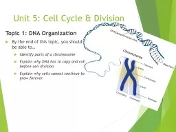
Topic 1 DNA Organization By the end of this topic you should be able to Identify parts of a chromosome Explain why DNA has to copy and coil before cell division Explain why cells cannot continue to grow forever ID: 760566

cells cell dna chromosomes cell cells chromosomes dna division meiosis mitosis chromosome cycle cancer daughter chromatids sex number checkpoint

Share:

Link:

Embed:

Download Presentation from below link

Download Presentation The PPT/PDF document "Unit 5: Cell Cycle & Division" is the property of its rightful owner. Permission is granted to download and print the materials on this web site for personal, non-commercial use only, and to display it on your personal computer provided you do not modify the materials and that you retain all copyright notices contained in the materials. By downloading content from our website, you accept the terms of this agreement.


Presentation Transcript

Slide1

Unit 5: Cell Cycle & Division

Topic 1: DNA Organization

By the end of this topic, you should be able to…

Identify parts of a chromosome

Explain why DNA has to copy and coil before cell division

Explain why cells cannot continue to grow forever

Slide2

1

. DNA is a double helix made of

nucleotides

Slide3

2. Histone proteins protect the DNA

Slide4

3. Nucleosomes are made of DNA and 8 histone proteins

Slide5

4. H1 Histone is added to make a chromatosome

Slide6

5

. These fold into

chromatin fibers

Slide7

6

.

The fibers make

loops

Slide8

7

.

The loops make

compressed chromatin

Slide9

8

. The chromatin is packed into a chromosome9. Chromosome consists of 2 sister chromatids connected by a centromere

9. Chromosome: 2 sister chromatids connected by centromere

Slide10

Organization of Genetic Material

All the DNA in a cell constitutes the cell’s genomeA genome can consist of a number of DNA molecules DNA molecules in a cell are packaged into chromosomes

Slide11

Organization Continued…

Eukaryotic chromosomes consist of chromatin, a complex of DNA and protein that condenses during cell divisionEvery eukaryotic species has a characteristic number of chromosomes in each cell nucleusNon-reproductive cells have two sets of chromosomes

Slide12

Slide13

DNA Terms

In preparation for cell division, DNA is replicated and the chromosomes condenseEach duplicated chromosome has two sister chromatids which separate during cell divisionThe centromere is where the two chromatids are most closely attached

Slide14

To reproduce or not to reproduce, that is the question.

-The Cell Cycle

Slide15

Growth vs. Division

When an animal or plant grows, what happens to its cells?

Does an animal get larger because each cell increases in size (grows) or because it produces more of them?

Slide16

Why can’t cells grow forever?

REASON 1: Not enough DNA! ...as the cell increases in size, it keeps the same amount of DNA. Eventually the cell will grow too much for the DNA to control all its activities

Every time a cell divides, the telomeres (ends of chromosomes) become smaller,

so organisms age!

DNA Overload:

when the DNA of a cell has too many tasks to do (like making proteins), and not enough DNA to get the job(s) done, the cell has “DNA overload” [cell size must be limited]

Slide17

Why Can’t Cells Grow Forever?

REASON #2: SURFACE AREA TO VOLUME RATIO:Surface area of membrane doesn’t increase as quickly as cell volume Too little membrane  not enough exchange of materials in and out of the cell

Slide18

The Solution?

Before a cell becomes too large, it divides to form 2 “daughter cells”. This process is called cell divisionCell division can only happen once a cell has made a copy of its DNA so that each daughter cell can have a full genetic library

Slide19

Why Divide?

Multicellular organisms depend on cell division forDevelopment from a fertilized cellGrowthRepairCell division is an integral part of the cell cycle, the life of a cell from formation to its own division

Slide20

Topic 2: Mitosis

By the end of this topic, you should be able to…

Explain why cells must divide

Draw and label the stages of mitosis

Compare and contrast cell division (cytokinesis) in plant and animal cells

Compare and contrast prokaryotic and eukaryotic division

Slide21

Purpose of Mitosis

To create two

identical daughter cells

from one parent cell

Cells begin

diploid

(2 sets of chromosomes) and end

diploid

Used

for somatic cell division and asexual reproduction

Slide22

Types of Asexual Reproduction

Binary fission

Sporulation

Regeneration

Vegetative propagation

Slide23

Vocab

Diploid Cells (2n) = 2 sets of chromosomes…one from each parent (Example: human body cell) --- SOMATIC CELLSHaploid Cells (n)= only have 1 set of chromosomes (Example: Sperm or Egg Cell) --- SEX CELLS

Slide24

Types of Chromosomes

Sex chromosomes = determine the sex of an organism; either X or Y, referred to as “23rd pair” Autosomes = all the other chromosomes in an organism (“1st – 22nd pair”)

Slide25

Cell Cycle

During the cycle, a cell grows, prepares for division, and divides to form 2 daughter cells, each of which then begins the cycle again2 Main Parts 1) Interphase 2) Cell Division (mitosis + cytokinesis)

The series of events that cells go through as the

grow

and

divide

Slide26

G1 SG2MitosisCytokinesis

Interphase

Stages of the Cell Cycle

Slide27

The Steps Prior to Cell Division

the cell doubles in size (

G1 Phase

)

chromosomes replicate (

S Phase

)

the number of organelles doubles (

G2 Phase)

most doubling is directed by the nucleus

Slide28

Replicated Chromosome

Sister chromatids

centromere

Slide29

What is DNA Replication?

A chromosome is unzipped and thus starts as one strand of

DNA

Each daughter cell

needs its own copy of the DNA strand

.

The DNA strand is duplicated and the two parts are

tied

together.

Slide30

Important Details

DNA replication

occurs during

S

phase

, NOT

mitosis

(S stands for Synthesis or making more DNA)

Mitosis and

cytokinesis

overlap

Slide31

Important Details

Cells can also enter a G

0

phase

in which they no longer

divide

Cells move to the next stage when enough “

trigger protein

has built up

Slide32

Cells of the adult central nervous system (brain and spinal cord) do NOT divide.

Slide33

Cassius Clay

Muhammad Ali

PARKINSON'S

Progressive nervous system

disorder

-

nerve cells break down and

die and cannot be replaced

Slide34

Christopher Reeve

Horseback injury left him paralyzed

Slide35

Mitosis is the process of dividing just the nucleus (not the whole cell.)

ProphaseMetaphaseAnaphaseTelophase

Slide36

PROPHASE

Slide37

Steps of MITOSIS

PROPHASE

Nuclear

membrane

is broken

down

2. Chromosomes appear

3. Centrioles migrate

(plants DON

T

have centrioles

)

4. Spindles form

Slide38

METAPHASE

Slide39

METAPHASEChromosomes align on the equatorial planeSpindle fibers attach to chromosomes

Steps of MITOSIS

Slide40

ANAPHASE

Slide41

ANAPHASEChromatids move to opposite ends of the cell with the help of spindle fibers

Steps of MITOSIS

Slide42

TELOPHASE

Slide43

TELOPHASEchromosomes stop moving and the nuclear membrane reforms

Steps of MITOSIS

Slide44

Which Stage?

Slide45

Which Stage?

Slide46

Which Stage?

Slide47

Which Stage?

Slide48

CYTOKINESIS

Division

of the entire

cell after the nucleus

divides

Differs for plants and

animals because plant cells

have

cell walls

Slide49

Slide50

Animal Cell Division

T

he

cell membrane

constricts to make a groove and

divide

This groove is referred to as the

cleavage furrow

Slide51

Plant Cell Division

Vesicles produced by

golgi

bodies

form a midline in the

cell

Vesicles fuse to make a

cell plate

which attaches to cell wall

Slide52

How does the beginning cell differ from the ending cells?

Slide53

Mitosis Rap

http://www.youtube.com/watch?v=pOsAbTi9tHw

Slide54

Binary Fission in Bacteria

Prokaryotes (bacteria and

archaea

) reproduce by a type of cell division called

binary fission

Slide55

Reproduction in Prokaryotes

Binary Fission

(

ASEXUAL

): cell parts reproduce and cell divides in

half

-The most

common form

of

reproduction for bacteria and

archae

-In

binary fission, the DNA

replicates,

and the two daughter chromosomes actively move

apart

-The

plasma membrane pinches inward, dividing the cell into

two

-Produces genetically identical daughter cells

Slide56

binary fission = bacteria divide

Slide57

Bacterial conjugation

with a conjugation bridge.

-One bacterium transfers the plasmid to the other bacterium through the conjugation bridge.-This produces genetic diversity in bacteria that may ensure their survival. Can pick up resistance to antibiotics this way!

Conjugation

(SEXUAL): exchange of genetic material (plasmid) between two bacterium

Reproduction in Prokaryotes

Slide58

Slide59

Topic 3: Meiosis

By the end of this topic, you should be able to…

Compare and contrast sexual and asexual reproduction

Illustrate meiosis I and meiosis II

Explain fertilization of eukaryotic cells

Explain production of egg and sperm cells

Explain nondisjunction and read a karyotype

Slide60

Warm-Up

Are somatic (body) cells haploid or diploid and what does this mean?

Haploid = one set of chromosomes (egg, sperm… gametes)

Diploid = two sets of chromosomes (body cells)

How many chromosomes are found in human body cells?

46 chromosomes (23 pairs – 1 set from mom, 1 set from dad)

Why is mitosis necessary and important?

Grow; repair; development

How do daughter cells differ from parent cells in mitosis?

Genetically identical (don’t differ)

How does mitosis differ from meiosis?

Slide61

Chromosome Structure, revisited

Slide62

Diploid Cell- where DNA comes from

Slide63

Meiosis does two things -

1) Meiosis takes a cell with two copies of every chromosome (diploid - 2n) and makes cells with a single copy of every chromosome (haploid – 1n).

In meiosis, one diploid cells produces

four haploid cells

.

Slide64

2) Meiosis scrambles the specific forms of each gene that each sex cell (egg or sperm) receives.

This makes for a lot of genetic diversity. This trick is accomplished through independent assortment and crossing-over.

Genetic diversity is important for the evolution of populations and species.

Slide65

Why do we need meiosis?

Meiosis is necessary to halve the number of chromosomes going into the sex cells

Why halve the chromosomes in gametes?

At fertilization the male and female sex cells will provide

½ of the chromosomes

each – so the offspring has genes from both

parents

that together total 100% of the genes the offspring should have

Slide66

Purpose

Meiosis is used to make special cells - sperm cells and egg cells - that have half the normal number of chromosomes. It reduces the number from 23 pairs of chromosomes to 23 single chromosomes. The cell copies its chromosomes, but then separates the 23 pairs to ensure that each daughter cell has only one copy of each chromosome. A second division that divides each daughter cell again to produce four daughter cells.

Slide67

Meiosis

Parent cell – chromosome pair

Chromosomes copied

1

st

division - pairs split

2

nd

division – produces 4 gamete cells with ½ the original no. of chromosomes

Slide68

Meiosis I : Separates Homologous (Matching) Chromosomes

Interphase

DNA is replicated

The result is two genetically identical sister chromatids which remain attached at their

centromeres

(just like in Mitosis)

Slide69

Prophase I

During this phase each pair of chromatids don’t move to the equator alone, they match up with their homologous pair and fasten together (synapsis) in a group of four called a tetrad.Extremely IMPORTANT!!! It is during this phase that crossing over can occur. Crossing Over is the exchange of segments during synapsis.

Slide70

Metaphase I

The chromosomes line up at the equator attached by their centromeres to spindle fibers from centrioles.Still in homologous pairs

Slide71

Anaphase I

The spindle guides the movement of the chromosomes toward the polesSister chromatids remain attachedMove as a unit towards the same poleThe homologous chromosome moves toward the opposite pole

Slide72

Telophase I

This is the end of the first meiotic cell division. The cytoplasm divides, forming two new daughter cells. Each of the newly formed cells has half the number of the parent cell’s chromosomes, but each chromosome is already replicated ready for the second meiotic cell division

Slide73

Cytokinesis

Occurs simultaneously with Telophase IForms 2 daughter cellsPlant cells – cell plateAnimal cells – cleavage furrowsNO FURTHER REPLICATION OF GENETIC MATERIAL PRIOR TO THE SECOND DIVISION OF MEIOSIS

Slide74

Slide75

Meiosis II : Separates sister chromatids

Proceeds

similar

to

mitosis

THERE IS NO INTERPHASE II

!

(no replication of DNA this time)

Slide76

Prophase II

Each of the daughter cells forms a spindle, and the double stranded chromosomes move toward the equator

Slide77

Metaphase II

The chromosomes are positioned on the metaphase plate in a mitosis-like fashion

Slide78

Anaphase II

The centromeres of sister chromatids finally separateThe sister chromatids of each pair move toward opposite polesNow individual chromosomes

Slide79

Telophase II and Cytokinesis

Nuclei form at opposite poles of the cell and cytokinesis occursAfter completion of cytokinesis there are four daughter cells All are haploid (n)

Slide80

Slide81

One Way Meiosis Makes Lots of Different Sex Cells (Gametes) – Independent Assortment

Independent assortment produces 2n distinct gametes, where n = the number of unique chromosomes.

In humans, n = 23 and 223 = 6,000,0000.

Slide82

Slide83

Another Way Meiosis Makes Lots of Different Sex Cells: Crossing-Over

Crossing-over multiplies the already huge number of different gamete types produced by independent assortment.

Slide84

Swapping genes is known as a crossing over

Slide85

Crossovers occur while the homologous chromosomes are paired in Prophase I.

Slide86

Meiosis

Sex cells

divide to produce

gametes

(sperm or egg)

.

Gametes

have

half

the # of

chromosomes

.

Occurs only in gonads (testes or ovaries).

Male: spermatogenesis

Female: oogenesis

Meiosis

is similar to

mitosis

with some chromosomal differences.

Slide87

Fertilization

The fusion of a sperm and egg to form a zygote.A zygote is a fertilized egg

n=23

egg

sperm

n=23

2n=46

zygote

Slide88

Nondisjunction

Occurs when chromosomes

fail

to

separate

.

Slide89

Nondisjunction

Can occur during

Anaphase I

or

Anaphase II

of

Meiosis

Result: eggs or sperm with

incorrect

number of

chromosomes

If the mutated egg or sperm is fertilized, the child will have abnormalities.

Note: It may also occur in anaphase of

Mitosis

, but usually the abnormal cells die and the whole organism is not affected.

Slide90

Slide91

Nondisjunction results in chromosomal abnormalities

Trisomy: Each cell has an extra chromosomeMonosomy: Each cell has one less chromosome

Slide92

Amniocentesis

amniotic fluid

fetus

14-16

weeks

Slide93

Slide94

Karyotypes can detect chromosomal abnormalities

Chromosomes are photographed, cut, and matched based on size

Slide95

Examples:

In humans, nondisjunction results in a person having more or less than

46

chromosomes.

Trisomy 21

Down Syndrome-

1 in 691 babies born in US are born with DS (alters course of development, low muscle tone)

Trisomy 13

Patau

Syndrome

(~1 in 9,500 births); many with this diagnosis will not make it to birth or will survive on average 10 days (clefts, improper brain formation, extra digits)

Monosomy –Turner Syndrome

 only has an X in pair 23 (missing another sex chromosome);

1 in 2,000 female births; delayed puberty, hearing/ear issues; infertility

Trisomy-

Klinefelter

Syndrome

 has XXY (an extra sex chromosome);

1 in 500 to 1 in 1,000 male births; small testes (less testosterone)

Slide96

Trisomy 21: Down Syndrome

Three copies of chromosome 21Occurrence: 1 in 700 births, increased chances when mother is over 40.Shorter average life span (35 yrs)Common facial characteristics

Slide97

Trisomy 13: Patau Syndrome

Extra copy of Chromosome 13Occurrence: 1 in 10,000 birthsCharacteristics:Cleft lip and palateMental disabilitiesPolydactylUsually on live about 3 months, 80% die within the first year

Slide98

Monosomy: Turner Syndrome

Missing a sex chromosome1 in 2,000 birthsUsually cannot tell before pubertySex organs do not fully developWebbed neck

Slide99

Trisomy: Klinefelter Syndrome (XXY)

Caused by an extra X chromosome1 in 1,000 malesUnderdeveloped testes, taller, may have breast development, sterile

Slide100

Stages of Meiosis

Meiosis I

chromosome reduction occurs

Slide101

Prophase I

homologous

chromosomes

tetrad

Slide102

Metaphase I

equator

(midline)

spindle

fibers

centromeres

Slide103

Anaphase I

homologous chromosomes

Slide104

Telophase I

n = 23

haploid

daughter cells

Slide105

homologous chromosomes

crossing over

crossing over helps to shuffle the genes

Slide106

Stages of Meiosis

Meiosis II

chromosomes separate as they do in mitosis

Slide107

Prophase II

spindle

nuclear membrane

Slide108

Metaphase II

centromere

equator

Slide109

Anaphase II

sister

chromatids

Slide110

Telophase II

n = 23

haploid

Slide111

Topic 4: Cell Cycle Regulation

By the end of this topic, you should be able to…

Explain the role of cell regulation checkpoints

Explain what happens when the cell cycle control fails

Slide112

Regulation of Cell Cycle

G1/S checkpoint

G2/M checkpoint

Tumor suppressor genes can control these checkpoints

T

umor suppressor genes turn off or decrease rate of cell division

Slide113

G

1 checkpoint

G1

G2

G2 checkpoint

M checkpoint

M

S

Controlsystem

Figure 12.15

Slide114

For many cells, the G1 checkpoint seems to be the most important If a cell receives a go-ahead signal at the G1 checkpoint, it will usually divideIf the cell does not receive the go-ahead signal, it will exit the cycle, switching into a non-dividing state called the G0 phase

M checkpoint

G

2

checkpoint

G

1 checkpoint

Slide115

Neighboring cells communicate with dividing cells to regulate their growth also.

Slide116

Some of the genes that control cell division are called Proto-oncogenes.

Proto-oncogenes encode proteins that function to stimulate cell divisionImportant for normal human development and for the maintenance of tissues and organs.

Slide117

Oncogenes and Cancer Cells

When

mutated, an

oncogene

may

produce a large amount of these growth proteins, resulting in

excessive

cell division.

When a cell can no longer regulate its rate of cell division it becomes a Cancer Cell.

Slide118

Sometimes a proto-oncogene undergoes a mutation and becomes an oncogene.

Slide119

Because there are several genes that act as proto-oncogenes it may take 3-4 mutations to cause harm.Anything that can damage genes can cause these harmful mutations. These are called carcinogens or mutagens.

Slide120

Cancer is a disease of the cell cycle. Some of the body cells divide uncontrollably and tumors form.

Tumors in Liver

Tumor in Colon

Slide121

DNA mutations disrupt the cell cycle.

Mutations may be caused by: 1. radiation 2. smoking 3. Pollutants 4. chemicals 5. viruses

Slide122

Due to DNA mutations, cancer cells ignore the chemical signals that start and stop the cell cycle.

Slide123

Due to DNA mutations, cancer cells cannot communicate with neighboring cells. Cells continue to grow and form tumors.

Skin cancer

Slide124

SUMMARY

Normal Cell DivisionDNA is replicated properly.Chemical signals start and stop the cell cycle.Cells communicate with each other to avoid becoming overcrowded.

Cancer Cells

Mutations occur in the DNA when it is replicated.

Chemical signals that start and stop the cell cycle are ignored.

Cells do not communicate with each other and tumors form.

Slide125

Cancer Vocabulary

Tumor

= loss of cell cycle control = abnormal growth of cells

Benign

= non-spreading

Malignant

= spreading

Metastasis

= spread rate of a malignant cancer to locations other than their origin

(tumor cells enter blood vessels and travel to other parts of the body)

Slide126

Environment and Cancer

Solid relationship exists between environmental factors and cancer

Cancer cluster:

Large number of cases in restricted area

Epidemiologists examine environment for link

Cancer cluster in Woburn, Massachusetts,

Environmental trigger, industrial solvents from contaminated well water

Slide127

Skin Cancer

~1 million new cases in U.S. per year

Almost all cases related to UV light exposure from sun or tanning lamps

Lightly pigmented people higher risk

genetic characteristics can affect the susceptibility

Slide128

Skin Cancer

Ozone depletion also contributes to increased UV exposure and risk

In spite of risk, some choose low

SPF

suntan lotions

and only 25% of Americans consistently use sunscreen

Slide129

Smoking

Related to cancers of oral cavity, larynx, esophagus, and lungs

Accounts for 30% of all cancer deaths

Most have very low survival rate (e.g. 13% lung cancer sufferers survive beyond 5

yrs

)

Slide130

Case Study Response

Hypothesis: If _______________________, then ___________

Cite

all

pieces of evidence used by Tina’s doctor in her diagnosis

Tie in information from page 1 (background info)

Use information written in the brainstorming section to put Tina’s case

all together