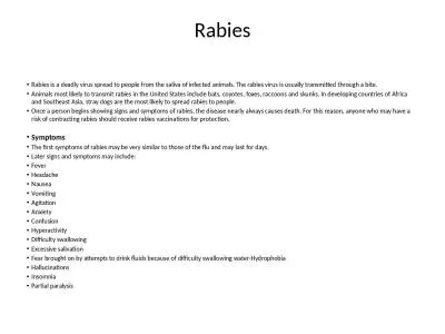
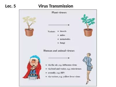
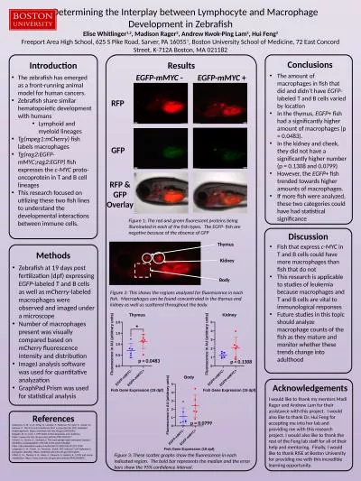
Nancy J Bigley PhD Microbiology and Immunology Program and Department of Neuroscience Cell Biology and Physiology College of Science and Mathematics and Boonshoft School of Medicine Wright State University Dayton Ohio ID: 774824
Download Presentation The PPT/PDF document " Differential Expression of SOCS1/SOCS3 ..." is the property of its rightful owner. Permission is granted to download and print the materials on this web site for personal, non-commercial use only, and to display it on your personal computer provided you do not modify the materials and that you retain all copyright notices contained in the materials. By downloading content from our website, you accept the terms of this agreement.
Slide1
Differential Expression of SOCS1/SOCS3 Ratios in Virus-Infected Macrophage Cell Lines
Nancy J. Bigley, Ph.D.Microbiology and Immunology Program, and Department of Neuroscience, Cell Biology, and Physiology ,College of Science and Mathematics and Boonshoft School of Medicine, Wright State University, Dayton, Ohio 2014
Slide2We previously noted that
murine keratinocyte
cell lines
(HEL-301 7 PAM-212) PRODUCED large
amounts of SOCS1 mRNA and
protein following infection with HSV-1 or treatment with interferon-gamma (IFN-
γ
). In contrast, murine
fibroblasts
(l929) exhibited
minimal increase in SOCS1 levels when treated with IFN-
following infection with
HSV-1
(
Frey et al. 2009).
An
antiviral state was induced in fibroblasts but not in keratinocytes. This resistance of keratinocytes to IFN-
corresponded to the
hyperinduction
of SOCS1 in these cells
.
The goal of the present study was to determine the effects of HSV-1 infection on morphology, CD14-CD86 expression, cell viability, and SOCS protein levels in
polarized M1
and M2
macrophage cell lines (J774A.1 and RAW 264.7)
during the first 24 hour of
infection. For comparison we examined these responses against the monocyte-macrophage trophic Dengue virus (DENV2) in the RAW 264.7).
Slide3Viruses
Herpes Simplex Virus-1 strain
Syn
17+ (HSV-1)
initially
obtained from Dr. Nancy
Sawtell
, Children’s Hospital Medical Center, Cincinnati, OH was propagated on confluent monolayers of Vero cells. After 4-5 days post infection or when CPE was evident, the cells were spun down, supernatant was
aliquoted
and stored at -80⁰C. Virus was quantified by infecting Vero cell monolayers with different dilutions of virus and plaque forming units were counted to calculate volume required for 0.1 multiplicity of infection (MOI).
Dengue Virus
DENV serotype 2 (DENV-2)
was provided by Dr
. Eric M. Vela,
Battelle Memorial Institute
Research
Center. DENV2 was propagated
on Vero 76 cells. Briefly, Vero 76 cells grown in 100 mm petri dishes to a confluence of approximately 85% at 37 °C, were infected with DENV-2 for 5-6 days or until CPE was evident. Cells were then scraped and centrifuged at 1500 rpm to eliminate cell debris.
The
supernatant
fluid was
aliquoted
and stored at −80°C until use. Dengue virus titers were determined by plaque assay
on
confluent monolayers of Vero 76 cells grown in 6-well plates
Murine Macrophage Cell Lines
264.7 (ATCC TIB-71) and J774A.1 (TIB-67) cells lines
were obtained from the American Type Culture Collection (ATCC) Manassas, VA.
Slide4DENV2 infection of RAW 264.7 macrophages at 3 days post infection
Slide5Macrophage Polarization Treatment
M0
No treatment
LPS (100
ng
/mL)IFN-γ (20 ng/mL)for 12-24 hours
M1
M2
CD86
SOCS1
SOCS3
20 ng/mL
IL-4 for 12-24 hours
Slide6J774A.1 Macrophages at 24 hours after polarization
LPS & IFN-
γ
IL-4
Slide7M1 J774A.1
M1 RAW 264.7
Vacuolated M1 Macrophages
Slide8J774A.1
Macrophages
Uninfected HSV-1-infected
Slide9RAW 264.7 Macrophages
Slide10J774A.1 Macrophages
RAW264.7 Macrophages
Slide111.
3
.
4.
2
.
Add virus (0.1 MOI)/cytokines
Release cells/centrifuge
Count
Virus Treatment of Macrophages
In order to accurately determine cell number
and
calculate
MOI accurately:
Cells
were released from culture
plate using
a non-enzymatic dissociation
reagent
Cell
count was taken using a TPP PCV
cell counting
tube, making it possible
to calculate MOI accurately
Slide12Monocyte/Macrophage Markers
Scavenger ReceptorsMembrane GlycoproteinCD14- LPS receptorsCD206 macrophage mannose receptor (MMR)CD200R- expressed mainly on monocytes and neutrophils. Interaction between CD200R and CD200 limit and suppress macrophage-induced inflammatory damage.CD80 (B7.1) co-receptor on antigen –presenting cells (APCs)CD163- hemoglobin-haptoglobin receptor; expressed on both monocytes and macrophages CD86 (B7.2) co-receptor on APSs
Slide13J774.1 macrophages polarized to M1 phenotype
Stained with FITC-labeled anti- mouse CD14
Stained with brilliant violet 421 -labeled anti-mouse CD86
Slide14J774a.1 Murine Macrophages
Slide15Jaguin
and colleagues
recently
used monocytes purified from the buffy coats of human peripheral blood cells to characterize phenotypic and genomic markers.
generated
macrophages from these primary human cells by treatment with M-CSF
polarized
them using the same inducers as used in the present study, LPS and IFN-γ to induce M1 phenotype and IL-4 to induce the M2
phenotype
the cell membrane marker unique to M1 cells was CD80 (B7.1
)
CD200R
expression was unique to the M2 polarized human
macrophages
As did we using M1 and M2 polarized murine macrophage cell lines (data not shown
),
they found that the mannose receptor CD 206 did not distinguish between M1 and M2 phenotypes of human macrophages.
Slide16Flow cytometry
summary of SOCS1 and SOCS3 expression by uninfected and infected J774A.1 macrophage subpopulations
Left Panel. Note Uninfected
cells at 24 h after polarization. M1 cells expressed higher levels of SOCS1 than SOCS3 with a
SOC1/SOCS3
ratio of 7:1.
Right Panel. Virus-infected
M1 cells expressed a SOCS1/SOCS3 ratio of 1:1 while M2-infected cells exhibited a SOCS1/SOCS3 ratio of
1:2
Slide17CD14/CD86 EXPRESSION IN RAW 264.7 Murine Macrophages
Slide18Slide19J774.A RAW 264.7 RatioRatioM0 1:21:2M0-HSV-1 1:21:2M1*7:11:3M1-HSV-11:11:1M21:21:2M2-HSV-11:21:2
Ratios determined by Flow cytometry (J774a.1) and Western Blot (RAW 267.4)
* Difference because of cell line or detection method,. Western Blot detects denatured antigenic fragments ; Flow cytometry detects native protein conformation. SOCS1:SOCS3 ratios in all DENV2-infected cells was 1:1.
Slide20HSV-1 infection led to morphological differences in all 3 experimental groupsHSV-1 infection decreased CD14/CD86 expression in all 3 experimental groupsM1 macrophages did not show an up regulation of SOCS1 following virus challenge, however, SOCS3 levels were increased HSV-1-infected unpolarized (M0) J774A.1 cells exhibited significant increases in expression levels of native SOCS1
Summation of Observations
Slide21At 24 h after infection, M0 control and M2 cells showed greater virus yield than did the M1 cells, presumably reflecting the loss of viable M1 cells.
Slide22Does up
regulation of SOCS3 expression in HSV-1-infected M1 macrophages over that seen in uninfected M1 cells
reflect the effects of M1 polarization or suggest
the cell’s attempt to counteract effects of
proinflammatory
molecules
?
Qasimi
and colleagues showed that different domains of SOCS3 protein are used to mediate interleukin-10 (IL-10) inhibition of TNF-α and nitric oxide production by this same macrophage cell line (
Qasimi
and others 2006). In this same macrophage cell
line (J774A.1) , Il-10
was responsible for the anti-inflammatory response to
Borrelia
burgdorferi
(Dennis and others 2006).
SOCS1/SOCS3
expression levels appeared relatively unchanged in virus-infected M2 macrophages when compared to their uninfected counterparts, suggesting microenvironment signals such as IL-4 play a greater role in SOCS expression levels than does HSV-1
infection.
We then hypothesized that the HSV-1-infected J774A.1 M1 macrophages were attempting to counteract the effects of inflammatory molecules induced by polarization.
Slide23Google Image
Note that only SOCS1 contains a nuclear localization sequence (NLS)
Structural domains of SOCS molecules
Slide24Overview of SOCS1 inhibition of cytokine-induced STATs
Slide25Pro-inflammatory and anti-inflammatory effects of SOCS3
Slide26LPS (100 ng/ml)
IFN-
γ (20 ng/ml)0.1 MOI HSV-1SOCS3 peptide or SOCS1 InhibitorIncubate 24 hours
Effects of SOCS1 and SOCS3 peptide mimetics and SOCS1 inhibitor (pJAK2) on polarized M1 J774A.1 macrophages.
p<0.001
when the SOCS1 groups were
compared
with the SOCS3 and
pJAK2 groups
Slide27Jo et al. (2005) used a recombinant cell-penetrating form of SOCS3 (CP-SCS3) to protect mice (C3H/
HeJ
) from the lethal effects of SEB and LPS by reducing production of inflammatory cytokines and attenuating apoptosis and hemorrhagic necrosis .
Within 2 hours after injection, CP-SOCS3 was distributed In multiple organs and persisted for at least 8 hours
The membrane-
translocating
motif (MTM) was composed of 12 amino acids from a hydrophobic signal sequence form fibroblast growth factor 4. The MTM was attached to either the N- terminal or C-terminal of SOCS3. Only these forms were capable of penetrating RAW cells.
Based on these observations, we tested whether the SOCS3 peptide mimetic could modify the cytotoxicity of the M1 polarization treatment or virus infection.
The peptide
mimetics
in this present study were provided by Dr. H.M. Johnson and his colleagues , University of Florida at Gainesville. These peptides contain an addition
of a
lipophilic group (
palmitoyl
-lysine) to the N terminus of the synthetic
peptide which provides them with the ability to penetrate cells.
Slide28P>0.03
P<0.03
P>0.03
SOC3 peptide
(35
μ
M/ml) or TC medium for 30 minutes prior to
LPS
(100 ng/ml)IFN-γ (20 ng/ml)0.1 MOI HSV-1Incubate 24 hours
SOCS3 Peptide Mimetic protects macrophages (RAW 264.7) from the lytic effect of HSV-1 and from the lytic effect of M1 polarization
Cell Viabilities of RAW 264.7 macrophages 24 hours after M1 polarization
Slide29CONCLUSIONS
S0CS3 peptide mimetic and the S0CS1 inhibitor (pJAK2) increased the viability of polarized M1 cells over SOCS1-treated M1 J 774A.1 macrophages similar to the observations in comparable cell groups infected with HSV-1 (p<0.001)
Prediction
: The
anti-inflammatory
effect
in these cells will be characterized by increased levels of IL-10
SOCS1 peptide mimetic decreases the viability of polarized M1 cells and HSV-1-infected M1 J774A.1 macrophages (p<0.001)
Prediction: The inflammatory effect in these cells will be characterized by increased levels of TNF-
α
.
SOCS3 Peptide Mimetic protects macrophages (RAW 264.7) from the lytic effect of HSV-1 and from the lytic effect of M1 polarization
These characterization are in progress at present.
Slide30Significance
Benefits of SOCS3 Peptide Mimetic
Neuro inflammation- already shown in microglial cells by
Benveniste’s
group (Qin et al, 2012).
Anti-inflammatory effects in inflammatory diseases including viral diseases such as Dengue fever and autoimmune tissue destruction.
Benefits of SOCS1 Peptide Mimetic
Convert the M2-type macrophage in solid tumors to an inflammatory M1 phenotype
Slide31References
Ahmed
C M,
Dabelic
R,
Waiboci
L W,
et al.
2009. SOCS-1
mimetics
protect mice against lethal poxvirus infection: Identification of a novel endogenous antiviral system.
J
Virol
83:1402-1415
.
Frey K.G., Ahmed C.H.I.,
Dabelic
R.,
et al.
2009. HSV-1-induced SOCS-1 expression in keratinocytes: Use of a SOCS-1 antagonist to block a novel mechanism of viral immune evasion.
J
Immunol
183:
1253-1262
.
Nowoslawski
Akhtar L., and
Benveniste
E.N. 2011. Viral Exploitation of Host
SOCS Protein
Functions.
J
Virol
85: 1912-1921.
Qin
H.,
Yeh
, W-I., De
Sarno
, P.,
et al
. 2012a. Signal transducer and activator of transcription-3/suppressor of cytokine signaling-3 (STAT3/SOCS3) axis in myeloid cells regulates
neuroinflammation
.
Proc
Natl
Acad
Sci
U S A
109 (13) 5004-5009.
Qin H.,
Holdbrooks
A.T., Liu Y.,
et al
. 2012b. SOCS3 deficiency promotes M1 macrophage polarization and inflammation.
J
Immunol
189: 3439-3448.
Bigley NJ. 2014.
Complexity
of Interferon-
Interactions with
HSV-1. Frontiers in Immunology/Immunotherapies and Vaccines. Feb 2014/
vol
5/article 15.
Reichard AC, Cheemarla NR, Bigley NJ.
SOCS1/3 Expression Levels in HSV-1-Infected, Cytokine-Polarized and -
Unpolarized
Macrophages.
J Interferon Cytokine Res. 2014 Jun 23. [
Epub
ahead of print]
Jaguin
M.,
Houlbert
N., Fardel O., et al. 2013. Polarization profiles of human M-CSF-generated macrophages and comparison of M1 markers in classically activated macrophages from GM-CSF and MS origin.
Cell
Immunol
281:51-61.
Slide32Qasimi
P., Ming-
Lum
A.,
Ghanipour
A.,
et al.
2006. Divergent mechanisms utilized by SOCS3 to mediate interleukin-10 inhibition of tumor necrosis factor α and nitric oxide production by macrophages.
J
Biol
Chem
281:6316-6324
.
Dennis V.A,. Jefferson A., Singh S.R., et al. 2006.Interleulin-10 anti-inflammatory response to
Borrelia
burgdorferi
,
the agent of Lyme Disease: a possible role for suppressor of cytokine signaling 1 and 3. Infect. Immun.. 74;5780-5789.
Jo D.,
Danya
l., Collins R.D.,
Hawiger
J. 2005, intracellular protein therapy with SOCS3 inhibits inflammation and apoptosis. Nature Med. 11:892-898.
Slide33Acknowledgements
Graduate Students
Adam C. Reichard
Nagarjuna Reddy Cheemarla
Sarah Al Sharif
Hind
Albershi
Kelley J. Williams
University of Florida Colleagues contributing the SOCS peptides and inhibitor
Drs. Howard M. Johnson,
Chulbul
M.I. Ahmed, and Joseph Larkin
Dr. Barbara E. Hull, Director of the Microbiology and Immunology Program, Wright State University