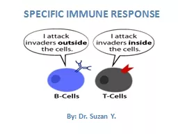
/
B cell response Macrophage and helper T cell involvement with initiating a B cell response: B cell response Macrophage and helper T cell involvement with initiating a B cell response:

B cell response Macrophage and helper T cell involvement with initiating a B cell response: - PowerPoint Presentation

marina-yarberry
marina-yarberry . @marina-yarberry
Follow
371 views
Uploaded On 2018-12-21

B cell response Macrophage and helper T cell involvement with initiating a B cell response: - PPT Presentation

Antigen fits with this B cell Different B cell clones Making antibodies Many plasma cells Some memory cells When specific B cells are activated they multiply Some cells become memory cells ID: 744768

cell cells killer antigen cells cell antigen killer response antibodies body virus immune blood allergies system surface ige specific

Share:

Link:

Embed:

Download Presentation from below link

Download Presentation The PPT/PDF document "B cell response Macrophage and helper T ..." is the property of its rightful owner. Permission is granted to download and print the materials on this web site for personal, non-commercial use only, and to display it on your personal computer provided you do not modify the materials and that you retain all copyright notices contained in the materials. By downloading content from our website, you accept the terms of this agreement.


Presentation Transcript

Slide1

B cell response

Macrophage and helper T cell involvement with initiating a B cell response:Slide2

Antigen fits with

this B cell

Different B cell clones

Making antibodies

Many plasma cells

Some memory cells

When specific B cells are activated, they multiply

Some cells become

memory cells

, stored in case of a

subsequent infection

B cell responseSlide3

Immunological memory from vaccines

Vaccines introduce antigen (dead or weakened) to induce production of memory B & T cells, antibodiesMemory cells are activated on real exposure to bacteria, virus. Antibodies already present to label

Lung cancer

vaccinationSlide4

Vaccines and the quest to eliminate infectious diseaseSlide5

What about T cells?

T cells recognize virus-infected

or

cancerous

body cells (cell-mediated)When triggered, T cells w/specific ‘self-antigen’ multiply. Killer T cells contact and release chemicals to kill the cells with the self-antigen markerSlide6

What triggers T cells?

Macrophages “wear what they eat” (in this case, self marker plus antigen from pathogen, or self markers on cancerous cells)

Helper T cells are triggered and activate killer T cells and memory T cellsSlide7

Self marker + antigen

Antigen-presenting cell

Body cell

Foreign microbe

Processed antigenAntigenMHC proteinSlide8

Helper T-cell activation and the B and T cells formed as a result Slide9

Protein coat

has antigen

Virus invades

host cell

Host cell

Virus

How Killer T-cells kill body cellsSlide10

Foreign viral antigen

Viral antigen is displayed on

surface of host cell

with self-antigen

Virus

invadedhost cell

Self-antigen

How Killer T-cells kill body cellsSlide11

Host cell

with virus

Killer T cell recognizes and binds

with a specific foreign

antigen complex

Killer T cell

Antigen complex

How Killer T-cells kill body cellsSlide12

Killer T cell releases chemicals that

destroy cell

How Killer T-cells kill body cellsSlide13

Helper T cells do not kill cells, but amplify effects of other WBCs:Enhance production of T and B cells, make

chemotaxins for phagocytes“Master switch” for immune response

Helper T cells

T-lymphocyteSlide14

The MHC

is a set of genes that code for glycoproteins on cell membranes and mark cells as “self”

So…what are these

self

markers? Slide15

Matching MHC markers is important when transplanting organs

So…what are these

self

markers? Slide16

Bacterial infection:

At first: phagocytes, histamine release, inflammatory responseInflammation brings phagocytes, plasma proteins (complement system, clotting proteins)Bacteria antigen stimulates helper T cells, B cells get activated: antibodies

Bacteria get labeled w/antibodies, killed by complement, macrophages, killer cells.

This slide is just another way to organize things for immune response to help study. I won’t use in lecture

Combining non-specific and adaptive immune responseSlide17

Viral infection:

Virus inside body cells do not trigger macrophages, B-cells, or complement.Virus-infected or cancerous cells release interferon, signaling neighboring cells and attracting natural killer cells, macrophages, complement. Virus ‘out in the open’ can be attacked.

Self-antigen combination triggers T-helpers, which help stimulate killer T cells (takes days) and attract macrophages to the area.

This slide is just another way to organize things for immune response to help study. I won’t use in lecture

Combining non-specific and adaptive immune responseSlide18

ABO blood types are named by antigens on the surface of RBCs: A, B, AB, or O (neither antigen).

People acquire antibodies for the blood antigens they do not have on their RBCs.

Blood type O : universal donor (no antigens).

Blood type AB : universal recipient (no antibodies)

Blood groupsSlide19

Reactivity to a harmless substance in environment

Common triggers: pollen, molds, bee stings, dust, fur, mites, penicillin

Allergies: adaptive immunity gone wrongSlide20

Hives - allergens on skin

Hay fever - allergens in nasal passagesAsthma - allergens in airway

Allergies: adaptive immunity gone wrongSlide21

Allergens

IgE antibodies

Allergies involve a particular type of antibody –

IgE

antibodies

IgE

antibodies trigger mast cells and basophils to produce histamine and other chemicals at the site of the allergens

SpecificB cellclonesAllergies: why in some people and not others?Slide22

Systemic anaphylaxis- when large amounts of histamine and inflammatory signals are released all at once to blood.

Widespread dilation - hypotension. Airway constriction. Victim can die within minutes. Often due to penicillin, bee venom.

Anaphylactic shockSlide23

Skin: Besides IgE response, can be a T-cell response to substance (ex:

urushiol oil)Airway: besides histamine, leukotrienes are released – airway constricted

Gut

: traditional food allergies are

IgE (egg, milk, wheat, nuts, shellfish, etc.) Histamine dilation, leukotriene constriction. Some are T-cell allergies w/delayed effects (gluten, milk).How does the immune system react differently for different allergies?Slide24

They generally reduce the histamine signalThey can be bronchodilators, reduce leukotrienes, decongestants (constrict capillaries), injectable epinephrine, anti IgE

Corticosteroids inhibit expression of cytokines and other signals of inflammation

What do allergy medications do?Slide25

Tolerance of substances develops early via clonal deletion of specific lymphocytesCentral tolerance

: B and T cells that respond to self-antigen are destroyed Error often results in autoimmune diseasePeripheral tolerance

: regulatory cells inhibit response to environmental antigens

Error often results in

allergiesAllergies can have late onset, even in adulthoodDevelopment of ‘tolerance’Slide26

Hygiene hypothesis

Recent study: Exposing ‘high-risk’ infants to peanuts reduced later allergies by 80%

Keeping a child’s environment ‘too clean’ may prevent proper development of immune systemSlide27

Immune system wrongly attacks body cells, often caused by production of autoantibodies

Rheumatoid arthritis – autoantibodies attach to joints and induce inflammation & attack by complement, and WBCs

What are autoimmune diseases?Slide28

A few immune related disorders:

Diabetes Type ICrohn’s DiseaseMultiple SclerosisPernicious anemia

Addison’s disease

LupusSlide29

AI diseases often have an environmental trigger, those with certain genotypes can be more susceptible to it

A trigger can be an infection w/antigen that is molecularly similar to markers on body cellsPossible environmental agents: silica, mercury, nitrates in drinking water, groundwater pollutants, drugs, many other chemicals…

Triggering of autoimmune diseaseSlide30

Respiratory system

External vs. cellular respirationSlide31

Respiratory system

Bronchioles can dilate and constrict

Airway: from nasal passages down to trachea, bronchioles and alveoli. The trachea and bronchi are reinforced with cartilage

Larynx (

voicebox

) has vocal

cordsSlide32

bronchioles

Alveolus

Smooth

muscle

Pulmonary

capillaries

Respiratory systemSlide33

chest cavitySlide34

Lung wall

Transmural pressure gradient

lungs will always expand to fill pleural cavity

Pleural cavity

Lungs

Chest wallSlide35

If lung pressure is less than atmospheric pressure, air enters the lungs.

Increasing cavity volume, air entersSlide36

Inspiration and expiration:

how we change chest volume Slide37

F = P

RMajor determinant of resistance is radius of bronchioles Disease can increase resistance (asthma, bronchitis)

What determines airflow?

same equation as blood flow!Slide38

Surface tension – whenever water layer meets air – water molecules are attracted to each other.

Surface tension along the lining of alveoli resists expansion of alveoli.

Surfactant

reduces surface tension.

Surface tension at alveoliSlide39

Air is a mixture of gases Nitrogen is 79% of air. Its partial pressure: 0.79 x 760 = 600.4

Gas exchange and partial pressure gradients

Alveoli

CapillariesPO2 100 40

PCO2 40 46Slide40

Most O2 is carried by

Hb - some is dissolved in plasma and determines partial pressure

Hb

saturation is high where P

O2 is high (lungs). Saturation remains high even PO2 is 60Small decrease in PO2 makes Hb unload much more O2 Hemoglobin saturation

Oxygen saturation curveSlide41

Increase in CO

2 from tissue shifts the saturation curve to the right

Increased acidity (H

+

, carbonic acid) and temperature has the same effect - Bohr effectShifting the curveSlide42

Most O

2

carried on hemoglobin

A teensy bit of O

2

is dissolved in plasmaSlide43

Carotid and aortic bodies send info to the medulla

PO2 and PCO2 and H

+

can be detected

O2 saturation is not detectedChemoreceptors sense O2 dissolved in blood Slide44

Measures airflow and volume of inspiration and expiration

Spirometry