/
 Invasive Aspergillosis 34-year-old woman  Invasive Aspergillosis 34-year-old woman

Invasive Aspergillosis 34-year-old woman - PowerPoint Presentation

luanne-stotts
luanne-stotts . @luanne-stotts
Follow
349 views
Uploaded On 2020-04-03

Invasive Aspergillosis 34-year-old woman - PPT Presentation

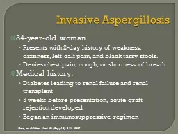
Presents with 2day history of weakness dizziness left calf pain and black tarry stools Denies chest pain cough or shortness of breath Medical history Diabetes leading to renal failure and renal transplant ID: 775063

aspergillosis invasive 2007 guha aspergillosis invasive 2007 guha infect med suppl acute infection diagnosis tissue septate therapy aspergillus hyphae

Share:

Link:

Embed:

Download Presentation from below link

Download Presentation The PPT/PDF document " Invasive Aspergillosis 34-year-old woma..." is the property of its rightful owner. Permission is granted to download and print the materials on this web site for personal, non-commercial use only, and to display it on your personal computer provided you do not modify the materials and that you retain all copyright notices contained in the materials. By downloading content from our website, you accept the terms of this agreement.


Presentation Transcript

Slide1

Invasive Aspergillosis

34-year-old womanPresents with 2-day history of weakness, dizziness, left calf pain, and black tarry stools.Denies chest pain, cough, or shortness of breathMedical history:Diabetes leading to renal failure and renal transplant3 weeks before presentation, acute graft rejection developedBegan an immunosuppressive regimen

Guha

, et al. Infect Med 24 (

Suppl

8): 8-11, 2007

Slide2

Invasive Aspergillosis

On admissionTachycardic, hypotensive and febrileInitial chest x-ray was normalLab results:AnemiaWBC = 4800/µl, 80% neutrophilsBlood cultures were positive for E. coliAntibiotic therapy initiated

Guha

, et al. Infect Med 24 (

Suppl

8): 8-11, 2007

Slide3

Invasive Aspergillosis

Day 6:Vesicular rash developed on buttocks and left calfCultures positive for HSV, antiviral therapy initiatedDay 8:Renal function continued to declineIntermittent hemodialysis startedDay 12:Decreased responsivenessIntubated for respiratory distress

Guha

, et al. Infect Med 24 (

Suppl

8): 8-11, 2007

Slide4

Invasive Aspergillosis

Chest x-ray:Diffuse bilateral lung nodulesCulture of BAL:Positive for Aspergillus spp.Immunesuppression decreasedLiposomal Amphotericin B startedCondition deteriorates:Acute MI, comatoseMulitple acute infarcts in frontal lobe and cerebellum by MRIMultiple skin nodules form on arms and trunk

Guha

, et al. Infect Med 24 (

Suppl

8): 8-11, 2007

Slide5

Invasive Aspergillosis

Culture of skin nodule biopsy:Aspergillus spp.

Guha

, et al. Infect Med 24 (

Suppl

8): 8-11, 2007

Slide6

Aspergillosis

Epidemiology:

Most common fungus worldwide – Ubiquitous

Hospital acquired infection - Major problem

Virulence factors and pathogenesis:

Thermo-tolerant to 50

C

Elastase

,

phospholipase

, protease and

catalase

Conidia bind to fibrinogen and

laminin

Invasive disease is dependent on impaired

neutrophil

function

Unable to generate the oxidative burst to kill

AT RISK:

Severe

neutropenia

, leukemia and lymphoma.

Slide7

Aspergillosis - Clinical Aspects

Clinical Manifestations: Route of infection: InhalationIncubation: days to weeksForms of infections: Allergic aspergillosisCavitary colonization - aspergilloma Primary pulmonary aspergillosisInvasive aspergillosis

Slide8

Types of

A

spergilloses

Slide9

ABPA – Allergic broncopulmonary aspergillosis (ABPA)

AsthmaPulmonary infiltratesPeripheral eosinopheliaElevated serum IgEHypersensitivity to Aspergillus antigenSkin test

Slide10

Aspergilloma

Colonization of paranasal sinuses and the lower airways

Obstructive bronchial aspergillosisOccurs in pre-formed cavitary lesionsCystic fibrosisChonic bronchitisTBNo tissue damage, asymptomatic

Slide11

Disseminated invasive aspergillosis

Slide12

Laboratory Diagnosis

: Monomorphic true mouldDifficult because of the universality of the fungusREPEAT ISOLATIONS ARE ESSENTIAL FOR DEFINITIVE DXSerum: galactomannan Ag +  invasive aspergillosisHistopathology: Septate hyphaedichotomous branching at ACUTE anglesMay see full conidial structures (i.e. fruiting bodies) In culture: A. fumigatus – “rapid grower” Septate, hyaline hyphaeconidiophores with phialidespointing upwards, bearing chains of conidia

Aspergillosis – Laboratory Diagnosis

Slide13

Direct prep from tissue specimen

Acute, dichotomous branching

Aspergillosis

Slide14

Septate hyphae

Aspergilloma

Conidiophore “fruiting body”

Aspergillosis

Slide15

Aspergillosis

Slide16

Aspergillosis

A. fumigatus

Slide17

Aspergillosis - Treatment

Treatment

:

Invasive disease is difficult to treat

Amphotericin

B,

caspofungin

(

echinocandins

),

voriconazole

Decrease

immunosuppression

or reconstitute immune defenses

Surgical debridement, if possible

Prevention in high-risk patients:

Neutropenic

: Filtered air to minimize exposure!

Slide18

Invasive Aspergillosis

Our patient:Expired on hospital day 23At autopsy, A. flavus was detected in multiple organs:Heart, lungs, adrenal galnd, thyroid, kidney, and liverExtreme example of disseminated aspergillosis in an immunocompromised host

Guha

, et al. Infect Med 24 (

Suppl

8): 8-11, 2007

Slide19

Opportunistic hyalohyphomycoses

Diverse agents

Many are ubiquitous – inhaled conidia

Many are resistant to antifungal agents

In tissue, they appear indistinguishable from

Aspergillus

!

(i.e. branching,

septate

hyphae)

Repeated isolation from multiple sites/multiple times

is best criteria to determine clinical significance.

BOTTOM LINE

: CULTURE IS CRITICAL FOR DX & TREATMENT

Slide20

Disseminated infection is increasing in incidence Some examples:Fusarium (R to ampB), immune reconst. + new triazolesScedosporium (R to ampB) – surgical resectionAcremonium (S unestablished)Paecilomyces – voriconazole…and many, many more.

Fus

Sce

Acr Pae

Opportunistic

hyalohyphomycoses

Slide21

Phaeohyphomycoses

Many are neurotropic: present as brain abscesses, sinusitis CNSBOTTOM LINE:Response to therapy is unpredictable between generaCulture is critical for diagnosis and therapy

Slide22

In tissue:Pigmented hyphae w/ or w/o yeast are presentDisseminated infection is increasing:Alternaria, Curvularia, Bipolaris, Cladosporium…and others

Alt

Cur

Bip

Cla

Phaeohyphomycoses

Slide23

Pneumocystosis

Etiology:

Pneumocystis

jirovecii

Most common opportunistic infection among individuals with AIDS

Incidence has decreased significantly with HAART

Reservoir in nature unknown

Pneumonia is clearly the most common presentation

Interstitial

pneumonitis

, mononuclear infiltrate

Onset insidious

Diagnosis based on microscopic examination of BAL