/
Structure of the lungs Structure of the lungs

Structure of the lungs - PowerPoint Presentation

luanne-stotts
luanne-stotts . @luanne-stotts
Follow
501 views
Uploaded On 2016-04-09

Structure of the lungs - PPT Presentation

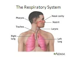
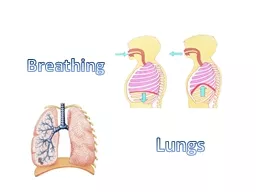
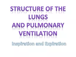
and Pulmonary Ventilation Inspiration and Expiration Structure of the R espiratory and System N ose hairs and mucus filter air A ir is warmed Pharynx ID: 277694

diaphragm muscles exercise alveoli muscles diaphragm alveoli exercise inspiration expiration contracting pressure diffusion lungs breathing increases relaxing blood rest

Share:

Link:

Embed:

Download Presentation from below link

Download Presentation The PPT/PDF document "Structure of the lungs" is the property of its rightful owner. Permission is granted to download and print the materials on this web site for personal, non-commercial use only, and to display it on your personal computer provided you do not modify the materials and that you retain all copyright notices contained in the materials. By downloading content from our website, you accept the terms of this agreement.


Presentation Transcript

Slide1

Structure of the lungs and Pulmonary Ventilation

Inspiration

and ExpirationSlide2

Structure of the Respiratory and SystemNose  hairs and mucus filter air. Air is warmedPharynx  Food and air pass through. At the bottom of the pharynx air is directed through the larynx (food

down

the

oesophagus

)

Larynx

The

opening

is

covered

by

the

epiglottis

(

made

of

cartilage

)

which

prevents

food

from

entering

the

lungs

.Slide3

Structure of the Respiratory and SystemTrachea  kept open and protected by C-shaped pieces of cartilage. Lined with mucus-secreting and ciliated cells. These cells remove foreign particles by pushing them back up towards

the

larynx

.

Bronchi

Enter

the

lungs

(

one

in

each

).

Reinforced

with

cartilage

.

Bronchioles

contain

smooth

muscle

.

If

this

muscle

contracts

(as

it

does

during

an

asthma

attack

),

it

can cause

severe

breathing

difficulties

.Slide4

Structure of the Respiratory and SystemAlveoli  approx. 300 million, which provide a surface area similar to that of a tennis court. Walls are extremely thin, aiding efficient gas exchangeSurrounded by capillaries

Surface

are

moist

to

aid

diffusion

and

disolve

oxygenSlide5

Keywords: Inhalation, Exhalation, Inter-coastal Muscles, Diaphragm, Alveoli, Diffusion, I know that lungs are specialised organs where oxygen from the air enters the blood and carbon dioxide in the blood passes into the alveoli. I know how the alveoli provide a large surface area for gas exchange.

I know the mechanism of breathing.

Cartilage (rings to prevent

collapse)

Cilia

(Hair like structures help move

mucus)

Mucus secreting cells

Smooth muscle

Trachea

Yes

Yes

Yes

Yes

Bronchus

Yes

Yes

Yes

YesLarge BronchiolesNoYesYesYesAlveolus NoNoNoNo

Structure

of

the

Respiratory

SystemSlide6

Keywords: Inhalation, Exhalation, Inter-coastal Muscles, Diaphragm, Alveoli, Diffusion, Slide7

Task 1: Complete the following table for breathing in only!Keywords: Inhalation, Exhalation, Inter-coastal Muscles, Diaphragm, Alveoli, Diffusion, I know the mechanism of brethg.Breathing INBreathing OUT

External Intercostal

muscles move

Ribs move

Volume of lungs

Diaphragm

Contract

Move OUT and UP

Increases

Contracts and moves DOWNSlide8

Keywords: Inhalation, Exhalation, Inter-coastal Muscles, Diaphragm, Alveoli, Diffusion, Slide9

Task 2: Complete the following table for breathing OUT.Keywords: Inhalation, Exhalation, Inter-coastal Muscles, Diaphragm, Alveoli, Diffusion, Breathing INBreathing OUTIntercostals muscle moveRibs move

Volume of lungs

Diaphragm

Contract

Move OUT and UP

Increases

Contracts and moves DOWN

Relax

Move IN and DOWN

Decreases

Relaxes and moves UPSlide10

Complete the following table:Muscles Contracting or relaxing?Muscles involvedChanges occurring to size and pressureInspiration at restExpiration at restInspiration during exerciseExpiration during exerciseUse page 31 of your text book to help.Slide11

Muscles Contracting or relaxing?Muscles involvedChanges occurring to size and pressureInspiration at restContractingExternal intercostals, diaphragmVolume of thoracic cavity increases, Pressure decreasesExpiration at restRelaxing (muscles are passive)External intercostals, diaphragmVolume of thoracic cavity decreases,

Pressure increases

Inspiration

during exercise

Expiration during exerciseSlide12

Muscles Contracting or relaxing?Muscles involvedChanges occurring to size and pressureInspiration at restExpiration at restInspiration during exerciseContractingDiaphragm, External intercostals,Scalenes, sternocleidomastoid and pectoralis minor.Volume of thoracic cavity increases, Pressure decreasesExpiration during exerciseSlide13

Strenocleidomastoid lifts the sternumScalenes and pectoralis minor lift the ribs furtherSlide14

Complete the following table:Muscles contracting or relaxing?Muscles involvedChanges occurring to size and pressureInspiration at restExpiration atrestInspiration during exerciseExpiration during exerciseContractingRelaxingAbdominals,Internal intercostals;Diaphragm

External

intercostals

Volume of thoracic cavity decreases,

Pressure increases.Slide15

External RespirationThe process of gas exchange between the lungs and the blood. Oxygen diffuses into the blood, while CO2 diffuses from the blood into the lungs.Slide16

Internal RespirationThe process of gas exchange between the blood, the fluids surrounding the cells, and the cells. Inside the cell, cellular respiration generates energy (ATP), using O2 and glucose and producing waste CO2.