/
An Bras Dermatol 2017921820Diabetes mellitus and the skin9 An Bras Dermatol 2017921820Diabetes mellitus and the skin9

An Bras Dermatol 2017921820Diabetes mellitus and the skin9 - PDF document

mackenzie
mackenzie . @mackenzie
Follow
342 views
Uploaded On 2022-09-03

An Bras Dermatol 2017921820Diabetes mellitus and the skin9 - PPT Presentation

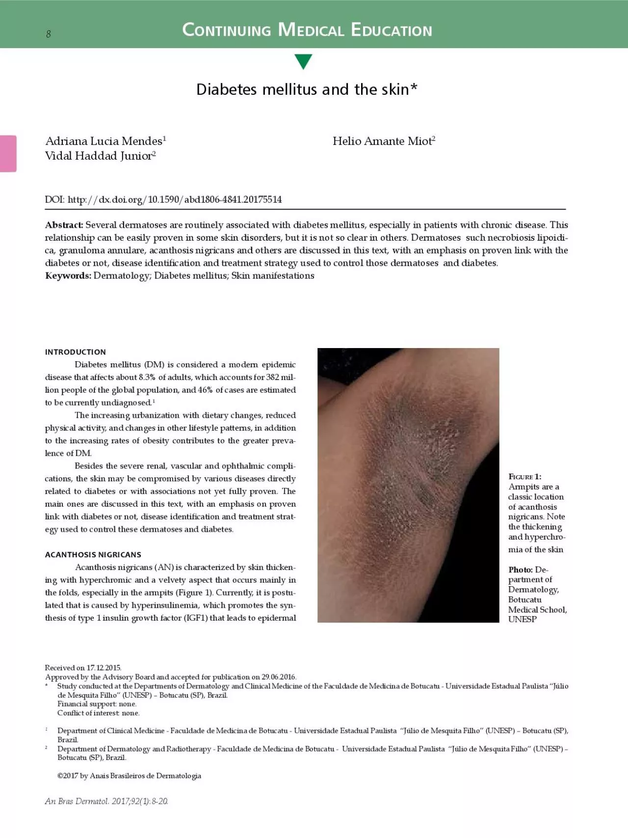
FI icorum blisters are asymptomDermatologyFurthermorecan be associated to skin tags achrocordonsscreeningresistance among general populationThe disease may also be associated with certain malign ID: 947597

diabetes dermatol mellitus diabetic dermatol diabetes diabetic mellitus skin patients scleredema lesions diabeticorum necrobiosis treatment arch lipoidica cutaneous clin

Share:

Link:

Embed:

Download Presentation from below link

Download Pdf The PPT/PDF document "An Bras Dermatol 2017921820Diabetes mell..." is the property of its rightful owner. Permission is granted to download and print the materials on this web site for personal, non-commercial use only, and to display it on your personal computer provided you do not modify the materials and that you retain all copyright notices contained in the materials. By downloading content from our website, you accept the terms of this agreement.


Presentation Transcript

An Bras Dermatol. 2017;92(1):8-20.Diabetes mellitus and the skin9 FI icorum blisters are asymptomDermatology,Furthermore,can be associated to skin tags (achrocordons)screeningresistance among general population.The disease may also be associated with certain malignanresistance,adrenal and thyroid disease may be associated.seems to evolve in association with syndrome.affectedareareolas,Topicaltreatmentlients with basis of urea and oral metformin can be used (due to resistance),effectivemeasuresareexercises,reversesBULLOSIS DIABETICORUMBullosis diabeticorumdiabetic blisters occurs in approximately 0.5% of diabetic patients.beticorum was proposed.sidered a distinct marker of DM and it is manifested in patients with nephropathyneuropathy,therearereportsof concomitant appearance to the initial presentation of DM.BD has been reported in patients aged 17 to 80 years with largerproportionpreferredareextremities,(FigurePathophysiology of the BD bullae is still unknown. The arelarge,neous onset in acral regions. The diameter of the blisters varies areclear,serous Other affected places are the back and side of the hands and the arms. These blisters are usually painless and non-pruritic and disappear sponta10,11Outbreaksoccur,aremicroangiopathyfailure.11-15Diagnosis is made on clinical basis and should be rememberedtherearelargeapparentin longstanding diabetic patients or those with chronic complicaHistologically,arethreedifferenttypes based on the level of cleavage. The most common type shows an subepidermal cleavage at the level of the lamina lucida without spontaneously, Blisters present hyaline content and are located at the frequentlyPatients with these clinical manifestations have good circulation in the affected limb and tend to present diabetic peripheral neuropathy.The second type is rarer and involves lesions that may be hemorrhagic including resolution with scars and atrophy.destructhirdtiple blisters associated with sun exposure and markedly tanned affectsfromporphyria cutanea tarda. differentialdrugeruptions and porphyria cutanea tarda.DIABETIC DERMOPATHYcircumscribedbrownishconsideredous manifestation of diabetic microangiopathy. Its incidence may range from 7% to 70% of diabetic pa moremoremore There is some controversy as to DD be a pathognomonic sign for diabetes since there are studies The origin of DD is unknown and there is no relation with decreased local perfusion.mild traumas that do not compromise wound healing. There ropathy.However,relationbetween DD and microvascular complications of diabetes. Studies strongnephropathy,retinopathyor neuropathy. observed increased incidence of DD in 52-ass

ociatedshowed that 42.9% of patients presented neuropathy associated no evidence of microangiopathy. The association between DD and cardiovascular disease has coronary 10Mendes AL, Miot HA, Haddad Jr VAn Bras Dermatol. 2017;92(1):8-20. DD had coronary artery disease.neuropathy,nephropathy,retinopathycoronaryprominences,ed that occur in response to sudden trauma.11,14,20,31frequentpresence of peripheral neuropathy.brownishpressions,atrophicresemblingthe lesions measure less than 1cm in diameter and present round(Figurereach2.5cm. Depressions are smooth and hyperpigmented and intensity of the pigment is related to the degree of atrophy. Generally asymp laterally in pretibial regions and distributed asymmetrically. More rarely,trunklocation and atrophic appearance causes many patients to consider DD as scars resulting from a possible trauma. The appearance of hardlydocumented,underreportedThe progression of DD is variable and does not appear to be affected by glycemic control.Individual lesions may persist inde�nitely.regresses,processatrophy.Cyclically,The diagnostic is clinically based: after careful history and presdemarcatedatrophicscars in the lower leg of a patient with diabetes is highly suggestive of DD. The presence of four or more typical lesions in diabetic Biopsy is not routinely perinterestingextremitiesHowever,atypical features or unusual locations may hinder the diagnosis and recommend the histopathological examination.atrophyhypertrophyerythrocytes,Therebroblast proliferation at the papillary dermis.The differential diagnosis of DD includes many diseases. brownishatrophicrequiredifferentiationSchamberg’s(progressivepapulonecrotic Many of these entities differentiatedtory.Treatment of DD is not recommended and is little effective.arespontaneous regression without treatment.requirerequirepresenceprevention,controlcontrolSCLERODERMIFORM DISORDERSPatients with diabetes may have thickening and hardening regionsclerosistend these places. These changes are more common in type 1 diabetes and occur in up to 50% of the patients. The cause seems to be the glicosylation of proteins that appears to cause hardening of the skin.sclerosisscleredemaadultorumwheresclerosisdiffuse,preferablycompromiseeven other regions. It is more common in men over 40 years with rare�bromucinousknown etiology resistant to therapy and without spontaneous resodiffusehardeningaffectingtrunk,(Figure The disease presentsgrouppreferences,however,more common in middle-aged men. DMis associated with about Its prevalence varies between 2.5 and 14% in diabetic areunderdiagprogressive loss of skin natural mark

s. In severe cases it can lead to neck Mobility is reduced and may lead to a restrictive respiratory syndrome due to the skin thickening. The affected area is painless and can present decreased sensitivity to touch. Viscer FI Diabetic dermopathy consists of small brownish-colored depressionsatrophiclike scars Dermatology, Diabetes mellitus and the skin11An Bras Dermatol. 2017;92(1):8-20. rare,affecting(pericardialeffuhepatosplenomegaly.groupdisorders,tes and other endocrine disorders. Three scleredema variants are Typemiddle-agedchildrenpresentsrespiratorystreptococcalcytomegalovirusresolution after several months or years.TypeThererelationly progressive and is associated with monoclonal gammopathy.This type tends to persist for years and may be at increased risk rheumatoidSjögren’ssyndrome,obstructivehyperparathyroidism,adrenocortical disease. Type It occurs generally in obese patients with long control,microangiopathyand need for insulin. It affects middle-aged men with history of longtime DM. This type also tends to persist and there is no clear relation to prognosis or glycemic control.resonance)extent or disease activity.scleredema,recomHistopathology shows marked thickening of the reticular bands of hyaline deposit mucin or hyaluronic acid best evidenced cally corresponds to an hyperintensity on magnetic resonance.Clinically,differentialscleroderma,scleromyxedema.lular matrix components appears to be represented by an abnormal expressionprotein�bronecdecreasingprocesses.deregulatedregardlesspresence�broblastareresponsetreatment,tic modalities are used: immunosuppressants (e.g. cyclosporine and methotrexate),prostaglandincorticosteroids,radiotherapy,(PUVA),recently,electroneffective,probablyupregulation�broblastsprognosis,cases is self-limitedhowever,therearesevereprogression,hardtreatmenttrollingIt is noteworthy that its chronicity can cause alterations in movement of the shoulders and impaired respiratory function.GRANULOMA ANNULAREannularerare,ed dermatitis of the pretibial regions and the extensor surfaces of the limbs. The cutaneous lesions are similar to necrobiosis lipoidica diabeticorum,atrophyresponseHIV,thyroiddiseases and malignancy.controversial and has been extensively studied. Samlaska controlstudy,revealedcorrelationretrospectiveaffectsaffectedareasareelbow, sometimes the affected(Figuresthe trunk is affected in almost all cases. arepresentcoloredway.annular,areerythematous-brownishly. The papules of annular shape grow slowly and can measure from 0.5 to 5.0cm.affectschildren FI scleredemadiffusehardeningcervical region and upper limbsDermatology,

Diabetes mellitus and the skin13An Bras Dermatol. 2017;92(1):8-20. thy. Spontaneous resolution is observed in 10% to 20% of cases.disorganizationunderlying subcutaneous fat. NL pathophysiology is still unclear. The presencearoundfeaturesleukocytoclastic vasculitis were not observed. Other studies suggest presencepalisades suggests delayed hypersensitivity reaction.Typical lesions of NL start in the pretibial areas with nonscaly erythematous papules that gradually enlarge and coalesce into large resultfromules and often develop atrophic center that corresponds to the dermal atrophyaremargins.reddishlow,may range from a few millimeters to several centimeters. When the chronic,sclerosisporcelaneous aspect. The metabolic control appears to have no proven eftherereportglucose control reduces the incidence of NL.areas,regionareforearms)whereatrophyrarelesions were observed in the heel and penis. NL also can develop sclerodermabody,ally the lower limbs are affected too.Diagnosis is made by clinical examination. HistopathologirequiredSarcoidosis,annulare,sclerosus et atrophicus and stasis dermatitis may be differential diagnosis of NL. Ulcerated lesions can resemble pyoderma gangrenoup to the knee or foam pads for protection.drugtreatment has little effect and should be reserved for symptomatic relief. Drugsarecorticosteroidsthreadschloroquine.arereportedfrom redness and warmth are important adverse events.Morerecently,thereare Strict glycemic control remains controversial in improving NL. treatmentthe prevention of secondary infection with systemic antibiotics and dressings. DIABETIC FOOTentity,treatedsurgery,surgery,dermatology.burdenshealth system and social security.Classically,chronicevolves after trauma or over a callus caused by changes in points alteredneuropathy(FigureproportionThe causes can coexist and about 25% of diabetics may pres FI Typical lesions of necrobiosis lipoidica begin in the pretibregionsgrowgrouplargeDermatology, FI necrobiosisatrophyDermatology, aresuppressionbordersresponsiblelimb amputations annually. The treatment is performed according to the etiology. If the areenergeticmeasuresbridement and dressing usually heal the wound in few weeks. On measuresareeffectivepresencesurgicalrevascularizationcrucialtreatmentcases. Secondary infections and osteomyelitis are factors that complicate the approach and systemic antibiotics should be evaluated Therearemultidisciplinary approach that surpass the scope of this text. fromfrequentlydependent diabetes and appear associated with microangiopathy both appear to be associated with skin xerosis seen in these patients.is a vascular erythema on

the face and neck presprobablyvasoconstrictorre�ectscontrolneuropathy.hyperglycemia can lead to a change in the microcirculation. It becomes clinically evident by facial venous dilatation. Rubeosismicroangiopathy,prudentcroangiopathy such as retinopathy and nephropathy. Tight glucose control is the mainstay of treatment for this disease.Yellowcarotenodermiarelatedcontrol,carotenemia,by increasing the glycosylation of collagen and dermal proteins.There is no treatment for this phenomenon. FI Diabetic foot may present a chronic ulcer on callus caused by changes in sensitivity associated with diabetic neuropathy and Dermatology,Other conditions not necessarily related to the presence of diabetes are the eruptive xanthomas. These lesions are observed when there is a marked exacerbation of triglyceride levels (greater factor,of the most common causes is the lack of DM control. The characterdiscretewith waxy yellow centers and an erythematous base. Lesions may itchy and even painful. Eruptive xanthomas should be faced as a threatingdisorderpancreatitisly resolved with proper correction of hypertriglyceridemia.but especially the decrease in leukocyte chemotaxis and phagocyreactionresponsetheir resolution.balanopreputial,indirectpresenceVulvovaginalduring periods of glycosuria. It comes with vulvar erythema and sometimes with white discharge. Treatment involves glycemic controltreatmentaresive pityriasis versicolor and dermatophytoses (e.g. which are associated to microangiopathy and poor glycemic control. Several opportunistic fungi infections are described in diabetics control.rare,mucormycosis,fromordernecroticprocessesprogression,Bacterial infections may be varied and severe as those severerentoccur,necrotizing/bullousarecommon among diabetics. External otitis by is also a Infections in diabetics have to be considered carefullyrequirecompromiseof immune response.disordersare areataVLTILIGOVitiligochronicor,pears as hypo/ achromic spots surrounded by healthy skin whose fromlargearoundregions, Withentirelyneurohumoral factors due to the impairment of nerve cells release toxic destructioncells while the pigment is forming.EnvironmentalMoretti found that the epidermis of vitiligo has a sigroundingproductionareinvolved in apoptosis of melanocytes process and depigmentation Vitiligodisordersetiology,disorders(thyroiditis,adrenalhipoparatathyreoidism)autoimmune syndrome whose clinical manifestations may appear differentprogressesadrenalinsuf�ciency,thyroiditisatrophicareata,fort and psychological stress can be considerable. Cosmetic treatimprovemicropigmentationconsidered,treatmentigo is

unsatisfactory in general. Patients should be advised to avoid sun exposure and use broad spectrum sunscreens. In small and locorticosteroidsaretreatment,widespreadtreatmentnarrowlet light B is more effective. chronicrecurrentstrongaffectscases it develops before 40 years of age and is rare in children.Its emergence or worsening can often be triggered by emopoorerreducedexpectancy,problems for the patient and family.fromwidespreadseverearea)syndrome,which is a set of risk factors for cardiovascular disease whose uniresistance,pro-in�ammatoryprothrombotic state.108-110Several studies have evidenced the association of psoriasis with cardiovascular diseases and components of the metabolic syndromeobesity,chronic107,110Psoriasis patients often are overweight or obese in greater propor111-114Furthermore,115wererencecomponentssyndrome,prematureatherosclerosisarepro�lemacrophages and monocytes are observed in both conditions.114-116revealTreatment of psoriasis depends on the clinical manifestapresented,fromications in mild cases to more complex treatments for more severe cases. The response to treatment also varies greatly from one patient to another and the emotional component should not be overlooked. stress,improvement. Moderate sun exposure is of great help as keeping the skin drugsaffectincreasingcardiovascularprove the response to treatments for psoriasis as well as reduce carcompleteremissionpropercontrolpromotesrehabilitationimprovingprobablydecreasingFINAL CONSIDERATIONSaredisordersfor the general practitioner.prevalentre�ectingcontrol,all prognosis of the disease.controlpreventionorganspromotedreinforcedassociated with DM are not necessarily related to glycemic levels ACKNOWLEDGEMENTAndrellaPretoFAMERP)their help at various stages of the preparation of the manuscript. Idf.org [Internet]. IDF Diabetes Atlas. 6th ed. [cited 2015 Jul 15]. Available from: http://www.idf.org/diabetesatlas/update2014. Callen JP, Jorizzo JL, Bolognia JL, Piette WW, Zone JJ, editors. Dermatological Signs of Internal Disease. 4th ed. Philadelphia: Saunders Elsevier; 2009.Barbato MT, Criado PR, Silva AK, Averbeck E, Guerine MB, Sá NB. Association of acanthosis nigricans and skin tags with insulin resistance. An Bras Dermatol. Tamega Ade A, Aranha AM, Guiotoku MM, Miot LD, Miot HA. Association between skin tags and insulin resistance. An Bras Dermatol. 2010;85:25-31.Papa CM. Niacinamide and acanthosis nigricans. Arch Dermatol. 1984;120:1281.Romano G, Moretti G, Di Benedetto A, Giofrè C, Di Cesare E, Russo G, et al. Skin lesions in diabetes mellitus: prevalence and clinical correlations. Diabete

s Res Clin Pract. 1998;39:101-6.Kramer DW. Early or warning signs of impending gangrene in diabetes. Med J Rec. Cantwell AR Jr, Martz W. Idiopathic bullae in diabetics. Bullosis diabeticorum. Arch Dermatol. 1967;96:42-4.Mendes AL, Haddad Jr V. Caso para diagnostico. Bullosis Diabeticorum. An Bras Dermatol. 2007;82:94-6.Basarab T, Munn SE, McGrath J, Russell Jones R. Bullosis diabeticorum. A case report and literature review. Clin Exp Dermatol. 1995;20:218-20.Paron NG, Lambert PW. Cutaneous manifestations of diabetes mellitus. Prim Care. Rocca FF, Pereyra E. Phlyctenar lesions in the feet of diabetic patients. Diabetes. Allen GE, Hadden DR. Bullous lesions of the skin in diabetes (bullosis diabeticorum). Br J Dermatol. 1970;82:216-20.Sehgal VN, Srivastava G, Aggarwal AK, Gupta M, Bhattacharya SN, Verma P. Noninsulindependent, type II diabetes mellitusrelated dermatoses: part II. Lipsky BA, Baker PD, Ahroni JH. Diabetic bullae: 12 cases of a purportedly rare cutaneous disorder. Int J Dermatol. 2000;39:196-200.Kurwa A, Roberts P, Whitehead R. Concurrence of bullous and atrophic skin lesions in diabetes mellitus. Arch Dermatol. 1971;103:670-5.Bernstein JE, Medenica M, Soltani K, Griem SF. Bullous eruption of diabetes mellitus. Arch Dermatol. 1979;115:324-5.Parex, I. Cutaneous manifestations of diabetes mellitus. J Am Acad Dermatol. Goodfield MJ, Millard LG, Harvey L, Jeffcoate WJ. Bullosis diabeticorum. J Am Acad Dermatol. 1986;15:1292-4.Shemer A, Bergman R, Linn S, Kantor Y, Friedman-Birnbaum R. Diabetic dermopathy and internal complications in diabetes mellitus. Int J Dermatol. Houck GM, Morgan MB. A reappraisal of the histologic findings of pigmented pretibial patches of diabetes mellitus. J Cutan Pathol. 2004;31:141-4.Binkley GW. Dermopathy in the diabetic syndrome. Arch Dermatol. 1965;92:625-Mahajan S, Koranne RV, Sharma SK. Cutaneous manifestation of diabetes mellitus. Indian J Dermatol Venereol Leprol. 2003;69:105-8.A Abdollahi A, Daneshpazhooh M, Amirchaghmaghi E, Sheikhi S, Eshrati B, Bastanhagh MH. Dermopathy and retinopathy in diabetes: is there an association? Dermatology. 2007;214:133-6.Danowski TS, Sabeh G, Sarver ME, Shelkrot J, Fisher ER. Skin spots and diabetes Lithner F. Cutaneous reactions of the extremities of diabetics to local thermal Wigington G, Ngo B, Rendell M. Skin blood flow in diabetic dermopathy. Arch Dermatol. 2004;140(:1248-50.Kiziltan ME, Benbir G, Akalin MA. Is diabetic dermopathy a sign for severe neuropathy in patients with diabetes mellitus? Nerve conduction studies and Jelinek JE. Skin disorders associated with diabetes mellitus. In: Rifkin H, Porte D, editors. Ellenberg

and Rifkin’s diabetes mellitus, theory and practice. New York: Elsevier; 1990: p. 838-49.Gouterman IH, Sibrack LA. Cutaneous manifestations of diabetes. Cutis. Stulberg DL, Clark N, Tovey D. Common hyperpigmentation disorders in adults, part II: melanoma, seborrheic keratoses, acanthosis nigricans, melasma, diabetic dermopathy, tinea versicolor, and postinflammatory hyperpigmentation. Am Fam Kroft EB, de Jong EM. Scleredema diabeticorum case series: successful treatment with UVA1. Arch Dermatol. 2008;144:947-8. Kobayashi T, Yamasaki Y, Watanabe T. Diabetic scleredema: a case report and biochemical analysis for glycosaminoglycan. J Dermatol. 1997;24:100-3.Cole GW, Headley J, Skowsky R. Scleredema diabeticorum: a common and distinct cutaneous manifestation of diabetes mellitus. Diabetes Care. 1983;6:189-Gervini RL, Lecompte SM, Pineda RC, Ruthner FG, Magnabosco EM, Silva LM. Escleredema de Buschke: relato de dois casos. An Bras Dermatol. 2002; 77:465-Emedicine.com [Internet]. PappasTaffer LK, Werth VP, Vinson RP, Libow LF, Elston DM. Scleredema. [cited 2015 Nov 01]. Available from: www.emedicine.com/derm/topic385.htm. Pitarch G, Torrijos A, Martínez-Aparicio A, Vilata JJ, Fortea JM. Escleredema de Buschke associado a diabetes mellitus. Estudio de cuatro casos. Actas Dermosifiliogr. 2005; 96:46-9.Sattar MA, Diab S, Sugathan TN, Sivanandasingham P, Fenech FF. Scleredema diabeticorum: a minor but often unrecognized complication of diabetes mellitus. Meguerditchian C, Jacquet P, Béliard S, Benderitter T, Valéro R, Carsuzza F, et al. Scleredema adultorum of Buschke: an under recognized skin complication of Carvalho JC, Costa TN, De Moura HH, Quintella LP, Carneiro S, Ramos-e-Silva M. Scleredema Adultorum of Buschke. Skinmed. 2014;12:337-40.Barde C, Masouyé I, Saurat JH, Le Gal FA. Scleroedema adultorum Buschke in a diabetic subject: intravenous immunoglobulin therapy. Ann Dermatol Venereol. Mokta J, Sharma B, Patial RK, Prasher BS, Prasher N. Systemic manifestations in Oyama N, Togashi A, Kaneko F, Yamamoto T. Two cases of scleredema with pituitaryadrenocortical neoplasms: an underrecognized skin complication. J Dermatol. 2012;39:193-5. Parmar RC, Bavdekar SB, Bansal S, Doraiswamy A, Khambadkone S. Scleredema adultorum. J Postgrad Med. 2000;46:91-3.Mattos e Dinato SM, Costa GL, Dinato MC, Sementilli A, Romiti N. Escleredema de Buschke associado ao diabetes melito tipo 2: relato de caso e revisão da literatura. Kövary PM, Vakilzadeh F, Macher E, Zaun H, Merk H, Goerz G. Monoclonal gammopathy in scleredema: observations in three cases. Arch Dermatol. Angeli-Besson C, Koeppel MC, Jacquet P, Andrac L, S

ayag J. Electronbeam therapy in scleredema adultorum with associated monoclonal hypergammaglobulinaemia. Br J Dermatol. 1994;130:394-7.Turchin I, Adams SP, Enta T. Answer to dermacase. Can Fam Physician. Ray V, Boisseau-Garsaud AM, Ray P, Pont F, Lin L, Hélénon R, et al. Obesity persistent scleredema: study of 49 cases. Ann Dermatol Venereol. 2002;129:281-Graff R. Discussion of scleredema adultorum. Arch Derm. 1968;98:319-20.Ulmer A, Schaumburg-Lever G, Bauer J, Kötter I, Fierlbeck G. Scleredema adultorum Buschke. Case report and review of the literature. Hautarzt. Cron RQ, Swetter SM. SM. Scleredema revisited. Clin Pediatr (Phila). 1994;33:606-Martín C, Requena L, Manrique K, Manzarbeitia FD, Rovira A. Scleredema diabeticorum in a patient with type 2 diabetes mellitus. Case Rep Endocrinol. Horger M, Fierlbeck G, Kuemmerle-Deschner J, Tzaribachev N, Wehrmann M, Claussen CD, et al. MRI findings in deep and generalized morphea (localized scleroderma). AJR Am J Roentgenol. 2008;190:32-9.Cole GW, Handler SJ, Burnett K. The ultrasonic evaluation of skin thickness in Kurihara Y, Kokuba H, Furue M. Case of diabetic scleredema: diagnostic value of magnetic resonance imaging. J Dermatol. 2011;38:693-6. Conde Fernandes I, Sanches M, Velho G, Lobo I, Alves R, Selores M.. Scleromyxedema vs scleredema: a diagnostic challenge. Eur J Dermatol. Gruson LM, Franks A Jr. Scleredema and diabetic sclerodactyly. Dermatol Online Thumpimukvatana N, Wongpraparut C, Lim HW. Scleredema diabeticorum successfully treated with ultra¬violet A1 phototherapy. J Dermatol. 2010;37:1036-Alsaeedi SH, Lee P. Treatment of scleredema diabeticorum with tamoxifen. J Dahl MV. Granuloma annulare. In: Freedberg IM, Eisen AZ, Wolff K, Austin KF, Goldsmith LA, Katz SL, editors. Fitzpatrick’s Dermatology in General Medicine. 5th ed. New York: McGrawHill; 1999. p. 1152-7.Dabski K, Winkelmann RK. Generalized granuloma annulare. Clinical and laboratory findings in 100 patients. J Am Acad Dermatol. 1989;20:39-47.Cohen PR. Granuloma annulare associated with malignancy. South Med J. Studer EM, Calza AM, Saurat JH. Precipitating factors and associated diseases in 84 patients with granuloma annulare: a retrospective study. Dermatology. Erkek E, Karaduman A, Bükülmez G, Sentürk N, Ozkaya O. An unusual form of generalized granuloma annulare in a patient with insulindependent diabetes mellitus. Acta Derm Venereol. 2001;81:48-50.Buendía-Eisman A, Ruiz-Villaverde R, Blasco-Melguizo J, Serrano-Ortega S. Generalized annular granuloma: response to isotretinoin. Int J Dermatol. Cyr PR. Diagnosis and management of granuloma annulare. Am Fam Physician. Sahi

n MT, Türel-Ermertcan A, Oztürkcan S, Türkdogan P. Generalized granuloma annulare in a patient with type II diabetes mellitus: successful treatment with isotretinoin. J Eur Acad Dermatol Venereol. 2006;20:111-4.Muller SA. Dermatologic disorders associated with diabetes mellitus. Mayo Clin Proc. 1966;41:689-703.Lowitt MH, Dover JS. Necrobiosis lipoidica. J Am Acad Dermatol. 1991;25:735-Muller SA, Winkelmann RK. Necrobiosis lipoidica diabeticorum: a clinical and pathological investigation of 171 cases. Arch Dermatol. 1966;93:272-81.Yosipovitch G, Hodak E, Vardi P, Shraga I, Karp M, Sprecher E. The prevalence of cutaneous manifestations in IDDM patients and their association with diabetes risk Garza-Elizondo MA, Diaz-Jouanen E, Franco-Casique JJ, Alarcón-Segovia D. Joint contractures and sclerodermalike skin changes in the hands of insulindependent Van Hattem S, Bootsma AH, Thio HB. Skin manifestations of diabetes. Cleve Clin Sibbald RG, Landolt SJ, Toth D. Skin and diabetes. Endocrinol Metab Clin North Petzelbauer P, Wolff K, Tappeiner G. Necrobiosis lipoidica: treatment with systemic corticoids. Br J Dermatol. 1992;126:542-5.Oumeish YO. Skin disorders in patients with diabetes. Clin Dermatol. 2008;26:235-Dahl MV, Ullman S, Fisher I. Direct immunofluorescence of granuloma annulare and necrobiosis lipoidica. Clin Res. 1976;24:95a.Quimby SR, Muller SA, Schroeter AL. The cutaneous immunopathology of necrobiosis lipoidica diabeticorum. Arch Dermatol. 1988;124:1364-71.Ullman S, Dahl MV. Necrobiosis lipoidica. An immunofluorescence study. Arch Dermatol. 1977;113:1671-3.Beattie PE, Dawe RS, Ibbotson SH, Ferguson J. UVA1 phototherapy for treatment of necrobiosis lipoidica. Clin Exp Dermatol. 2006;31:235-8.Nieboer C, Kalsbeek GL. Direct immunofluorescence studies in granuloma annulare, necrobiosis lipoidica and granulomatosis disciformis Miescher. Dermatologica. 1979;158:427-32.Dwyer CM, Dick D. Ulceration in necrobiosis lipoidica: a case report and study. Clin Exp Dermatol. 1993;18:366-9.Bauer M, Levan NE. Diabetic dermangiopathy. A spectrum including pretibial pigmented patches and necrobiosis lipoidica diabeticorum. Br J Dermatol. Ahmed I, Goldstein B. Diabetes mellitus. Clin Dermatol. 2006;24:237-46.Ferringer T, Miller F 3rd. Cutaneous manifestations of diabetes mellitus. Dermatol Cohen O, Yaniv R, Karasik A, Trau H. Necrobiosis lipoidica and diabetic control Tokura Y, Mizushima Y, Hata M, Takigawa M. Necrobiosis lipoidica of the glans penis. J Am Acad Dermatol. 2003;49:921-4.Nguyen K, Washenik K, Shupack J. Necrobiosis lipoidica diabeticorum treated with chloroquine. J Am Acad Dermatol. 2002;46:S34-6.Alex

is AF, Strober BE. Offlabel dermatologic uses of antiTNFa therapies. J Cutan Tabor CA, Parlette EC. Cutaneous manifestations of diabetes. Signs of poor glicemy control or newonset disease. Postgrad Med. 2006;119:38-44.Ngo BT, Hayes KD, DiMiao DJ, Srinivasan SK, Huerter CJ, Rendell MS. Cutaneous manifestations of diabetic microangiopathy. Am J Clin Dermatol. 2005;6:225-37.Frykberg RG, Zgonis T, Armstrong DG, Driver VR, Giurini JM, Kravitz SR, et al. American College of Foot and Ankle Surgeons. Diabetic foot disorders. A clinical practice guideline (2006 revision). J Foot Ankle Surg. 2006;45:S1-66.Geerlings SE, Hoepelman AI. Immune dysfunction in patients with diabetes Arnáiz-García ME, Alonso-Peña D, González-Vela Mdel C, García-Palomo JD, Sanz-Giménez-Rico JR, Arnáiz-García AM. Cutaneous mucormycosis: report of five cases and review of the literature. J Plast Reconstr Aesthet Surg. Passeron T, Ortonne JP: Physiopathology and genetics of vitiligo. J Autoimmun. Moretti S, Spallanzani A, Amato L, Hautmann G, Gallerani I, Fabiani M, et al. New insights into the pathogenesis of vitiligo: imbalance of epidermal cytokines at sites of lesion. Pigment Cell Res. 2002;15:87-92.Olasode A. Why vitiligo in diabetes? Egypt Dermatol Online J. 2005;1:8.Huang CL, Nordlund JJ, Boissy R. Vitiligo: a manifestation of apoptosis? Am J Clin Dermatol. 2002;3:301-8.Dittmar M, Kahaly GJ. Polyglandular autoimmune syndromes: Immunogenetics and longterm followup. J Clin Endocrinol Metab. 2003;88:2983-92.Forschner T, Buchholtz S, Stockfleth E. Current state of vitiligo therapyevidencebased analysis of the literature. J Dtsch Dermatol Ges. 2007;5:467-75.Tsoi LC, Spain SL, Knight J, Ellinghaus E, Stuart PE, Capon F, et al. Identification of 15 new psoriasis susceptibility loci highlights the role of innate immunity. Nat Gisondi P, Galvan A, Idolazzi L, Girolomoni G. Management of moderate to severe psoriasis in patients with metabolic comorbidities. Front Med (Lausanne). 2015 Gisondi P, Girolomoni G. Impact of TNFε antagonists on the quality of life in selected skin diseases. G Ital Dermatol Venereol. 2013;148:243-8.Sociedade Brasileira de Dermatologia. Consenso Brasileiro de Psoríase 2012. Guias de Avaliação e Tratamento. 2. ed. Rio de Janeiro: Sociedade Brasileira de Dermatologia; 2012.Grundy SM, Brewer HB Jr, Cleeman JI, Smith SC Jr, Lenfant C; American Heart Association; National Heart, Lung, and Blood Institute. Definition of metabolic syndrome: report of the National Heart, Lung, and Blood Institute/American Heart Association conference on scientific issues related to definition. Circulation. Gisondi P, Girolomoni G. C

ardiometabolic comorbidities and the approach to patients with psoriasis. Actas Actas Dermosifiliogr. 2009;100:14-21.Sommer DM, Jenisch S, Suchan M, Christophers E, Weichenthal M. Increased prevalence of the metabolic syndrome in patients with moderate to severe psoriasis. Arch Dermatol Res. 2006;298:321-8.Armstrong AW, Harskamp CT, Armstrong EJ. The association between psoriasis and obesity: a systematic review and metaanalysis of observational studies. Nutr Setty AR, Curhan G, Choi HK. Obesity, waist circumference, weight change, and the risk of psoriasis in women: nurses’ health study II. Arch Intern Med. How to cite this article: V. MAILING ADDRESS: Vidal Haddad JuniorDepartamento de Dermatologia 970 Botucatu, SP. Tel: 14 3880 1259 Gelfand JM, Troxel AB, Lewis JD, Kurd SK, Shin DB, Wang X, et al. The risk of mortality in patients with psoriasis: results from a populationbased study. Arch Dermatol. 2007;143:1493-9.Davidovici BB, Sattar N, Prinz J, Puig L, Emery P, Barker JN, et al. Psoriasis and systemic inflammatory diseases: potential mechanistic links between skin disease and comorbid conditions. J Invest Dermatol. 2010;130:1785-96.Boehncke WH, Boehncke S, Tobin AM, Kirby B. The ‘psoriatic march’: a concept of how severe psoriasis may drive cardiovascular comorbidity. Exp Dermatol. Hansson GK, Hermansson A. The immune system in atherosclerosis. Nat Diabetes mellitus and the skin19An Bras Dermatol. 2017;92(1):8-20. QUESTIONS Diabetes mellitus is considered a modern epidemic. The disresistancediabetic blisters occurs in approximately 0.5% of diabetic papreferredarethe blisters are usually painless and nonpruritic associated with cardiovascular diseasetypically located bilaterally in pretibial regions and distributed 5. The differential diagnosis of diabetic dermopathy includes (seSchamberg’s(progressivediffusehardening of the skinaffectstrunk, affects twice as many men than womencated papules of about 4mm most commonly seen in children disappeared lesions have about 40% chance to reappearNecrobiosisdiabeticorumwrongis not exclusive to diabetics because up to a third of cases occur approximately 35% of the lesions progress to ulcerationtypical lesions start up in the pretibial areas9. The diabetic foot is a severe complication that (select the wrong should be treated only by the dermatologistis a chronic ulcer that evolves after trauma or over a callus caused by changes in points with altered sensitivity caused by diabetes neuropathyresponsibleareenergeticmeasuresthe debridement and dressing usually heal the wound in a few re�ectscontrolthe tight glucose control is

not important for the treatment of are observed when there is a marked exacerbation of triglycer(greaterdiscrete do not resolve with proper correction of hypertriglyceridemia.balanopreputial,vulvovaginal candidiasis is almost universal among diabetic women in the long term and is a common cause of vulvar itchtreatmentcontrol,temic treatment for are alopecia areata thyroiditishipoparatathyreoidismalopecia areata15. Patients with diabetes may have thickening and hardening of of the dorsal region of the toes as well as the epidermis overlythese changes are more common in type 1 patientsthe cause seems to be the glicosylation of proteins16. The etiology of the granuloma annulare is unknown, but apthyroid diseases18. Bacterial infections in diabetes may be varied and severe. are mainly caused by recurrentoccur,necrotizing/bullouserysipelas are commoner among diabetics.the bacterial infections in diabetics have to be considered carerequirecompromiseof immune response19. Which drug is not used in the treatment of the necrobiosis licorticosteroidsthreads under occlusionDiabetes mellitus is a high prevalent systemic metabolic disorder whose cutaneous manifestations can help in early diagnore�ectcontrol,organiccompromiseor overall disease prognosis.Proper glycemic control and primary prevention of organreinforcedmany dermatologic manifestations during diabetes are not re all the statements are correcthyperhidrosis: questionnaireDermatology: www.anaisdedermatologia.org.br. The deadquestionnairefrom CONTINUING EDI DU VidalDOI: http://dx.doi.org/10.1590/abd1806-4841.20175514areroutinelychronicrelationship ApprovedBoard Study conducted at the Departments of Dermatology and Clinical Medicine of the Faculdade de Medicina de Botucatu - Universidade Estadual Paulista “Júlio Con�ictinterest: FI areand hyperchroDermatology, 12Mendes AL, Miot HA, Haddad Jr VAn Bras Dermatol. 2017;92(1):8-20. abetes but, in adults with diabetes, a disseminated form can occur, which is expressed in about 0.5% to 10% of these patients. generalized perforating form is characterized by umbilicated papules of about 4mm located at the extremities and it is most commonly seen in children and young adults.The probable pathophysiology is a stimulus that triggers the release of lymphokines by previously activated lymphocytes. These lymphokines stimulate the synthesis and activity of collage FIGURE Granuloma annulare manifests by ery�rm dermal papules that expand gradually, hyperpigmen FIGURE Detail of the granuloma annulare, showing in�ltration at Department of Dermatology, Botucatu Medical Sch