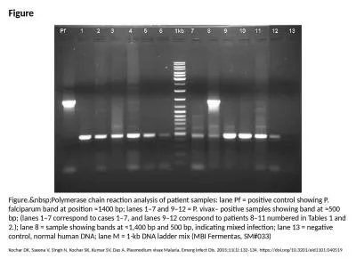
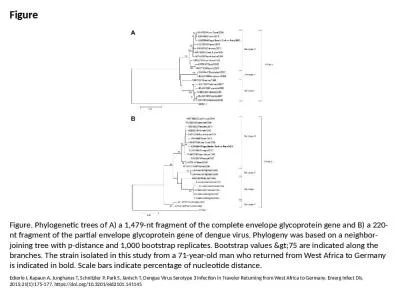
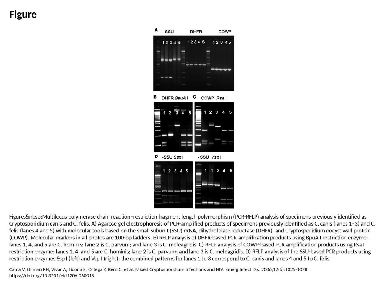
/
Figure Figure. Multilocus polymerase chain reaction–restriction fragment length Figure Figure. Multilocus polymerase chain reaction–restriction fragment length

Figure Figure. Multilocus polymerase chain reaction–restriction fragment length - PowerPoint Presentation

mackenzie
mackenzie . @mackenzie
Follow
64 views
Uploaded On 2024-01-13

Figure Figure. Multilocus polymerase chain reaction–restriction fragment length - PPT Presentation

Cama V Gilman RH Vivar A Ticona E Ortega Y Bern C et al Mixed Cryptosporidium Infections and HIV Emerg Infect Dis 200612610251028 httpsdoiorg103201eid1206060015 ID: 1040746

pcr lanes products lane lanes pcr lane products based analysis restriction rflp cryptosporidium canis felis ssu cowp amplification enzyme

Share:

Link:

Embed:

Download Presentation from below link

Download Presentation The PPT/PDF document "Figure Figure. Multilocus polym..." is the property of its rightful owner. Permission is granted to download and print the materials on this web site for personal, non-commercial use only, and to display it on your personal computer provided you do not modify the materials and that you retain all copyright notices contained in the materials. By downloading content from our website, you accept the terms of this agreement.


Presentation Transcript

1. FigureFigure. Multilocus polymerase chain reaction–restriction fragment length polymorphism (PCR-RFLP) analysis of specimens previously identified as Cryptosporidium canis and C. felis. A) Agarose gel electrophoresis of PCR-amplified products of specimens previously identified as C. canis (lanes 1–3) and C. felis (lanes 4 and 5) with molecular tools based on the small subunit (SSU) rRNA, dihydrofolate reductase (DHFR), and Cryptosporidium oocyst wall protein (COWP). Molecular markers in all photos are 100-bp ladders. B) RFLP analysis of DHFR-based PCR amplification products using BpuA I restriction enzyme; lanes 1, 4, and 5 are C. hominis; lane 2 is C. parvum; and lane 3 is C. meleagridis. C) RFLP analysis of COWP-based PCR amplification products using Rsa I restriction enzyme; lanes 1, 4, and 5 are C. hominis; lane 2 is C. parvum; and lane 3 is C. meleagridis. D) RFLP analysis of the SSU-based PCR products using restriction enzymes Ssp I (left) and Vsp I (right); the combined patterns for lanes 1 to 3 correspond to C. canis and lanes 4 and 5 to C. felis.Cama V, Gilman RH, Vivar A, Ticona E, Ortega Y, Bern C, et al. Mixed Cryptosporidium Infections and HIV. Emerg Infect Dis. 2006;12(6):1025-1028. https://doi.org/10.3201/eid1206.060015