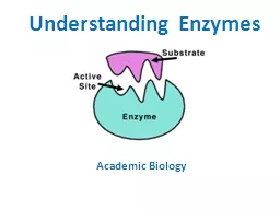
/
5  E’sy  Ways to Investigate enzymes! 5  E’sy  Ways to Investigate enzymes!

5 E’sy Ways to Investigate enzymes! - PowerPoint Presentation

marina-yarberry
marina-yarberry . @marina-yarberry
Follow
348 views
Uploaded On 2018-10-30

5 E’sy Ways to Investigate enzymes! - PPT Presentation

Learning Objectives Educators attending this workshop will Utilize tools that support NGSS student learning outcomes of three dimensional lesson design including Molymods to model basic amino acid structure and build a dipeptide ID: 703295

structure protein modeling amino protein structure amino modeling www twins beery acids chains org https side acid beery

Share:

Link:

Embed:

Download Presentation from below link

Download Presentation The PPT/PDF document "5 E’sy Ways to Investigate enzymes!" is the property of its rightful owner. Permission is granted to download and print the materials on this web site for personal, non-commercial use only, and to display it on your personal computer provided you do not modify the materials and that you retain all copyright notices contained in the materials. By downloading content from our website, you accept the terms of this agreement.


Presentation Transcript

Slide1

5 E’sy Ways to Investigate enzymes!Slide2

Learning Objectives

Educators attending this workshop will:

Utilize tools that support NGSS student learning outcomes of three dimensional lesson design including -

Molymods

to model basic amino acid structure and build a dipeptide

Amino Acid Starter Kit

©

to simulate protein folding

Enzymes in Action

©

to demonstrate enzymatic specificity and actions

Acetylcholinesterase Active Site Cube

©

to apply knowledge of protein structure/function to an observed environmental phenomenon

Landscapes of David

Goodsell

©

to apply multiple representations to a concept

Learn something new and interesting for your own professional development.

H

A

V

E

F

U

N

!Slide3

Learning Objectives

What NGSS dimensions will be targeted?

SEPs

CCCs

DCIs

Asking questions

Patterns

PS1.A: Structure and properties of matter

Developing and using models

Cause and effect

PS1.B: Chemical reactions

Constructing explanations

Scale, proportion, and quantity

LS1.A: Structure and function


Structure and function

LS2.C: Ecosystem dynamics, functioning, and resilienceSlide4

the Mosquito

Protein Structure versus . . .

Engage!

https://www.orkin.com/other/mosquitoes/

https://cdn.rcsb.org/pdb101/learn/resources/what-is-a-protein/what-is-a-protein.pdfSlide5

Amino Acids:

Building Blocks for Proteins

Explore!

Explain!Slide6

Amino Acids:

D and L Forms of Amino Acids

Extend!

http://www.phschool.com/science/biology_place/biocoach/bioprop/landd.htmlSlide7

Building a DipeptideSlide8

The Protein Folding Problem

Beware student preconceptions about protein folding!

Amino acid #1

Amino acid #2

http://www.biochem.ucl.ac.uk/~martin/c40/peptide.htmlSlide9

Modeling Protein Structure

Elaborate!Slide10

Modeling the Primary Structure

2 cysteine side chains (

green

).

2 acidic side chains (

red

).

2 basic side chains (

blue

).

Your favorite 3 hydrophilic amino acids (white).

Your favorite 6 hydrophobic amino acids (

yellow

).

Randomly distribute them on the TOOBER. (Space them about three inches apart to get an even distribution). 

You will need 15 total side chains:Slide11

Modeling Tertiary Structure

Fold your protein according to basic principles of chemistry and physics:


In an aqueous environment, where do you expect the

hydrophobic

side chains to fold? Why?

Likewise, consider where the

hydrophilic

side chains would fold.

What interaction may occur between

acidic

and

basic

amino acids?

What interaction may occur between

cysteine

amino acids?Slide12

Modeling Secondary Structure

Beta Sheets

Alpha Helices

Why

are

secondary structures important to protein structure/function?Slide13

Modeling Quaternary Structure

HemoglobinSlide14

Modeling Enzyme Action: Catabolism

Extend!

https://www.khanacademy.org/science/biology/energy-and-enzymes/introduction-to-enzymes/a/activation-energySlide15

Modeling Enzyme Action: Anabolism

Extend!Slide16

Modeling Enzyme Action: Specificity

Extend!Slide17

Competitive Inhibition

Noncompetitive Inhibition

Modeling Inhibition

Extend!Slide18

the Mosquito

Protein Structure versus . . .

Evaluate!

https://www.orkin.com/other/mosquitoes/

https://cdn.rcsb.org/pdb101/learn/resources/what-is-a-protein/what-is-a-protein.pdfSlide19

Neuromuscular Signaling

A Landscape from David

Goodsell

https://www.education.com/Slide20

A Molecular Story:

Acetylcholinesterase!Slide21

Resources!

http://www.rcsb.orgSlide22

http://pdb101.rcsb.org/

Resources!Slide23

THANK YOU!

For more molecular stories. . .

Come visit us at booths 419 and 421!

We hope to see you soon!Slide24

Another Molecular Story:

The Beery Twins!Slide25

ENZYMES!A key enzyme in the Beery twins molecular story is

sepiapterin

reductase

.

Sepiapterin

reductase is necessary to make a cofactor called

tetrahydrobiopterin

.

What’s going on with the Beery twins?Slide26

What’s going on with the Beery twins

sepiapterin

reductase?Slide27

Joe Beery’s Contribution:

A single amino acid change!

Arginine

 Glycine

What’s going on with the Beery twins

sepiapterin

reductase?Slide28

Retta Beery’s Contribution:

A missing piece of the terminal end of the protein.

What’s going on with the Beery twins

sepiapterin

reductase?Slide29

Analyzing Data:

The

Beery’s

Pedigree

How did the Beery twins get the variant forms?Slide30

Joe

Beery’s

Mutation – Missense at 498

DNA base change from adenine to guanine

Retta Beery’s Mutation – Nonsense at 801

DNA base change from adenine to thymine resulting in an early stop codon.

What are the implications of these variant forms?Slide31

The Beery Twins Today! Slide32

Resources!

http://www.raregenomics.org/e-books/Slide33

THANK YOU!

For more molecular stories. . .

Come visit us at booths 419 and 421!

We hope to see you soon!