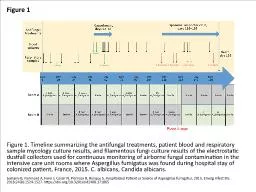
IPA Invasive pulmonary aspergillosis ABPA ABPA pathophysiology conidia of Aspergillus trapped in mucous and narrowed airways of asthmaticsCF germinate to form hyphae release of solubleparticulate antigens ID: 775064
Download Presentation The PPT/PDF document " Aspergillus & ABPA Disease spectru..." is the property of its rightful owner. Permission is granted to download and print the materials on this web site for personal, non-commercial use only, and to display it on your personal computer provided you do not modify the materials and that you retain all copyright notices contained in the materials. By downloading content from our website, you accept the terms of this agreement.
Slide1
Aspergillus & ABPA
Slide2Disease spectrum
IPA Invasive pulmonary
aspergillosis
Slide3ABPA
Slide4ABPA – pathophysiology
conidia of
Aspergillus
trapped in mucous and narrowed airways of asthmatics/CF
germinate to form hyphae
release of soluble/particulate antigens
airway inflammation
epithelial damage
Antigen diffuses into
interstitium
inflammatory mediators released
influx of inflammatory cells esp. E
& Ag presented to Th2 cells
IL-4, IL-5 & IL-13 cytokines synthesized and secreted
IgE
synthesis
mast cell degranulation
tissue injury
bronchial obstruction, inflammation, mucous plugging
bronchiectasis
fibrosis
respiratory failure
Slide5Slide6Diagnostic algorithm
Slide7Clinical Presentation
Slide8Physical Exam
Normal
Other findings include:
Polyphonic wheezing
Clubbing (16%)
Coarse crackles (15%)
Sx
of pulmonary HTN and/or respiratory failure
Slide9Differential Diagnosis of ABPA
Slide10Asthma vs. ABPA vs. AH
Slide11Immunological Findings
Cutaneous hypersensitivity
Intradermal test > Skin prick test
Type 1 immediate +
ve
&
Type 3 delayed +
ve
positive in virtually all ABPA
40% of asthmatics without ABPA also +
ve
Slide12Immunological Findings
Total
IgE
Cutoff
>1000
IU/mL
With Rx
Reduction of often
>
35% = REMISSION
levels do NOT reach normal values
repeated measurements needed to determine ‘new baseline’
Doubling of baseline = relapse
Slide13Radiographic Features - CXR
Parenchymal infiltrates (generally of upper lobes)
Transient vs. fixed
Atelectasis due to mucous plugging
Findings consistent with bronchiectasis
“Tram line” shadows d/t thickened non-dilated bronchial walls
“Parallel lines” d/t
ectactic
bronchi
Ring shadows d/t mucous filled bronchi or small abscesses
“Toothpaste shadows” d/t mucous plugging in 2
nd
to 4
th
order bronchi
“Gloved finger shadows” (branched tubular
radiodensities
, 2-3 cm long, 5-8 cm wide, extending from the hilum) d/t
intrabronchial
exudates w/ bronchial wall thickening
Slide14Radiographic Findings - HRCT
Widespread proximal cylindrical bronchiectasis (mostly of upper lobes)
Bronchial wall thickening
Mucous plugging
Atelectasis
Peripheral airspace consolidation
Ground-glass attenuation
Mosaic perfusion
Air trapping
Slide15Diagnosis of ABPA
Slide16Diagnostic algorithm
Slide17Slide18Diagnosis
6/8 criteria
If not confirmed i.e. less than full compliment of
dx
criteria
FU with 6/52
IgE
if increasing or >1000 IU/ml Rx
ABPA-S (mild) vs. APBA-CB (moderate) vs. ABPA-CB-ORF
Recurrent episodes of remissions and relapses
Slide19Staging of ABPA
Slide20Remission
No established definition
Reduction of often
>
35
%
Usually, by end of 6-9 months with a significant fall in
IgE
, patient said to be in remission i.e. Stage II (Remission)
‘
Complete remission
’ if no exacerbation for next 03 months after stopping Rx
FU with
IgE
every 6 months for 1
st
year, then annually
Slide21Tx - Goals
1) Early control of immunologic activity / inflammation to try to prevent progression to bronchiectasis and fibrosis
2) Monitoring for response and early detection of relapses
3) ?? Decrease fungal burden in airways
Slide22Tx - Steroids
Doses vary depending on stage and prescriber preference. Higher dosages for longer durations may be more effective for tx’g flares.
Stages 1 & 3 – Prednisone 0.5-1.0 mg/kg Qday x 14 days, then QOD x 6-8 wks, then taper by 5-10 mg q 2 weeks until d/c’d
Should see resolution of infiltrates and 35-50% dec in serum total IgE (measured q1-2 months during acute treatment)
Stage 2 – Steroids not needed. Monitor IgE q6 months x 1 year then q 1-2 years. Doubling of baseline IgE indicates relapse (stage 3)
Stage 4 – Steroid dependent. Aim for lowest possible dose
Stage 5 – Steroids not helpful
Steroid “prophylaxis” – Ca, Vit D, bisphosphonate
Slide23Slide24DoseCalculated doseDuration0.5 mg/kg/daymg/day2/520.5 mg/kg/alternate daysmg/alternate days6-8/52Reduce by 5mgEvery 2/52FU every 6/52 with CXR & IgETotal duration of treatment 6 – 12 months
DoseCalculated doseDuration0.75 mg/kg/daymg/day6/520.5 mg/kg/daymg/day6/52Reduce by 5mgEvery 6/52FU every 6/52 with CXR & IgETotal duration of treatment 6 – 12 months
Regime 1
Regime 2
Slide25Total IgE levels
Date
Description
Load
IU/ml
IU/ml
IU/ml
IU/ml
IU/ml
IU/ml
IU/ml
IU/ml
IU/ml
IU/ml
IU/ml
Slide26Tx - Itraconazole
Decreases
antigenic stimulus for bronchial inflammation, possibly by
decreasing
specific
Aspergillus
IgG
Decreased
metabolism of steroids, so may be able to use lower dosages
16 week course + steroids
significant increased likelihood of clinical response (46
vs
19%)
200 TID x 3 days, then 200 BID x 16
wks
, +/-
once day
x 16
wks
LFTs: Twice weekly for 3 months Monthly
Slide27Slide28Miscellaneous
Slide29SAM
The
presence of concomitant allergic fungal sinusitis (AFS) and allergic
bronchopulmonary
mycosis in the same patient represents an expression of the same process of fungal hypersensitivity in the upper and lower airways.
SAM syndrome: an
acronym for
sinobronchial
allergic
mycosis
Slide30Diagnostic Criteria
chronic
sinusitis involving multiple
sinuses
asthma
total serum
IgE
levels are usually elevated as well
immediate
cutaneous reactivity to fungal
allergens
peripheral eosinophilia
radiographic
evidence of bronchiectasis.
variety
of chest radiographic abnormalities may
occur:
ranging from mass lesions to diffuse pulmonary infiltrates and even normal findings on chest radiographs.