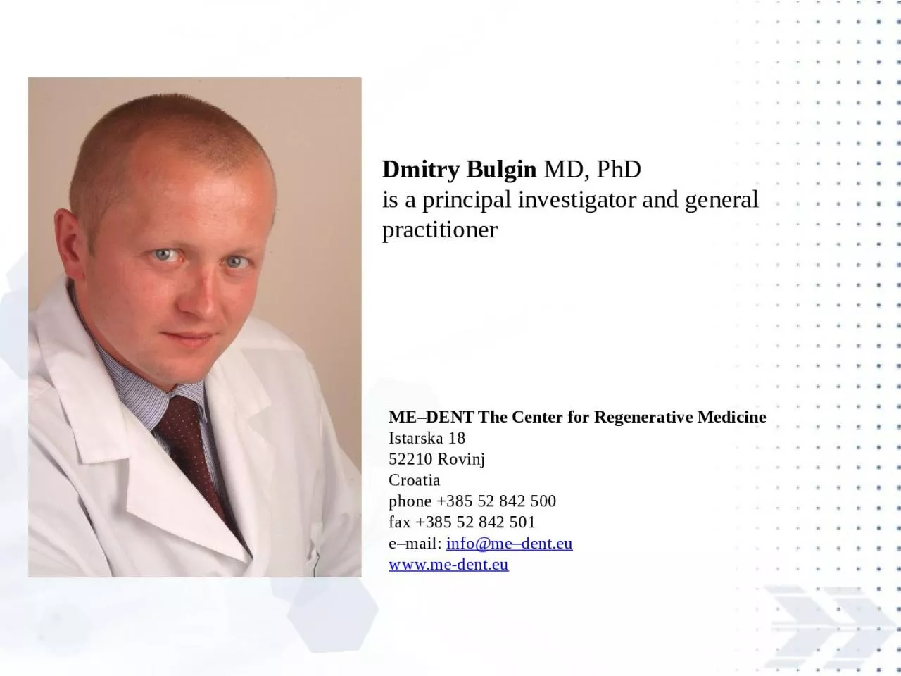
is a principal investigator and general practitioner MEDENT The Center for Regenerative Medicine Istarska 18 52210 Rovinj Croatia phone 385 52 842 500 fax 385 52 842 501 ID: 927935
Download Presentation The PPT/PDF document "Dmitry Bulgin MD, PhD" is the property of its rightful owner. Permission is granted to download and print the materials on this web site for personal, non-commercial use only, and to display it on your personal computer provided you do not modify the materials and that you retain all copyright notices contained in the materials. By downloading content from our website, you accept the terms of this agreement.
Slide1
Dmitry Bulgin
MD, PhD is a principal investigator and general practitioner
ME–DENT
The Center for Regenerative Medicine
Istarska 18
52210 Rovinj
Croatia
phone
+385 52 842 500
fax +385 52 842
501
e–mail
:
info
@me–
dent
.eu
www.me-dent.eu
Slide2Dr. Dmitry Bulgin
applies cell–based technologies (autologous bone marrow–derived and adipose tissue–derived mesenchymal stem cells) and other proprietary methods in the field
of:
Reconstructive
Dentistry
Aesthetic
&
Reconstructive
Surgery
T
raumatology
&
Orthopeadics
Vascular
(
angiology
) Medicine
Wound Care
Sports
Medicine.
Slide3The main research interest:
The development of methods to
accelerate
the healing
processes
of tissues.
The
tools
and technologies for tissue and
metabolic
engineering to
enable
regenerative medicine.
Biological
approaches for
maintaining
the human’s
performance
and
capabilities
in the face of
harsh
accident
conditions
.
Slide4The research results and clinical
outcomes have been presented at conferences (as a speaker):
The
First
Symposium
of
Yangming
and Nagasaki
Universities
, Nagasaki,
Japan,
November
29, 2004.
The International
Symposium
«
Young
Scientists
Organizing
Nagasaki
Symposium
of International
Consortium
for
Medical
Care of
Hibakusha
and
Radiation
Life
Science
», Nagasaki,
Japan.
March
7-8, 2005
.
Russian
-
American
Conference
in
Hematology
,
Saint
-
Petersburg
,
Russia
, June 21-23, 2006.
International
Scientific
Conference
in Stem Cells Technologies,
Moscow
,
Russia
,
May
30-31, 2007.
III-rd
Congress
of
Russian
Pathologist
Society
, Samara,
Russia
,
May
27-30, 2009.
International
Scientific
Symposium
: “Stem Cells,
Umbilical
Cord
Blood
and Placenta in Regenerative Medicine”,
Ljubljana,
Slovenia
,
May
27, 2011.
2012
eCM
XIII
Conference
: Bone
Fixation
,
Repair
& Regeneration (
Focus
CMF,
Spine
, Trauma,
Vet
),
Davos
,
Switzerland
, June 24 – 26, 2012.
5
th
Vienna
Biomaterial
symposium
,
Vienna
University of
Technology
,
Vienna
,
Austria
,
November
19 – 21, 2012.
UAE International Dental
Conference
&
Arab
Dental
Exhibition
AEEDC,
Dubai
,
UAE,
February
5 -7, 2013.
1
st
International
Congress
of
Plastic
Surgery
-
Fellows
in
Science
, Ljubljana,
Slovenia
,
September
18 – 21, 2013
.
Bredent
group days SKY Meeting 2014, Berlin,
Germany, May 22 - 24, 2014.
Slide5The research results and clinical outcomes have been published
:
Podtcheko
A,
Takakura
S,
Bulgin D
,
Namba
H,
Saenko
V,
Ohtsuru
A,
Yamashta
S.
Differ
role of JNK and p38
kinase in
ionizing
radiation
-
induced
thyroid
cell radio-
sensitization
.
The International
Symposium
«
Young
Scientists
Organizing
Nagasaki
Symposium
of International
Consortium
for
Medical
Care of
Hibakusha
and
Radiation
Life
Science
»
,
Abstracts
p.13, 2005.
Podtcheko
Alexei
,
Bulgin Dmitry
,
Takakura
Osamu,
Namba
Hiroyuki
,
Ohtsuru
Akira
,
Saenko
Vladimir,
Yamashita
Shunichi
.
Inhibition
of c-Jun NH2-Terminal Kinase and
Thyroid
Cell Terminal Growth
Arrest
.
Folia
Endocrinologica
Japonica
,
Volume
81,
Number
1, 2005, p. 139.
Bulgin Dmitry
,
Podtcheko
Alexei
,
Takakura
Shu
,
Mitsutake
Norisato
,
Namba
Hiroyuki
,
Saenko
Vladimir,
Ohtsuru
Akira
,
Rogounovitch
Tatiana
,
Palona
Iryna
, and
Yamashita
Shunichi
.
Selective
Pharmacologic
Inhibition
of c-Jun NH2-Terminal Kinase
Radiosensitizes
Thyroid
Anaplastic
Cancer
Cell
Lines
via
Induction
of Terminal Growth
Arrest
.
Thyroid
:
Volume
16,
Number
3, 2006, p. 217-224.
Bulgin Dmitry,
Hodzic Enes, Komljenovic-Blitva Danijela.
Advanced
and
Prospective
Technologies for
Potential
Use
in Craniofacial Tissues Regeneration
by
Stem Cells and Growth Factors.
Journal
of
Craniofacial
Surgery
:
January
2011 -
Volume
22 -
Issue
1 –
pp
. 342-348.
Bulgin Dmitry,
Hodzic Enes. Autologous Bone Marrow-Derived
Mononuclear
Cells
Combined
With β-Tricalcium
Phosphate
(β -TCP) for Maxillary Bone Augmentation in
Implantation
Procedures
.
Journal
of
Craniofacial
Surgery
:
November
2012 -
Volume
23-
Issue
6 -
pp
. 1728-1732.
Bulgin Dmitry
.
Prospective
Technologies in Dental Tissues Regeneration.
Journal
of Oral
Hygiene
&
Health
: June 2013 -
Volume
1-
Issue
2 -
pp
.1-2.
Bulgin Dmitry
,
Irha
Ernst
, Hodzic Enes,
Nemec
Boris. Autologous bone marrow derived
mononuclear
cells
combined
with β-tricalcium
phosphate
and
absorbable
atelocollagen
for a
treatment
of
aneurysmal
bone
cyst
of the
humerus
in
child
.
Journal
of Biomaterials
Applicatios
:
September
2013 -
Volume
28 -
Issue
3-
pp
. 343-353 [
Epub
ahead
of
print
2012 Jun 12].
Bulgin Dmitry
,
Vrabic
Erik
, Hodzic Enes. Autologous bone-marrow-derived-
mononuclear
-cells-
enriched
fat
transplantation
in
breast
augmentation:
evaluation
of clinical
outcomes
and
aesthetic
results
in a 30-
year
-
old
female
.
Case
Reports
in
Surgery
. 2013;
2013
;2013:782069.
doi
: 10.1
155/2013/782069.
Epub
2013
Aug
19.
Slide6Prospective
Technologies in Dental Tissues RegenerationInterest in applications for dental tissues regeneration continues to increase as clinically relevant methods alternative to traditional treatments.
Dental tissues engineering is an opportunity that dentistry
cannot afford to miss.
Recent progress in the studies of molecular
basis of tooth development, adult stem cell biology, and regeneration
will provide fundamental knowledge for that.
The impact of dental tissues engineering extends beyond clinical practice. Dental tissues engineering could not have advanced to the current stage without the incorporation of interdisciplinary skill sets of stem cell biology, bioengineering, polymer chemistry, mechanical engineering, robotics, etc. Thus, dental tissues engineering and regenerative dental medicine are integral components of regenerative medicine.
Citation:
Bulgin D (2013) Prospective Technologies in Dental Tissues
Regeneration.
Oral
Hyg
Health
1: e102.
doi
:10.4172/2332-0702.1000e102
Slide7Replacing
missing bone or adding mass to existing bone is often essential to the success of a dental implant.
Slide8An implant needs a critical mass of bone surrounding it in order to bind to it and deliver sufficient strength and stability
.
Slide9If in the location where the implants are intended there is low mass of bone (width or height) a bone graft must be applied in order to maintain this critical bone mass.
Slide10A large variety of graft materials have been used for maxillary and mandibular atrophy to fill bone defects
: AUTOGENOUS
HOMOGENOUS
(allograft)
HETEROGENEOUS
(xenograft)
SYNTHETIC
(predominantly osteo-conductive ) substitutes.
Slide11OSTEOCONDUCTIVE MATRIX
, which acts as scaffold to new bone growth Bone-graft materials usually have one or more components:
OSTEOINDUCTIVE PROTEINS
, which support mitogenesis of undifferentiated cells
OSTEOGENIC CELLS
, which are capable of forming bone in the appropriate environment
Slide12Many
clinicians consider harvested autologous bone (i.e.
taken
from the same
individual
) as the
“
gold
standard”
material for the
reconstruction of osseous defects. autological bone fragment from posterior iliac crestAutologous bone grafts by
their
very
nature are
able
to
deliver
a
physiologically
optimized
combination
of osteogenic cells and growth factors in a
mineralized
scaffold
.
Slide13However,
limited availability
of
donor
sites
,
requirement
for an
additional
surgery to obtain the graft material, and extra chair time are the limitations of this technique. A large amount of harvested bone raises the risk of postoperative functional or cosmetic morbidities at the donor site.
Slide14The autologous application of human bone marrow cells which are
not
expanded
ex
vivo
has medico-
legal
advantages in clinical applications.
Defect
repair and bone ingrowth, maturation, and modeling
are cell-
mediated
processes
.
Most clinical trials
report
successful bone regeneration
after the application of mixed cell populations from bone marrow.
Slide16Bone marrow derived
mononuclear cells (
BMMNCs
)
secrete
many
kinds
of growth factors and
represent a potential key component in autologous graft for bone regeneration. Growth factors that encourage the
formation
of new bone have been
identified
and
applied
to heal bone defects
around
medical
and dental implants and
without
implant
placement.
Slide17To reduce bone
harvesting
,
various
synthetic bone void fillers with improved biocompatibility have been developed
.
Slide18The
extracellular matrix of bone has been described as a composite material composed of
collagen
type
I
fibrils
mineralized
with
nanocrystals of hydroxyapatite. Bernhardt A, Lode A, Boxberger S, Pompe W, Gelinsky M. J Mater Sci Mater Med 2008;19:269.
Slide19S
ynthetic bone replacement
materials
are
osteoconductive
scaffolds
that
encourage
the growth of new bone
by apposition from adjacent bone surfaces or from bone-forming cells. After sufficient bone has grown throughout the defect site, the
scaffold
no
longer
plays
a role in the
reparative
process
and,
ideally
, is
resorbed
.
Slide20These materials are made from
calcium salts:
CALCIUM SULFATE
CALCIUM CARBONATE
CALCIUM PHOSPHATE
BIOACTIVE GLASSES CONTAINING CALCIUM OXIDE
Highly biocompatible material
s
with
retention of the natural porous trabeculation,
architecture, and natural mineral content -
closest components of human
cancellous
bone.
HUMAN BONE
CALCIUM SALT
Slide21The class of
calcium phosphate bone replacements is composed: porous ß-tricalcium phosphate (ß-TCP)
dense hydroxyapatite
(HA)
porous HA
that has retained the structure of coral
(the exoskeleton of which has been chemically converted, in full or in part, from calcium carbonate into HA)
biphasic mixture of TCP and HA
Slide22Recently highly pure porous
β-tricalcium phosphate (β-TCP) represents a very interesting practical tool for bone regeneration Scanning electron micrograph
of highl
y pure porous
β
-TCP:
(a)
Macrostructure
has a macrodiameter of 0.5
-
1.5 mm, (b) micropores of 100-400 μ
m, various size of micropores are seen
.
The open and interconnected porosity of
β-TCP
allows body fluids to circulate throughout its entire extent.
The range of pore sizes encourages tissue ingrowth
.
β-TCP
shows resorbable characters during bone regeneration, and can be completely substituted for the bone tissue after stimulation of bone formation.
β-TCP
is biocompatible porous material, new bone formation takes place along the surface in their micropores.
Slide23Hydroxyapatite can be
produced with either
a
dense
or
macroporous
morphology
, and is
typically
sintered at temperatures above 1000 °C in granular or
block
forms
.
The
high
heat
of
sintering
produces
a material that
cannot
be
reshaped
to
fit
into a bone
defect
(i.e.
if
in
block
form
) and is
nonresorbable
.
H
ydroxyapatite
is a
brittle
material with
low
fracture
resistance
.
H
ydroxyapatite
is
biocompatible
,
osteoconductive
, and
bioactive
(i.e.
it
develop
s
a
direct
,
adherent
bond
with bone).
Slide24Biphasic
calcium phosphate
products
which
contain
hydroxyapatite
and
β-TCP in various ratios are aimed at the provision of a bone grafting material which is able to degrade within a physiologically optimized time frame, while
providing
some
measure
of
mechanical
stability
until
sufficient
bone in-growth has
occurred
.
While the ß-TCP exhibits quick osseous organization and is replaced by newly formed bone within a short period, the HA content ensures that the volume remains unchanged.
Slide25Collagen as a material for scaffold-based therapy
Scanning electron micrograph of porous atelocollagen
sponge
:
macrostructure
micropores
of 50
-
500
μ
m, various size of micropores are seen. The atelocollagen is a
cross
-
linked
collagen
material with
abundant
micropores
.
It
has large
pores
that
permit
cellular
entry
and is
degraded
in
vivo
.
These
characteristics
suggest
that
this
material
may
be a
good
candidate
for use as
scaffolding
for
implantation
of cells.
Slide26Slide27Synthetic
materials
require
the
addition
of
bioactive
factors or cells to
promote
tissue regeneration.
Slide28Recently
, the transplantation of autogenous
BMMNCs
combined
with
synthetic
grafting
materials has been
inaugurated and introduced as a clinical procedure in oral and maxillofacial surgery.
Slide29To date there has been no graft material which can be regarded as completely satisfactory
.
M
y experience with freshly isolated autologous bone marrow derived mononuclear cells combined with synthetic biphasic calcium phosphate
ceramic
and absorbable
atelocollagen
for augmentation of the extremely atrophic maxilla and mandible is presented.
The techniques are based on stimulation of natural events continuously present in living bone, that is, the process of bone remodeling and offering both
osteoinduction
and
osteoconductive
features.
Slide30A
57-year-old man. Panoramic
X-
ray
image
before
augmentation
procedures
(h
orizontal and vertical augmentation, left maxillary sinus lifting).A 46-year-old woman. Panoramic X-ray image before augmentation procedures (horizontal and vertical augmentation, left and right maxillary sinus lifting).
A
48-
year
-
old
man
.
Panoramic
X-
ray
image
before
augmentation
procedures
(h
orizontal
and vertical augmentation).
Cases breakdown
Preoperative r
adiological
evaluation
Slide31Maxillary
bone augmentation with the aim of embedding implants was performed under deep sedation
in three healthy
patients
with
advanced
atroph
y of the alveolar bone of
maxilla.
Materials and methods: Patients
Slide32the
bone marrow harvesting
from
posterior
iliac
crest
c
ollection of bone marrow in plastic bagthe bone marrow processing by using Cell
Separation
System
SEPAX S-100
qualitative
assessment
of
BMMNCs
population
by
haematoxylin
and
eosin
cytological
staining
(
magnification x
400
).
B
one marrow derived mononuclear cells preparation:
Slide33At the time of surgery, BMMNCs and
synthetic biphasic calcium phosphate ceramic granules were mixed, and excess fluid volume was removed using filtration and low-grade vacuum.Freshly isolated autologous
BMMNCs
in combination with
synthetic biphasic calcium phosphate
ceramic
Slide34Micro- and
macroporosity (magnification 25x)nano
structure
(
magnification
1000x)
•
60% hydroxyapatite
(HA)
• 40 % ß-tricalcium phosphate (ß-TCP)A fully synthetic, two-phase calcium phosphate ceramic consisting of
Slide35Surgical
grafting and dental implants procedures
A
, M
axillary
bone augmemtation site preparation.
B
, Implants placement.
C
, Sinus filled by synthetic biphasic calcium phosphate ceramic combined with BMMNCs. D
, S
urgical
sites
are
closing
with
silk
non
-
absorbable
sutures
.
M
axillary
bone augmentation
with simultaneous implants placement.
Slide36A
, Alveolar crest incision at the beginning of the bone augmentationprocedure. B, The planned maxillary bone augmentation site with advanced atrophy. C, Sinus lift procedure: window preparation and sinus membrane elevation. D, The BMMNCs and
Ossceram
nano
®
mixture grafting. The graft material isolation from the soft tissues by membrane.
Sinus lift and bone augmentation
procedures
Slide37Approximately 7 months later
, dental implants were inserted into the augmented maxilla. A, Embedding of the implantation material into the augmented maxilla
(7 months after augmentation procedure).
B
, Surgical site reopening for revision of implant integration
after
3 months from the date of
implants placement
.
C, Histological condition of graft tissue 7 months after augmentation procedure. Hematoxylin and eosin stain method. Original magnification x200.
Slide38Advanced Maxillary Atrophy
before augmentation Patient
S
. L
.
, 46
y. o., female
(Case 2)
in 10 months after augmentation reopening for revision of implant integration
Slide39Patient
S
. L
.
, 46
y. o., female
(Case 2)
PREOPERATIVE24 months of follow-upPOSTOPERATIVE 1 month
POSTOPERATIVE 12 months
24 months of
follow
-up
24 months of
follow
-up
24 months of
follow
-up
Slide40Favorable bone bonding was seen in the embedded implantation
material in all of the casesA, Before augmentation procedure. B, At one month after operation
.
C
, At twelve months after operation.
A
57-year-old man.
A
46-year-old woman.
A 48-year-old man.After a recovery period of approximately 12 months the upper structures were mounted. Since
dental implant placement, patients have maintained good
peri
-implant
health
(no bleeding on probing) and oral hygiene
.
Slide41Radiological evaluation of
healing periodThe panoramic radiographic imaging was taken at 1 and 12 months after operation to evaluate the extent of reconstruction at grafted sites by using x-ray image analysis
Planmeca
Romexis
software (
Planmeca
Oy
, Helsinki, Finland).
The mean increasing height of the graft site was 16.7 mm at 1 month and 15.3 mm at 12 months; that is, the height of the graft site showed approximately 8.4% reduction from 1 to 12 months.
Slide42Isolation of
synthetic biphasic calcium phosphate
ceramic
b
y
absorbable
atelocollagen
membrane
from the oral cavity.
Slide43Patient :
D. M., 52 y. o., female
Diagnose
:
Advanced Maxillary and Mandibular Atrophy,
bone
resorbtion
,
Parodontosis
, gum recession.
Slide44I
solation of synthetic biphasic calcium phosphate
ceramic
b
y
absorbable
atelocollagen
membrane
from the oral cavity
Slide45A
fter a recovery period of approximately 12 months the upper structures were mounted.
Slide46Patient : E. F., 59 y. o., female
Diagnose: Advanced Maxillary and Mandibular Atrophy, bone resorbtion
,
Parodontosis
, gum recession.
Slide47Window preparation
and sinus membrane
elevation
Slide48I
solation of synthetic biphasic calcium phosphate ceramic
b
y
absorbable
atelocollagen
membrane
from the oral cavity
Slide49A
fter a recovery period of approximately 12 months the upper structures were mounted.
Slide50Within the constraints of our clinical outcomes,
freshly isolated autologous BMMNCs in combination with synthetic biphasic calcium phosphate ceramic and
a
bsorbable
ateloco
llagen
membrane
(a
s a barrier membrane) led to significant improvements clinically as well as radiographically and, hence, can be successfully used in the treatment of extremely atrophied maxilla and mandible.CONCLUSIONS
Slide51This
method does not require cell culturing
, and
lacks
any
risk
of
immune
reaction as the grafts are autologous in nature. The adjunctive clinical benefit of the BMMNCs preparation can be explained on the basis of tissue engineering, i.e., tissue engineering generally combines three
key
elements
for regeneration:
scaffolds
or
matrices
signaling
molecules
or growth factors
cells
.
Slide52The advantages of this method for clinical use:
the application of the cells immediately
after the bone marrow is collected, consequently the surgery can be performed the same
day
;
the cells do not need to be expanded
in vitro
, they preserve their osteogenic potential to form bone and promote the proper bone
defect healing.
Slide53Dentistry Related Journals
JBR Journal of Interdisciplinary Medicine and Dental
Science
Journal of Oral Hygiene &
Health
Oral Health and Dental Management