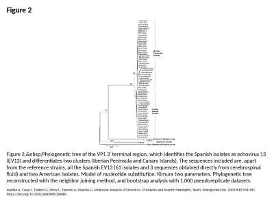
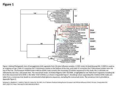
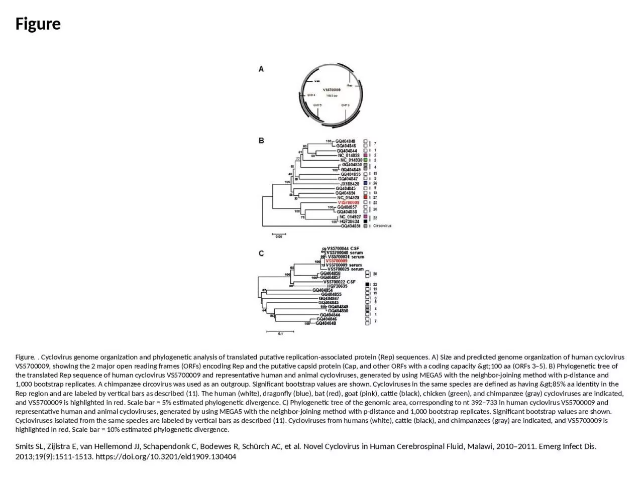
Smits SL Zijlstra E van Hellemond JJ Schapendonk C Bodewes R Schürch AC et al Novel Cyclovirus in Human Cerebrospinal Fluid Malawi 20102011 Emerg Infect Dis 201319915111513 httpsdoiorg103201eid1909130404 ID: 1042580
Download Presentation The PPT/PDF document "Figure Figure. . Cyclovirus genome organ..." is the property of its rightful owner. Permission is granted to download and print the materials on this web site for personal, non-commercial use only, and to display it on your personal computer provided you do not modify the materials and that you retain all copyright notices contained in the materials. By downloading content from our website, you accept the terms of this agreement.
1. FigureFigure. . Cyclovirus genome organization and phylogenetic analysis of translated putative replication-associated protein (Rep) sequences. A) Size and predicted genome organization of human cyclovirus VS5700009, showing the 2 major open reading frames (ORFs) encoding Rep and the putative capsid protein (Cap, and other ORFs with a coding capacity >100 aa (ORFs 3–5). B) Phylogenetic tree of the translated Rep sequence of human cyclovirus VS5700009 and representative human and animal cycloviruses, generated by using MEGA5 with the neighbor-joining method with p-distance and 1,000 bootstrap replicates. A chimpanzee circovirus was used as an outgroup. Significant bootstrap values are shown. Cycloviruses in the same species are defined as having >85% aa identity in the Rep region and are labeled by vertical bars as described (11). The human (white), dragonfly (blue), bat (red), goat (pink), cattle (black), chicken (green), and chimpanzee (gray) cycloviruses are indicated, and VS5700009 is highlighted in red. Scale bar = 5% estimated phylogenetic divergence. C) Phylogenetic tree of the genomic area, corresponding to nt 392–733 in human cyclovirus VS5700009 and representative human and animal cycloviruses, generated by using MEGA5 with the neighbor-joining method with p-distance and 1,000 bootstrap replicates. Significant bootstrap values are shown. Cycloviruses isolated from the same species are labeled by vertical bars as described (11). Cycloviruses from humans (white), cattle (black), and chimpanzees (gray) are indicated, and VS5700009 is highlighted in red. Scale bar = 10% estimated phylogenetic divergence.Smits SL, Zijlstra E, van Hellemond JJ, Schapendonk C, Bodewes R, Schürch AC, et al. Novel Cyclovirus in Human Cerebrospinal Fluid, Malawi, 2010–2011. Emerg Infect Dis. 2013;19(9):1511-1513. https://doi.org/10.3201/eid1909.130404