/
IRANIAN JOURNAL OF PATHOLOGYVol6 No2 Spring 2011 IRANIAN JOURNAL OF PATHOLOGYVol6 No2 Spring 2011

IRANIAN JOURNAL OF PATHOLOGYVol6 No2 Spring 2011 - PDF document

mila-milly
mila-milly . @mila-milly
Follow
342 views
Uploaded On 2022-10-27

IRANIAN JOURNAL OF PATHOLOGYVol6 No2 Spring 2011 - PPT Presentation

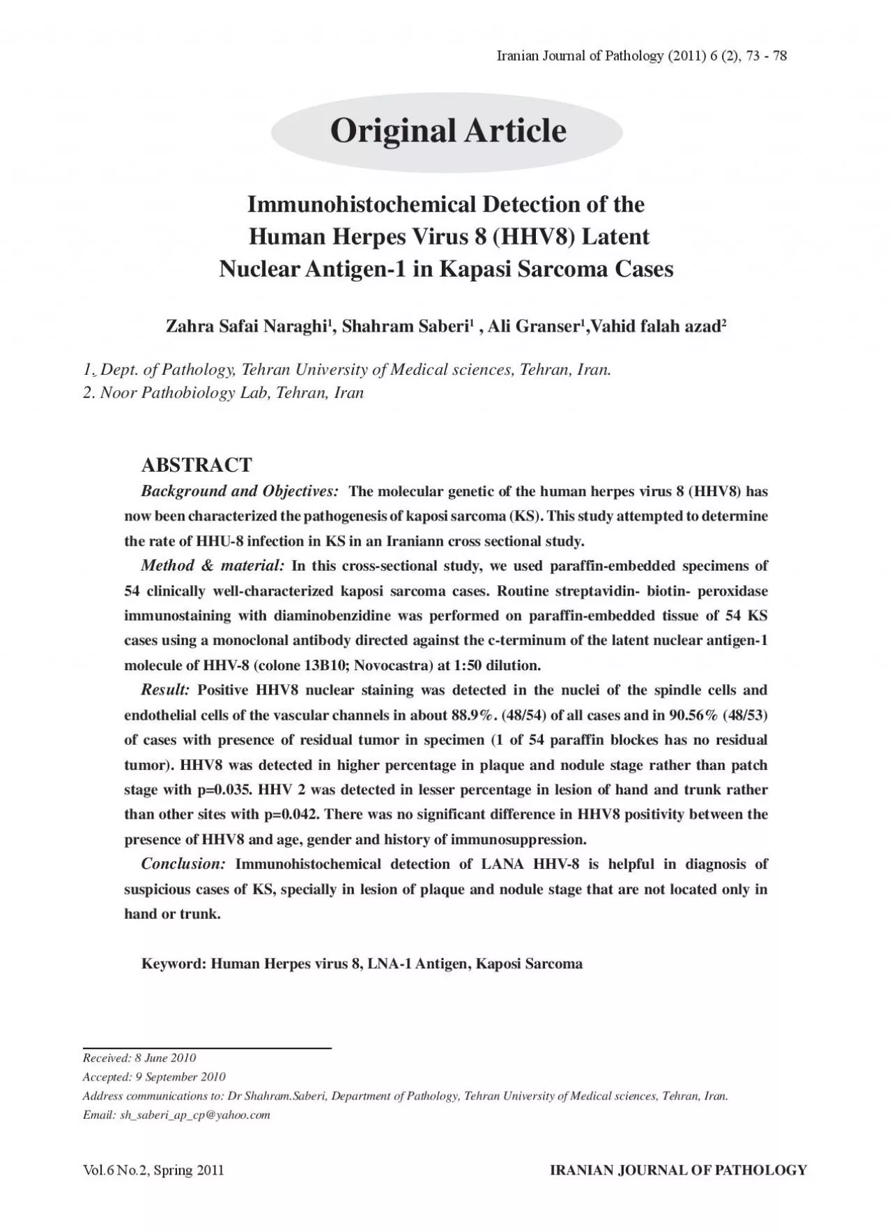
Original ArticleReceived 8 June 2010Accepted 9 September 2010 Human Herpes Virus 8 HHV8 Latent Nuclear Antigen1 in Kapasi Sarcoma Cases Ali GranserVahid falah azad Dept of Pathology Tehran ID: 960733

sarcoma hhv virus kaposi hhv sarcoma kaposi virus human stage cases herpes study nuclear hhv8 journal detected staining detection

Share:

Link:

Embed:

Download Presentation from below link

Download Pdf The PPT/PDF document "IRANIAN JOURNAL OF PATHOLOGYVol6 No2 Spr..." is the property of its rightful owner. Permission is granted to download and print the materials on this web site for personal, non-commercial use only, and to display it on your personal computer provided you do not modify the materials and that you retain all copyright notices contained in the materials. By downloading content from our website, you accept the terms of this agreement.


Presentation Transcript

IRANIAN JOURNAL OF PATHOLOGYVol.6 No.2, Spring 2011 Original ArticleReceived: 8 June 2010Accepted: 9 September 2010 Human Herpes Virus 8 (HHV8) Latent Nuclear Antigen-1 in Kapasi Sarcoma Cases , Ali Granser,Vahid falah azad. Dept. of Pathology, Tehran University of Medical sciences, Tehran, Iran.2. Noor Pathobiology Lab, Tehran, Iran The molecular genetic of the human herpes virus 8 (HHV8) has now been characterized the pathogenesis of kaposi sarcoma (KS). This study attempted to determine the rate of HHU-8 infection in KS in an Iraniann cross sectional study. In this cross-sectional study, we used paraffin-embedded specimens of 54 clinically well-characterized kaposi sarcoma cases. Routine streptavidin- biotin- peroxidase cases using a monoclonal antibody directed against the c-terminum of the latent nuclear antigen-1 molecule of HHV-8 (colone 13B10; Novocastra) at 1:50 dilution. Positive HHV8 nuclear staining was detected in the nuclei of the spindle cells and endothelial cells of the vascular channels in about 88.9%. (48/54) of all cases and in 90.56% (48/53) of cases with presence of residual tumor in specimen (1 of 54 paraffin blockes has no residual tumor). HHV8 was detected in higher percentage in plaque and nodule stage rather than patch stage with p=0.035. HHV 2 was de

tected in lesser percentage in lesion of hand and trunk rather than other sites with p=0.042. There was no significant difference in HHV8 positivity between the presence of HHV8 and age, gender and history of immunosuppression. Immunohistochemical detection of LANA HHV-8 is helpful in diagnosis of suspicious cases of KS, specially in lesion of plaque and nodule stage that are not located only in hand or trunk.Keyword: Human Herpes virus 8, LNA-1 Antigen, Kaposi Sarcoma Iranian Journal of Pathology (2011) 6 (2), 73 - 78 Vol.6 No.2, Spring 2011IRANIAN JOURNAL OF PATHOLOGYIntroduction It classified as classic, African, Iatrogenic and AI starting on the feet and leg. The histologic features with a largely spindle cell is seen, and in the nodular sieve-like appearance(6). The most typical feature . identified DNA belonging to a novel virus in tissue affected by KS. His virus , human herpes virus type-8 (HHV-8) was originally As whit other cell-transforming DNA viruses, infection with HHV-8 alone is probably not sufficient for the development of KS, additional cofactor are probably required (9). Further, the explosive incidence of HIV-associated KS in early 1980s result from colling epidemics of HIV and HHV-8 infection in homosexual and bisexual It is believed to be necessary but not s

ufficient contributory role. Parenthetically, HHV-8 is Castleman’s disease and primary effusion lymphoma (11-14). The most important recent development in herpes virus (HHV-8) is present in almost 100% of KS lesions, whether I-IIV-related, classic, endemic, HHV 8 infection in an Iranian series of KS cases available antibody against the HHV 8 latent nuclear : Paraffin embedded specimens of : Paraffin sections were Citrate buffer (2.1 g/1000 ml; pH 6.01) for antigen the sections were washed in phosphate- buffer of HHV 8 clone 13B10; novocastra of 1:50 dilution. Cells were classified as positive for HHV 8 when Correlation between HHV 8 immunostainig and HHV 8 was detected in the nuclei of the spindle of infected KS lesions(Fig.1,2). The lesions that are Immunohistochemical Detection of the Human Herpes Virus 8 ... IRANIAN JOURNAL OF PATHOLOGYVol.6 No.2, Spring 2011lesional cells. Positive HHV 8 nuclear staining was all of which showing positive result. The median respectively.There was significant difference between stage tochemical staining for HHV 8.HHV 8 was detected in 69.23% of patch stage, value= 0.035) (Table 1).HHV 8 was detected in 66.67% of the lesions that that present only in lower extremity, 50% of lesions and lower extremity, with significant lesser HHV value= 0.042

). However there was no significant difference in HHV 8 positivity between the presence of HHV and age, gender and Table 1: Immunohistochemical detection of HHV 8 in different stages HHV 8 positivenegative13942120120191Table 2: HHV 8 immunodetectionpositivenegative4137413112Upper extremityLower extremity Trunk32136342422541Upper or lower extremityImmunosuppression 8 Vol.6 No.2, Spring 2011IRANIAN JOURNAL OF PATHOLOGY H & E staining of kaposi sarcoma × Immunohistochemical staining of spindle cell nuclei with HHV 8- LANA-1 Immunohistochemical Detection of the Human Herpes Virus 8 ... In this study we have shown that HHV-8 infection majority of KS lesions as other studies (2-4),(Table 1). The plaque and nodule stage have significant staining for HHV-8 than patch stage, also sites(Table 2). that none of the angiosarcoma cases and none of the with the LANA-1 antibody. In contrast, HHV-8 cases of KS and multicentric Castleman’s disease. DNA level (5). Angela . detected HHV-8 in 78 immunohistochemical staining for LANA of HHV-8 is sensitive to confirmation the diagnosis of KS in patch stage, and limits the study only to we limits the site of study to upper extremity, lower extremity, whole of body , trunk and upper and We do not access to PCR method to confirmed our nega

tive result in patch stage about HHV-8 compared to Angela use this method, that limits our study. There is no significant difference about the result of our lesion involving only hand or trunk). There was of HHV8 and age, gender, and history of studies (15-17).We could not find any study that call in question the pathogenesis of HHV-8 in KS detection of latent nuclear antigen-1 of HHV-8 was IRANIAN JOURNAL OF PATHOLOGYVol.6 No.2, Spring 2011Thanks to Dr.Zahra Safai Naraghi for financial archive of Razi Hospital. The authors declare that ReferencesDer Hut. Arch Dermatol Syphiol 1827;(4):265-73.3. Pellett PE, Wright DJ, Engels EA, Ablashi DV, Dollard SC, Forghani B, 8 seroprevalence among US blood donors. Transfusion 4. Cheuk W, Wong KO, Wong CS, Dinkel JE, Kaposi sarcoma from its mimickers. Am J Clin Pathol 6. Hong A, Davies S, Lee CS. Immun-ohistochemical nuclear antigen-1 in Kaposi’s sarcoma. Pathology 7. Uccini S, Scarpino S, Ballarini F, Soriani A, Chilosi chemokine-receptor expression in Kaposi sarcoma. Am 8. Serfling U, Lukowsky A, Audring H, Sterry W. Search for Kaposi’s sarcoma-associated virus DNA 9. Iscovich J, Boffetta P, Franceschi S, Azizi E, Sarid 10. Antman K, Chang Y. Kaposi’s sarcoma. N Engl J 11. Kavak A, Akman RY, Alper M, Buyukbabani N. Penile Kaposi’s sarcoma

in a human immunodeficiency 12. Ablashi DV, Chatlynne LG, Whitman JE, Jr., Cesarman E. Spectrum of Kaposi’s sarcoma-associated 13. Geraminejad P, Memar O, Aronson I, Rady PL, Hengge U, Tyring SK. Kaposi’s sarcoma and other manifestations of human herpesvirus 8. J Am Acad . Differential viral protein expression in Kaposi’s sarcoma-associated herpesvirus-infected diseases: Kaposi’s sarcoma, primary effusion lymphoma, and multicentric Castleman’s disease. Am J 15. Tappero JW, Conant MA, Wolfe SF, Berger TG. Kaposi’s sarcoma. Epidemiology, pathogenesis, histology, clinical spectrum, staging criteria and therapy. J Am Acad Dermatol 1993,28(3):371-95.16. Beral V, Peterman TA, Berkelman RL, Jaffe HW. Kaposi’s sarcoma among persons with AIDS, a sexually 17. Montagnino G, Bencini PL, Tarantino A, Caputo R, Ponticelli C. Clinical features and course of Kaposi’s 18. Pyo R, Jensen KK, Wiekowski MT, Manfra D, Alcami A, Taubman MB, the chemokine-binding protein M3. Am J Pathol 2004; 19. Masini C, Abeni DD, Cattaruzza MS, Capuano Vol.6 No.2, Spring 2011IRANIAN JOURNAL OF PATHOLOGYImmunohistochemical Detection of the Human Herpes Virus 8 ...M, Pedicelli C, Cerimele F, D. Reactivation of Kaposi’s sarcoma-associated ORF 50 transactivator, a homolog of the EBV R protein. Virology 1998;252(2):304-12