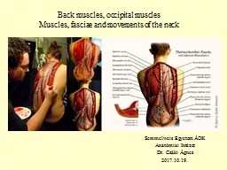
/
Muscles and  fascial  plane of the neck Muscles and  fascial  plane of the neck

Muscles and fascial plane of the neck - PowerPoint Presentation

mila-milly
mila-milly . @mila-milly
Follow
64 views
Uploaded On 2024-01-13

Muscles and fascial plane of the neck - PPT Presentation

Structures Palpated in the Midline body of the hyoid bone thyroid cartilage cricoid cartilage Trachea isthmus of the thyroid gland suprasternal notc h Structures Palpated Posteriorly ID: 1040015

hyoid neck fascia muscles neck hyoid muscles fascia carotid bone muscle belly artery platysma head insertionactionnerve anterior lateral posterior

Share:

Link:

Embed:

Download Presentation from below link

Download Presentation The PPT/PDF document "Muscles and fascial plane of the neck" is the property of its rightful owner. Permission is granted to download and print the materials on this web site for personal, non-commercial use only, and to display it on your personal computer provided you do not modify the materials and that you retain all copyright notices contained in the materials. By downloading content from our website, you accept the terms of this agreement.


Presentation Transcript

1. Muscles and fascial plane of the neck

2. Structures Palpated in the Midline body of the hyoid bonethyroid cartilagecricoid cartilageTracheaisthmus of the thyroid glandsuprasternal notch

3.

4. Structures Palpated Posteriorly InionNuchal grooveVertebra prominenceLigamentum nuchae which covers spines of C2 to C6

5.

6. Structures Palpated LaterallySternocleido-mastoid muscleTrapezius muscleExternal jugular veins

7.

8. Muscles of the neck

9.

10. Anterior view of neck muscles

11. PLATYSMA•Platysma is a broad sheet of muscle of varying prominence.•arises from the fascia covering the upper parts of pectoralis major and deltoid. •Its fibers cross the clavicle and ascend medially in the side of the neck.•Anterior fibers interlace across the midline with the fibers of the contra lateral muscle, below and behind the symphysis menti. •Other fibers attach to the lower border of the mandible or to the lower lip or cross the mandible to attach to skin and subcutaneous tissue of the lower face.

12. •Vascular supply •Platysma receives its blood supply from–the submental branch of the facial artery –and the suprascapular artery from the thyrocervical trunk of the subclavian artery. •Innervation -Platysma is innervated by the cervical branch of the facial nerve which descends on the deep surface of the muscle close to the angle of the mandible. Action-Contraction diminishes the concavity between the jaw and the side of the neck and produces tense oblique ridges in the skin of the neck.

13. Platysma muscle

14.

15. Sternocleidomastoid MuscleOrigin InsertionActionNerve SupplyTwo heads:Sternal head –manubrium sterniClavicular head- Medial 2/3 of the clavicleMastoid process of the of the temporal bone and lateral half of the superior nuchal line.Bilateral:Flex the neckSingle:Flex the head and laterally rotate to the opposite side1. Spinal accesory nerve2. Anterior primary rami of spinal nerves C2 and C3.15

16. sternocleidomastoid

17. Trapezius MuscleOrigin InsertionActionNerve SupplyExternal occipital protruberance, the medial half of the superior nuchal line, the ligamentum nuchae, and the lower cervical and all the thoracic spinesFibers originating from the head insert into the lateral third of the clavicle, and the remaining fibers insert into the acromion process and spine of the scapulaContract to rotate and elevate the scapula. Its cervical portion, acting bilaterally, can extend the head and singly, it can rotate the head and face to the opposite side 1. Spinal accessory nerve2. Anterior primary rami of the spinal nerves C3 and C4.17

18. Trapezius muscle

19. Trapezius muscle

20. I. Infrahyoid MusclesMuscleOrigin InsertionActionNerve SupplyOmohyoidInferior belly:Superior border of the scapulaSuperior belly:Intermediate tendonInferior belly:Intermediate tendonSuperior belly:Lower border of the body of the hyoid boneDepresses the hyoid bone and larynxAnsa cervicalis (APR of C1, C2, C3)SternohyoidManubrium of the sternum, posterior aspectLower border of the body of the hyoid boneDepresses the hyoid bone and larynxAnsa cervicalis (APR of C1, C2, C3)20

21. Infrahyoid MusclesMuscleOrigin InsertionActionNerve SupplySternothyroidManubrium of the sternum, posterior aspectOblique line of thyroid cartilageDepresses the larynxAnsa cervicalis (APR of C1, C2, C3)ThyrohyoidOblique line of thyroid cartilageBody and greater horn of hyoid bone, lower borderDepresses the hyoid boneThyrohyoid branch of the hypoglossal nerve21

22. Infra-hyoid muscles

23. Infra-hyoid muscles

24. Suprahyoid MusclesMuscleOrigin InsertionActionNerve SupplyMylohyoid Mylohyoid line on the medial aspect of mandibular bodyMedian raphe and body of hyoidElevates the hyoid bone, base of the tongue, and floor of the mouthNerve to mylohyoid muscleGeniohyoid muscleInferior border genial tubercle of the mandibleBody of hyoid boneElevates hyoid bone, protracts hyoid boneAPR of C124

25. II. Suprahyoid MusclesMuscleOrigin InsertionActionNerve SupplyStylohyoidStyloid processGreater horn of hyoid boneElevates hyoid boneFacial nerveDigastric(Posterior belly)Digastric notch of the temporal bone at the base of the skullIntermediate tendonRaises the hyoid boneFacial nerveDigastric(anterior belly)Intermediate tendonDigastric fossa of the mandibleElevates the hyoid boneNerve to the mylohyoid muscle25

26.

27. Muscles that form the floor of the oral cavity, superior view

28. Suprahyoid muscles, lateral and inferior view

29. Muscles in the anterior triangle of the neck

30. Carotid system

31. Branches of the external carotid artery

32.

33. Muscles associated with the posterior triangle of the neck

34. Muscles of the posterior triangle of the neck

35. Prevertebral and lateral muscles

36. Prevertebral and lateral muscles

37. Fascial planes of the neck

38. The fascia of the neck has a number of unique features.•The superficial fascia in the neck contains a thin sheet of muscle (the platysma), •Platysma begins in the superficial fascia of the thorax, •runs upwards to attach to the mandible •and blend with the muscles on the face, •is innervated by the cervical branch of the facial nerve [VII], and is only found in this location.

39. Fascia of neck, transverse view

40. Fascia of the neck, sagittal view

41.

42. :Deep to the superficial fascia•is deep cervical fascia which is organized into several distinct layers1.an investing layer: which surrounds all structures in the neck; 2.the prevertebral layer: which surrounds the vertebral column and the deep muscles associated with the back; 3.the pretracheal layer: which encloses the viscera of the neck.4.the carotid sheaths: which receive a contribution from the other three fascial layers and surround the two major neurovascular bundles on either side of the neck.

43. Carotid sheath•Each carotid sheath is a column of fascia that surrounds the common carotid artery, the internal carotid artery, the internal jugular vein, and the vagus nerve as these structures pass through the neck.•It receives contributions from the investing, prevertebral, and pretracheal layers, though the extent of each component's contribution varies

44. Contents of carotid sheath