/
The Physical Stimulus for Vision The Physical Stimulus for Vision

The Physical Stimulus for Vision - PowerPoint Presentation

mila-milly
mila-milly . @mila-milly
Follow
365 views
Uploaded On 2022-05-17

The Physical Stimulus for Vision - PPT Presentation

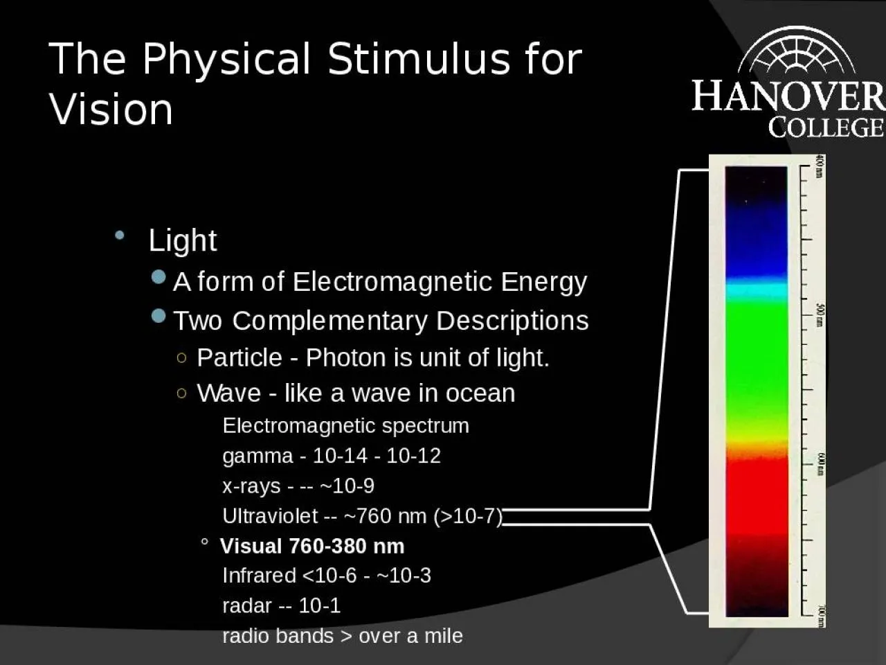
Light A form of Electromagnetic Energy Two Complementary Descriptions Particle Photon is unit of light Wave like a wave in ocean Electromagnetic spectrum gamma 1014 1012 xrays 109 ID: 911605

vision color contrast depth color vision depth contrast eye light sensitivity acuity luminance bars matching cones visual size object

Share:

Link:

Embed:

Download Presentation from below link

Download Presentation The PPT/PDF document "The Physical Stimulus for Vision" is the property of its rightful owner. Permission is granted to download and print the materials on this web site for personal, non-commercial use only, and to display it on your personal computer provided you do not modify the materials and that you retain all copyright notices contained in the materials. By downloading content from our website, you accept the terms of this agreement.


Presentation Transcript

Slide1

The Physical Stimulus for Vision

LightA form of Electromagnetic EnergyTwo Complementary DescriptionsParticle - Photon is unit of light.Wave - like a wave in oceanElectromagnetic spectrumgamma - 10-14 - 10-12x-rays - -- ~10-9Ultraviolet -- ~760 nm (>10-7)° Visual 760-380 nmInfrared <10-6 - ~10-3radar -- 10-1radio bands > over a mile

Slide2

Measures of Light

Wavelength relates to color, e.g., the spectrumUsually measured in billionths of a meter - nanometers or nm

Slide3

Measures of Light - 2

Amplitude = Intensity relates to brightnessMeasures use only that light that is effective in stimulating the human eye.Important types of measures of Amplitude1. Illuminance - light falling on a surface2. Luminance - light coming off a surface3. Reflectance = luminance/illuminance4. Contrast Ratio = luminance of brightest area/luminance of darkest area

Slide4

Structures of the Eye

1. Sclera2. Cornea3. Aqueous Humor4. Iris5. Pupil6. Lens7. Ciliary muscle8. Retina

9. fovea (pit)10. blind spot/optic disc

11. Pigment Epithelium

Slide5

The Retina

Two Types of ReceptorsRods~120 million/eyenight visionno colornot in fovea most about 20deg in peripheryCones~7 million/eyeday visionthree types so color visionmost in foveafrom Lewis, Zeevi, & Werblin (1969)

Slide6

The Retina - 2

RodsCones

Data from

Osterberg (1935).

Slide7

Simultaneous

Contrast

Slide8

Mach Bands

Click to add successively lighter bars. Watch the edge to

the right side of the last bar

.

Slide9

Craik-Cornsweet

: Filling In

Slide10

Craik-Cornsweet: Described

The figure above is an exaggerated map indicating the light levels across the image on the previous slide. Note how the center and edges have identical luminance. That can be seen by sitting far enough away from the screen

Slide11

Minimal Contours

Slide12

Minimal Contours Described

There are two circles below. Both circles have the same luminance

(intensity level) at the center. Click on your mouse and

This one changes abruptly watch as the edges are blurred

to the level at the center. and the circle disappears.

Slide13

Accommodation

DEFINITION: The adjusting of the lens thickness to focus at different distances.Necessary because can only see clearly one distance at a timeGoes very rapidlyCloses can focus in Near PointFarthest can focus is Far PointLoose ability to focus as age - moves towards far pointIn dark accommodation moves to ~1 meter from faceAs fatigue, accommodation moves to this dark focus.

Slide14

Acuity

DEFINITION: ability to resolve or see fine details.Visual Angle: DEFINITION: Angle formed by object on retina.Types of Acuity: what is meant by acuity depends upon the stimulus used to measure it.Detection: black bar on white fieldResolution: a gratingRecognition: e.g. Snellen, where you read letters.

Slide15

Acuity - 2

Measures of Acuity20/20: can see at 20’ what a normal person can see at 20’.This is normal, not perfect, vision. 20/200: can see at 20’ what a normal person can see at 200’.Visual angle of the critical feature in a test, e.g. the width of the bars in a grating. A typical population average is 1 arcmin (1/60 degree).Acuity and Retinal Location:Best at fovea. Falls off rapidly in periphery. Is tied to density of cones.

Slide16

Contrast Sensitivity

DEFINITION: the minimum contrast ratio that can be detected.Indicates the smallest difference between shades of gray that can be detected.Depends on Spatial FrequencyDEFINITION: how many pairs of white and black bars fit into 1 deg. of visual angle.Low spatial frequencyfew bars/deg.High spatial frequencymany bars/deg.

Slide17

The Contrast Sensitivity Function

Our sensitivity to contrast depends on this spatial frequency.Peak sensitivity is 4-6 cycles/degree.The highest spatial frequency we can see at any contrast is limited by our acuity.

Slide18

Contrast Sensitivity and Performance

Increasing contrast above threshold will allow for faster identification, up to a pointBeyond a certain contrast ratio - about 3 to 1 or 4 to 1 - increasing contrast ratio has no effect (Krantz, Silverstein, & Yeh, 1992)

Slide19

Fundamental Limits of Vision

Operating Range of Vision - 1:1014if: 1 cent (threshold) =100 Most incomes are between $10,000 and $100,000 = 106 to 107 GNP of U.S.A. for 1 year = ~1015

Slide20

Fundamental Limits of Vision - 2

Duplex Theory of VisionTwo eyes in one. One for day. One for night.

Slide21

Spectral Sensitivity

Slide22

Dark/Light

AdaptationDark AdaptationThe term applied to the increased sensitivity that occurs when we enter a region of lowered sensitivity.Two phases:early rapid phase - lasts ~7 minutes and due to cones.later slower phase - complete in about 30 to 40 minutes due to rods.Light Adaptation Takes 2-3 minutes

Slide23

So Why are Blue Headlights Stupid?

Slide24

So Why do Nighttime Briefings in Red Light?

Slide25

Eye Movements

We move our eyes because of the limited field with good acuity.There are 6 eye muscles4 rectus2 obliqueTypes of Eye MovementsVersion: Both eyes move togetherVergence: Eye move in opposite directionsfrom Kaiser, 1997, http://www.yorku.ca/research/vision/eye/thejoy.htm

Slide26

Eye Movements - 2

VersionSaccades, most common (link to ESP)places object on fovea.can be > 400 deg/sec.Takes ~ 200 msec to beginSmooth Pursuittrack moving objectsrelatively slow ~30 deg/sec.Vergence: Convergence (together) and divergence (apart)Allows us to look at closer and farther objects.relatively slow and also takes about 200 msec to begin

Slide27

Color Vision I: Color Matching

Dimensions of ColorHue: refers to the color name we apply.Saturation: purity of color, to desaturate add whiteBrightness: Trichromatic Theory of VisionWe have three classes of conesL or red: peak at 564 nmM or green: peak at 534 nmS or blue: peak at 420 nm

Slide28

Color Vision I: Color Matching - 2

Color Matching in the Trichromatic TheoryTwo patches of light will appear the same if the activity across the three cones is the same, regardless of wavelengths making up the two patches.In general, can match any one color with three other colorsThis is where we get three primaries.Neutral colors - whites and graysa balance of activity across the three receptors.

Slide29

Color Vision I: Color Matching - 3

Formalization of the Trichromatic TheoryBy CIE originally in 1931Based on 300 observers to develop standard observer.A set of Equations that allow predictions of matching.Used in photo printing, TV and film.Updates in 1960, 1976

Slide30

Color Vision II:

Color DeficienciesMost can be understood using Trichromatic TheoryDichromatism: Missing one of the three cones (link)Dichromats tend to see through camouflage better than thrichromats

Other TypesMonochromatism: One cone or only rods

Anomalous Trichromats: Three cones but one is different.

Slide31

Color Vision III: Color Appearance

Color Opponent TheoryFour Primaries: red-green, blue-yellowArranged in opposition pairsRed vs. GreenBlue vs. YellowAdd on to other get neutral colorNever see a mixture of opposition pairs.Evidence:complimentary colors, color aftereffect, simultaneous contrast, color naming - try it with just red, green blue and yellowCells in visual system respond this way.

Slide32

Color Aftereffect

Slide33

Test Color Aftereffect

Slide34

Color Vision IV: Resolution

modified from Kaiser, 1997, http://www.yorku.ca/research/vision/ eye/thejoy.htm

Slide35

Depth Perception

If retinas are flat (2 dimensional) how do we see depth (the 3rd dimension)?We use cues: sources of information about depth.Monocular or one eye cues

Slide36

Depth Perception - 2

Binocular or two eye cuesVergence (Only cue to give absolute depth information): muscular feedback from effort to converge or diverge gives information about depth.works only for relatively near objects: <20’Stereopsis (link)Binocular Disparity: measure of difference of position of an object on the two retinasDEFINITION: ability to use binocular disparity to see depth.Basis of 3-D moviesAccuracy of Depth Judgments:In general, more cues more accurate.

Slide37

Depth Perception - 3

Size ConstancyDEFINITION: seeing objects as a relatively constant size despite change in retinal image size.Sretinal image a 1/distance to object (a mean proportional to)Sperceived = ConstantCan be quite useful in object recognitionA Variation is Emmert’s Law for after effectsSretinal image = Constant

Sperceived a distanceAlso applies to depth generated by stereopsis

Slide38

Depth Perception - 4

To experience Emmert’s Law fixate the center of the dot below for about 45 seconds. Then quickly view the next slide and note the size. Then look at surfaces of different distances, also noting the size.

Slide39

Depth Perception - 5