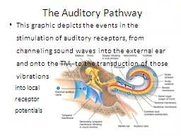
/
 The  Auditory Pathway This graphic depicts the events in the stimulation of auditory  The  Auditory Pathway This graphic depicts the events in the stimulation of auditory

The Auditory Pathway This graphic depicts the events in the stimulation of auditory - PowerPoint Presentation

mitsue-stanley
mitsue-stanley . @mitsue-stanley
Follow
350 views
Uploaded On 2020-04-02

The Auditory Pathway This graphic depicts the events in the stimulation of auditory - PPT Presentation

into local receptor potentials Scala vestibuli Cochlear duct contains endolymph Scala tympani Perilymph Basilar membrane Cochlea Sound waves Helicotrema Stapes vibrating in oval window ID: 774681

membrane auditory organ window membrane auditory organ window scala vibrating tympanic ear cochlear sound vestibular waves external tectorial spiral

Share:

Link:

Embed:

Download Presentation from below link

Download Presentation The PPT/PDF document " The Auditory Pathway This graphic depi..." is the property of its rightful owner. Permission is granted to download and print the materials on this web site for personal, non-commercial use only, and to display it on your personal computer provided you do not modify the materials and that you retain all copyright notices contained in the materials. By downloading content from our website, you accept the terms of this agreement.


Presentation Transcript

Slide1

The

Auditory Pathway

This graphic depicts the events in the stimulation of auditory receptors, from channeling sound waves into the external ear and onto the TM, to the transduction of those vibrations

into local

receptor

potentials

Slide2

Scala

vestibuli

Cochlear duct

(contains endolymph)

Scalatympani

Perilymph

Basilarmembrane

Cochlea

Sound waves

Helicotrema

Stapes vibratingin oval window

Malleus

Incus

External auditorycanal

Tympanicmembrane

Secondary tympanicmembrane vibratingin round window

Auditory tube

Vestibular membrane

Middle ear

Tectorial membrane

Spiral organ(organ of Corti)

1

Scala

vestibuli

Cochlear duct

(contains endolymph)

Scala

tympani

Perilymph

Basilar

membrane

Cochlea

Sound waves

Helicotrema

Stapes vibrating

in oval window

Malleus

Incus

External auditory

canal

Tympanic

membrane

Secondary tympanic

membrane vibrating

in round window

Auditory tube

Vestibular membrane

Middle ear

Tectorial membrane

Spiral organ

(organ of Corti)

1

2

Scala

vestibuli

Cochlear duct

(contains endolymph)

Scala

tympani

Perilymph

Basilar

membrane

Cochlea

Sound waves

Helicotrema

Stapes vibrating

in oval window

Malleus

Incus

External auditory

canal

Tympanic

membrane

Secondary tympanic

membrane vibrating

in round window

Auditory tube

Vestibular membrane

Middle ear

Tectorial membrane

Spiral organ

(organ of Corti)

1

2

3

Scala

vestibuli

Cochlear duct

(contains endolymph)

Scala

tympani

Perilymph

Basilar

membrane

Cochlea

Sound waves

Helicotrema

Stapes vibrating

in oval window

Malleus

Incus

External auditory

canal

Tympanic

membrane

Secondary tympanic

membrane vibrating

in round window

Auditory tube

Vestibular membrane

Middle ear

Tectorial membrane

Spiral organ

(organ of Corti)

1

2

3

4

Scala

vestibuli

Cochlear duct

(contains endolymph)

Scala

tympani

Perilymph

Basilar

membrane

Cochlea

Sound waves

Helicotrema

Stapes vibrating

in oval window

Malleus

Incus

External auditory

canal

Tympanic

membrane

Secondary tympanic

membrane vibrating

in round window

Auditory tube

Vestibular membrane

Middle ear

Tectorial membrane

Spiral organ

(organ of Corti)

1

2

3

4

5

Scala

vestibuli

Cochlear duct

(contains endolymph)

Scala

tympani

Perilymph

Basilar

membrane

Cochlea

Sound waves

Helicotrema

Stapes vibrating

in oval window

Malleus

Incus

External auditory

canal

Tympanic

membrane

Secondary tympanic

membrane vibrating

in round window

Auditory tube

Vestibular membrane

Middle ear

Tectorial membrane

Spiral organ

(organ of Corti)

1

2

3

4

5

6

Scala

vestibuli

Cochlear duct

(contains endolymph)

Scala

tympani

Perilymph

Basilar

membrane

Cochlea

Sound waves

Helicotrema

Stapes vibrating

in oval window

Malleus

Incus

External auditory

canal

Tympanic

membrane

Secondary tympanic

membrane vibrating

in round window

Auditory tube

Vestibular membrane

Middle ear

Tectorial membrane

Spiral organ

(organ of Corti)

1

2

3

4

5

6

7

Scala

vestibuli

Cochlear duct

(contains endolymph)

Scala

tympani

Perilymph

Basilar

membrane

Cochlea

Sound waves

Helicotrema

Stapes vibrating

in oval window

Malleus

Incus

External auditory

canal

Tympanic

membrane

Secondary tympanic

membrane vibrating

in round window

Auditory tube

Vestibular membrane

Middle ear

Tectorial membrane

Spiral organ

(organ of Corti)

1

2

3

4

5

6

7

8

8

Scala

vestibuli

Cochlear duct

(contains endolymph)

Scala

tympani

Perilymph

Basilar

membrane

Cochlea

Sound waves

Helicotrema

Stapes vibrating

in oval window

Malleus

Incus

External auditory

canal

Tympanic

membrane

Secondary tympanic

membrane vibrating

in round window

Auditory tube

Vestibular membrane

Middle ear

Tectorial membrane

Spiral organ

(organ of Corti)

1

2

3

4

5

6

7

8

8

9

Slide3

The Auditory Pathway

The cell bodies of the sensory neurons are located in the

spiral ganglia.

Nerve impulses pass along the axons of these neurons, which

form the cochlear

branch of the

vestibulo-

cochlear (VIII)

nerve

Slide4

The Auditory Pathway

The nerve impulses follow CN VIII en route to the medulla, pons, midbrain, and thalamus, and finally to the primary auditory cortex in the temporal lobe. Slight differences in the timing of nerve

impulses arriving from the

two ears at the superior

olivary nuclei in the

pons allow us to

locate the source

of a sound

Slide5

Equilibrium

Equilibrium is another function of the inner ear - controlled by the

vestibular apparatus

(the saccule and utricle of the vestibule, and the 3 semicircular canals)

Static equilibrium

refers to a

state of balance relative to

the force of gravity

Dynamic equilibrium

involves the maintenance

of balance during sudden movements

Slide6

Static Equilibrium

Static equilibrium is controlled by the

sensory hairs within the macula of the utricle and saccule

Slide7

The Inner Ear

Movements of the hair cells in contact with the tectorial membrane

transduce mechanical vibrations

into electrical signals which generate nerve impulses along the cochlear branch of CN VIII

Slide8

Static Equilibrium

An

otolithic membrane,

studded with dense calcium carbonate crystals (otoliths), responds to gravity when head position is changed

This movement opens

transduction channels

in the hair cells,

producing local potentials

which summate to

form nerve AP