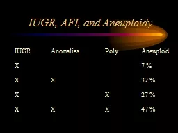
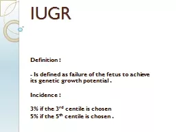
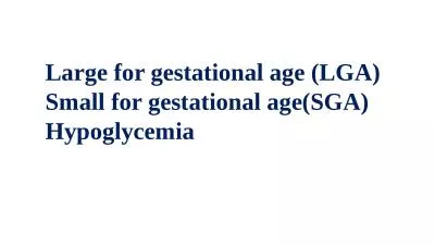
Phases of fetal growth First 16 weeks mostly cellular hyperplasia 1632 weeks both hyperplasia and hypertrophy gt32 weeks mostly hypertrophy Thus early growth restriction will affect cell numbers and have a global symmetrical IUGR effect Later cell size will be affected asymme ID: 929304
Download Presentation The PPT/PDF document "IUGR Definition : Is a fetal weight that..." is the property of its rightful owner. Permission is granted to download and print the materials on this web site for personal, non-commercial use only, and to display it on your personal computer provided you do not modify the materials and that you retain all copyright notices contained in the materials. By downloading content from our website, you accept the terms of this agreement.
Slide1
IUGR
Slide2Definition
:
Is a fetal weight that is below the 10th percentile for gestational age as determined through an ultrasound. This can also be called small-for gestational age (SGA) or fetal growth restriction.
Slide3Slide4Phases
of fetal growth
First 16 weeks:
mostly cellular hyperplasia
16-32 weeks:
both hyperplasia and hypertrophy
>32 weeks:
mostly hypertrophy
Thus: early growth restriction will affect cell numbers and have a global (symmetrical IUGR) effect. Later cell size will be affected (asymmetrical IUGR)
Etiology of IUGR
Maternal Factors
Placental Factors
Fetal Factors
Slide7Maternal Factors
1. Genetic size
2. Demographics:
Age (extremes of reproductive age)
Socioeconomic status
3. Underweight before pregnancy or malnutrition
4. Exposure to teratogens (drugs, radiation, etc.)
Slide85
. Factors that interfere with placental flow and function:
-Heart disease
-Renal disease
-Hypertension
-Pulmonary disease
-
Hemoglobinopathies
-Collagen-vascular disease
-Diabetes
-
Postmaturity
Slide9-
Multiple gestation
-Uterine anomalies
-Thrombotic disease
-High altitude environment
-Smoking
-Cocaine
Slide10Placental
Factors
-Malformations
-Abruption
-Previa
-Abnormal trophoblast invasion
Slide11Fetal
Factors
-Constitutional – genetically small, but genetically normal
-Chromosomal abnormality – only about 5% of SGA babies
-Malformations – CNS, skeletal
-Congenital infections – CMV, rubella
Slide12Characteristics of IUGR
symmetric:
1. Early onset
2. Head circumference, length &weight are all proportionally reduced for gestational age(below 10
th
percentile)
3. Could be constitutional or normal small.
4. Examples:
-Genetic causes, chromosomal
-TORCH infections
Slide13Asymmetrical
:
1. Late onset 2
nd
-3
rd
trimester.
2. Growth arrest.
3.Brain sparing ,which means transfer of oxygen & nutrients to the brain .This allows normal brain and head growth while diminished glucose transfer and hepatic storage would primarily affect cell size and not number, and fetal abdominal circumference—which reflects liver
size—would be reduced.
Slide144
. Examples:
-
Chronic hypoxia
-Preeclampsia, chronic hypertension
-malnutrition
Diagnosis
of IUGR
Abdominal examination:
Serial Fundal height measurement in centimeters usually corresponds with the number of weeks of pregnancy after the 20th week. If the measurement is low for the number of weeks (2-3 cm), the baby may be smaller than expected.
Slide17Ultrasound
more accurate method of estimating fetal size. Measurements can be taken of the fetus' head and abdomen and compared with a growth chart to estimate fetal weight. The fetal abdominal circumference is a helpful indicator of fetal nutrition. , obtaining a second growth assessment over a 2- to 4-week interval is important unless strong supportive data or risk factors warrant an immediate change in management plans
.
Slide18Amniotic
fluid volume
An association between pathological fetal-growth restriction and oligohydramnios has long been recognized. Hypoxia and diminished renal blood flow explain oligohydramnios
Slide19Slide20Doppler
flow
Doppler velocimetry is considered standard in the evaluation
and management of the growth-restricted fetus. abnormal umbilical artery Doppler velocimetry
findings—characterized by absent or reversed end-diastolic
flow—have been uniquely linked with fetal-growth restriction. Other
modality of Doppler
velocimetry, include middle cerebral arteries & ductus
venosus
assessment
Slide21Slide22MANAGEMENT
Slide23Important
points regarding managements:
1.If fetal-growth restriction is suspected, then efforts are made
to confirm the diagnosis, assess fetal condition, and search for
possible causes.
2. antepartum fetal surveillance should include Daily fetal heart rate tracings, weekly Doppler velocimetry, and sonographic assessment
of fetal growth every 3 to 4 weeks are initiated
Slide243
. growth-restricted fetuses
may not
tolerate the metabolic effects of corticosteroids in the
same way
as an unstressed fetus, increased surveillance
during administration.
4. timing of delivery is crucial, and the risks of fetal death
versus the hazards of preterm delivery must be considered.
5. delivery between 34
and 37
weeks when there are concurrent conditions such as oligohydramnios. With a reassuring fetal heart rate pattern,
vaginal delivery
is planned
Slide25However
, some of these fetuses do not tolerate labor, necessitating cesarean
delivery,why
?
Answer:
- Fetal-growth restriction is commonly the result of placental
insufficiency , this condition is likely aggravated by labor
- diminished
amnionic
fluid volume increases the likelihood of cord compression during labor
Slide26Slide27Early
neonatal morbidities in IUGR
Birth asphyxia
Meconium aspiration
Hypoglycemia
Hypocalcemia
Hypothermia
Polycythemia,hyperbilirubiemia
Slide28Thrombocytopenia
Pulmonary
haemorrhage
Malformation
Sepsis
Respiratory distress syndrome
Necrotizing
enterocolitis
.
Later in life
Hypertension and atherosclerosis.
Slide29