/
X-rays;   Scattered from electrons X-rays;   Scattered from electrons

X-rays; Scattered from electrons - PowerPoint Presentation

motivatorprada
motivatorprada . @motivatorprada
Follow
342 views
Uploaded On 2020-11-06

X-rays; Scattered from electrons - PPT Presentation

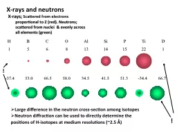
proportional to Z red Neutrons scattered from nuclei amp evenly across all elements green H B C O Al Si P Ti D 1 5 6 8 13 14 15 22 1 ID: 816151

ladi neutron lobster cianci neutron ladi cianci lobster neutrons source amp crustacyanin final protonation concanavalin heartily upgrade resolving sources

Share:

Link:

Embed:

Download Presentation from below link

Download The PPT/PDF document "X-rays; Scattered from electrons" is the property of its rightful owner. Permission is granted to download and print the materials on this web site for personal, non-commercial use only, and to display it on your personal computer provided you do not modify the materials and that you retain all copyright notices contained in the materials. By downloading content from our website, you accept the terms of this agreement.


Presentation Transcript

Slide1

X-rays;

Scattered from electrons proportional to Z (red). Neutrons; scattered from nuclei & evenly across all elements (green)

H B C O Al Si P Ti D 1 5 6 8 13 14 15 22 1

-37.4 53.0 66.5 58.0 34.5 41.5 51.3 -34.4 66.7

X-rays and neutrons

Large difference in the neutron cross-section among isotopes

Neutron diffraction can be used to directly determine the positions of H-isotopes at medium resolutions (~2.5 Ǻ)

!

!

Slide2

Concanavalin

A studied with small molecule accuracy at 0.94Å resolution captures the Chemists’ imagination

Ashley Deac

on et al Faraday Transactions, 93 (24), 4305–4312.Diffraction data measured at MacCHESS on their MPW.

Resolving protonation states

James Raftery

Slide3

Resolving histidine protonation states with neutrons; concanavalin A at LADI

Based on data measured at the Institut Laue Langevin LAue

DIffractometer

LADI courtesy of the LADI Instrument responsible Dr Matthew BlakeleyFor a review see Blakeley Crystallography Reviews Vol. 15, 2009, 157–218Neutron macromolecular crystallography

Slide4

Cianci

et al (2001) Acta

Cryst

D57, 1219 “Structure of apocrustacyanin A1

using softer X-rays”

Naomi Chayen

Mike Cianci

β-Crustacyanin

crystalApocrustacyanin A1

Cianci et al (2002) PNAS USA 99, 9795

‘The molecular basis of the coloration mechanism in lobster shell:

β-crustacyanin

at 3.2 A resolution’

A final highlight Marine colouration in lobster shell

.

Slide5

Lobster crustacyanin hits the media! starting from work at SRS in the early 2000s

From the Liverpool Daily Post but articles also in The Times,

The Guardian, The Independent also Radio, TV, Scientific American,

New Scientist, Physics Today etc.

Slide6

Research directions for today?New SR sources: PETRA III the most brilliant ie

the ‘ultimate storage ring’ ; also soon NSLS II, MAX IVDiamond Light Source is a cutting edge source for UK with a vibrant research and development programmeALBA likewise is a cutting edge SR source for Spain

ESRF has a new upgrade programme for

nanofocus beams; also APS at advanced planning with its upgrade The X-ray laser: towards nanoclusters of molecules New neutron sources such as the SNS and ESS

Slide7

Final acknowledgementsI heartily thank my PhD students and PostDocs

over the last 35 years Daresbury Laboratory & the Universities of York, Oxford, Keele & Manchester

I heartily thank

all the SR and neutron facilities for their collaborationI especially thank my wife Dr Madeleine

Helliwell

Slide8

Thank you