/
Color what we know: Color what we know:

Color what we know: - PowerPoint Presentation

myesha-ticknor
myesha-ticknor . @myesha-ticknor
Follow
387 views
Uploaded On 2016-03-03

Color what we know: - PPT Presentation

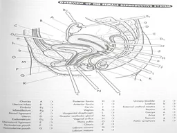
Grey Urethra b drains bladder Brown Rectum d fecal storage Anus f expels fecal matter Black Urinary bladder a stores urine Yellow Coccyx e aka tail bone of vertebrae Femal e Reproductive System ID: 240397

labia color amp uterus color labia uterus amp pubis minoria perineum glands structures reproductive female fallopian tubes bone yellow

Share:

Link:

Embed:

Download Presentation from below link

Download Presentation The PPT/PDF document "Color what we know:" is the property of its rightful owner. Permission is granted to download and print the materials on this web site for personal, non-commercial use only, and to display it on your personal computer provided you do not modify the materials and that you retain all copyright notices contained in the materials. By downloading content from our website, you accept the terms of this agreement.


Presentation Transcript

Slide1
Slide2

Color what we know:

Grey

Urethra (b) drains bladder

Brown

Rectum (d) fecal storage

Anus (f) expels fecal matter

Black

Urinary bladder (a) stores urine

Yellow

Coccyx (e) aka tail bone of vertebraeSlide3

Femal

e Reproductive System

Internal Structures (Female Gonads( Consists

of

:OvariesFallopian tubesUterusVaginaBartholin’s glandsSlide4

Ovaries

Almond shape

Located in the pelvic cavity & attached to the uterus by ligaments

Q 28d,

an ovum is produced (ovulation).Produces hormones that aid in the development of the reproductive organs and produce secondary sexual characteristics.Color the ovaries (A) pink.Slide5
Slide6

Fallopian Tubes

About 4 inches long

Attached

to the upper part of the uterus and above the ovaries.

Serves as a passageway for the ovum (ovary to the uterus).Movement by peristalsis & ciliaFertilization usually takes place in the Fallopian tubes.Color the fallopian tubes (B) greenSlide7
Slide8

Uterus

A hollow, muscular, pear-shaped organ located behind the bladder.

2

sections: Body, Cervix.Allows for the development and growth of the fetusContracts to aid in expulsion of the fetus during birthInner layer of the uterus – EndometriumImplantation & aids in the development of the fetusMenstruation

Color the Uterus (D) red

Color the Cervix (J) orangeSlide9
Slide10

Vagina

A muscular tube that connects the cervix of the uterus to the outside of the body.

Functions:

Passageway for the menstrual flow

Receives sperm and semenBirth Canal during delivery of an infantBartholin’s Glands

Located on B sides of the vagina

Secrete

mucous for lubrication during intercourseSlide11
Slide12

Femal

e Reproductive System

External

Structures (Vulva) Consists

of :Mons PubisSymphysis PubisLabia MajoraLabia Minora

Clitoris

PerineumSlide13

Mons Pubis

Pad of fatty tissue that lies over the lower portion of the pelvic bone.

Color the Mons Pubis (O) purple

Symphysis Pubis

Bone on the lower portion of the pelvisColor the symphysis pubis yellowSlide14

Labia

Majoria

2

large fold of fatty

tissue that contain glandsColor the labia majoria (R) light blueLabia

Minoria

2

smaller hairless folds

that join the labia

majoria

anteriorly

Color the labia

minoria

(Q) dark blueSlide15

Vestibule

Area

of the vulva

between

the labia minoria and clitorisColor this area with a pink highlighter and include it on the list of structures on your coloring sheet.ClitorisErectile tissue that is just behind the anterior juncture of the labia

minoria

Color the

clitoris (P) with a yellow highlighter.

Perineum

: between vagina & anus

Mammary Glands

To secrete milk (lactate) after childbirth.Slide16

Perineum

Between vaginal opening & the anus

Add the perineum to your list of structures on the coloring sheet, highlight the perineum green.