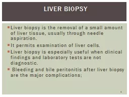
/
Liver biopsy is the removal of a small amount of liver tiss Liver biopsy is the removal of a small amount of liver tiss

Liver biopsy is the removal of a small amount of liver tiss - PowerPoint Presentation

myesha-ticknor
myesha-ticknor . @myesha-ticknor
Follow
425 views
Uploaded On 2017-10-24

Liver biopsy is the removal of a small amount of liver tiss - PPT Presentation

It permits examination of liver cells Liver biopsy is especially useful when clinical findings and laboratory tests are not diagnostic Bleeding and bile peritonitis after liver biopsy are the major complications ID: 598946

fluid liver jaundice patient liver fluid patient jaundice blood ascites procedure pressure biopsy bilirubin bile patients results sodium obstruction

Share:

Link:

Embed:

Download Presentation from below link

Download Presentation The PPT/PDF document "Liver biopsy is the removal of a small a..." is the property of its rightful owner. Permission is granted to download and print the materials on this web site for personal, non-commercial use only, and to display it on your personal computer provided you do not modify the materials and that you retain all copyright notices contained in the materials. By downloading content from our website, you accept the terms of this agreement.


Presentation Transcript

Slide1

Liver biopsy is the removal of a small amount of liver tissue, usually through needle aspiration. It permits examination of liver cells. Liver biopsy is especially useful when clinical findings and laboratory tests are not diagnostic. Bleeding and bile peritonitis after liver biopsy are the major complications;

1

LIVER BIOPSYSlide2

2LIVER BIOPSYSlide3

PREPROCEDURE1.Ascertain that results of coagulation tests (prothrombin time, partial thromboplastin time, and platelet count) are available and that compatible donor blood is available.

2. Check for signed consent; confirm that informed consent has been provided.

1. Many patients with liver disease have clotting defects and are at risk for bleeding.

2. Procedure should be done with agreement of patient

3

NURSING ACTIVITIES RATIONALESlide4

3. Measure and record the patient’s pulse, respirations, and blood pressure immediately before biopsy.4. Describe to the patient in advance: steps of the procedure;

3. Pre-biopsy values provide a basis on which to compare the patient’s vital signs and evaluate status after the procedure.

4. Explanations allay fears and ensure cooperation.

4Slide5

5. Support the patient during the procedure.6. Expose the right side of the patient’s upper abdomen (right hypochondriac).7. Instruct the patient to inhale and exhale deeply several times, finally to exhale, and to hold breath at the end of expiration. The physician promptly introduces the biopsy needle by way of the transthoracic

(intercostal) transabdominal

(

subcostal

) route, penetrates the liver, aspirates, and withdraws.

Encouragement and support of the nurse enhance comfort and promote a sense of security.

The skin at the site of penetration will be cleansed and a local anesthetic will be infiltrated.

Holding the breath immobilizes the chest wall and the diaphragm; penetration of the diaphragm thereby is avoided, and the risk of lacerating the liver is minimized.5DURING PROCEDURESlide6

9. Immediately after the biopsy, assist the patient to turn onto the right side; place a pillow under the costal margin, and caution the patient to remain in this position and immobile, for several hours. Instruct the patient to avoid coughing or straining.10. Measure and record the patient’s vital sings at a15-minute intervals for the first hour, then every 30 minutes for the next 1 to 2 hours or until the patient’s condition stabilizes.

Complications:PneumothoraxPeritonitis

Hemorrhage

6

POSTPROCEDURESlide7

Ultrasonography, computed tomography (CT), and magnetic resonance imaging (MRI) are used to identify normal structures and abnormalities of the liver and biliary tree. A radioisotope liver scan may be performed to assess liver size and hepatic blood flow and obstruction. Laparoscopy (insertion of a fiber-optic endoscope through a small abdominal incision) is used to examine the liver and other pelvic structures.

7

OTHER DIAGNOSTIC TESTSSlide8

Results from damage to the liver’s parenchymal cells, either directly from primary liver diseases or indirectly from obstruction of bile flow. Liver dysfunction may be acute or chronic; chronic dysfunction is more common than acute.The most common cause of

parenchymal damage is malnutrition, especially that related to alcoholism.

The

parenchymal

cells respond to most noxious agents by replacing glycogen with lipids, producing fatty infiltration with or without cell death or necrosis.

This is commonly associated with inflammatory cell infiltration and growth of fibrous tissue.

8

Hepatic DysfunctionSlide9

Jaundice, resulting from increased bilirubin concentration in the blood Portal hypertension, ascites, and

varices, resulting from circulatory changes within the diseased liver and producing severe GI hemorrhages and marked sodium and fluid retention

Nutritional deficiencies, which result from the inability of the damaged liver cells to metabolize certain vitamins;

Hepatic encephalopathy or coma, reflecting accumulation of ammonia in the serum due to impaired protein metabolism by the diseased liver

9

Among the most common and significant symptoms of liver disease are the following:Slide10

When the bilirubin concentration in the blood is abnormally elevated, all the body tissues, including the sclera and the skin, become yellow-tinged or greenish-yellow, a condition called jaundice.becomes clinically evident when the serum bilirubin

level exceeds 2.5 mg/dL.

There are several types of jaundice:

Hemolytic

Hepatocellular

Obstructive

.

10JAUNDICESlide11

Results from increased destruction of the red blood cells, too much bilirubin reaches the liver, although functioning normally, cannot excrete the bilirubin as quickly as it is formed. Occurs with patients with hemolytic transfusion reactions and other hemolytic disorders.

Prolonged jaundice, however, even if mild, predisposes to the formation of pigment stones in the gallbladder, and extremely severe jaundice (levels of free bilirubin exceeding 20 to 25 mg/

dL

) poses a risk for brain stem damage.

11

Hemolytic JaundiceSlide12

Caused by the inability of damaged liver cells to clear normal amounts of bilirubin from the blood. The cellular damage may be from infection, such as in viral hepatitis or other viruses that affect the liver (eg, yellow fever virus, Epstein-Barr virus), from medication or chemical toxicity (eg, carbon tetrachloride, chloroform, phosphorus, certain medications), or from alcohol.

Cirrhosis of the liver is a form of hepatocellular disease that may produce jaundice. It is usually associated with excessive alcohol

12

Hepatocellular JaundiceSlide13

Caused by occlusion of the bile duct by a gallstone, an inflammatory process, a tumor, or pressure from an enlarged organ. The obstruction may also involve the small bile ducts within the liver (ie, intrahepatic obstruction), caused, for example, by pressure on these channels from inflammatory swelling of the liver or by an inflammatory

exudate within the ducts themselves. Intrahepatic

obstruction resulting from stasis and

inspissation

(thickening) of bile within the

canaliculi

may occur after the ingestion of certain medications.13Obstructive JaundiceSlide14

Examples for medications: phenothiazines, antithyroid, sulfonylureas, tricyclic antidepressant agents, nitrofurantoin

, androgens, and estrogens.

Whether the obstruction is

intrahepatic

or

extrahepatic

, and whatever its cause may be, bile cannot flow normally into the intestine but is backed up into the liver substance. It is then reabsorbed into the blood and carried throughout the entire body, staining the skin, mucous membranes, and

sclerae. It is excreted in the urine, which becomes deep orange and foamy. 14Obstructive JaundiceSlide15

Because of the decreased amount of bile in the intestinal tract, the stools become light or clay-colored. The skin may itch intensely, requiring repeated soothing baths. Dyspepsia and intolerance to fatty foods may develop because of impaired fat digestion in the absence of intestinal bile. AST, ALT, and GGT levels generally rise only moderately, but bilirubin and alkaline

phosphatase levels are elevated.

15

Obstructive JaundiceSlide16

Results from several inherited disorders can also produce jaundice. Gilbert’s syndrome is a familial disorder characterized by an increased level of unconjugated bilirubin that causes jaundice. Other conditions that are probably caused by inborn errors of biliary metabolism include

Dubin–Johnson syndrome (chronic idiopathic jaundice, with pigment in the liver) and Rotor’s syndrome (chronic familial conjugated hyperbilirubinemia

without pigment in the liver);

16

Hereditary

HyperbilirubinemiaSlide17

Obstructed blood flow through the damaged liver results in increased blood pressure (portal hypertension) throughout the portal venous system. It is commonly associated with hepatic cirrhosis, but can also occur with noncirrhotic liver disease.

Portal hypertnesion leads to:

Splenomegaly

(enlarged spleen)

Ascites

Varices

.17PORTAL HYPERTENSIONSlide18

18III. ASCITESSlide19

Caused by portal hypertension and the resulting increase in capillary pressure and obstruction of venous blood flow through the damaged liver. The failure of the liver to metabolize aldosterone increases sodium and water retention by the kidney. Sodium and water retention, increases intravascular fluid volume, and decreased synthesis of albumin by the damaged liver all contribute to fluid moving from the vascular system into the peritoneal space

19

ASCITES -

PathophysiologySlide20

Loss of fluid into the peritoneal space causes further sodium and water retention by the kidney in an effort to maintain the vascular fluid volume, and the process becomes self-perpetuating. As a result of liver damage, large amounts of albumin-rich fluid, 15 L or more, may accumulate in the peritoneal cavity as ascites. With the movement of albumin from the serum to the peritoneal cavity, the osmotic pressure of the serum decreases.

This, combined with increased portal pressure, results in movement of fluid into the peritoneal cavity.

20Slide21

21Slide22

Increased abdominal girth and rapid weight gain are common presenting symptoms of ascites. The patient may be short of breath and uncomfortable from the enlarged abdomen, and Striae and distended veins may be visible over the abdominal wall. Fluid and electrolyte imbalances are common.

22

Clinical ManifestationsSlide23

The presence and extent of ascites are assessed by percussion of the abdomen. When fluid has accumulated in the peritoneal cavity, the flanks bulge when the patient assumes a supine position. The presence of fluid can be confirmed either by percussing for shifting dullness or by detecting a fluid wave. Daily measurement and recording of abdominal girth and body weight are essential to assess the progression of ascites and its response to treatment.

23

Assessment and Diagnostic EvaluationSlide24

24Slide25

The goal of treatment for the patient with ascites is a negative sodium balance to reduce fluid retention. Table salt, salty foods, salted butter and margarine, and all ordinary canned and frozen foods should be avoided. In the meantime, the taste of unsalted foods can be improved by using salt substitutes such as lemon juice, oregano, and thyme.

25

Medical Management

A)

DIETARY MODIFICATIONSlide26

Use of diuretics along with sodium restriction is successful in 90% of patients with ascites. Spironolactone (Aldactone), an aldosterone blocking

agent, is most often the first-line therapy. When used with other diuretics, it helps prevent potassium loss.Daily weight loss should not exceed 1 to 2 kg.

Possible complications of diuretic therapy include fluid and electrolyte disturbances (including

hypovolemia

,

hypokalemia

,

hyponatremia). 26B) DIURETICSSlide27

In patients with ascites, an upright posture is associated with activation of the renin-angiotensin-aldosterone system and sympathetic nervous system. This results in reduced renal glomerular filtration and sodium excretion and a decreased response to loop diuretics.

Bed rest may be a useful therapy, especially for patients whose condition is refractory to diuretics.

27

C) BED RESTSlide28

Paracentesis is the removal of fluid (ascites) from the peritoneal cavity through a small surgical incision or puncture made through the abdominal wall under sterile conditions. Ultrasound guidance may be indicated in some patients at high risk for bleedingUse of large-volume (5 to 6 liters) paracentesis has been shown to be a safe method for treating patients with severe ascites

. This technique, in combination with the intravenous infusion of saltpoor albumin or other colloid,

Salt-poor albumin helps to reduce edema by causing the

ascitic

fluid to be drawn back into the bloodstream and ultimately excreted by the kidneys.

28

D) PARACENTESISSlide29

29

Paracentesis

ParacentesisSlide30

Prepare the patient by providing the information and instructions about the procedure 2. Instruct the patient to void.3. Gather appropriate sterile equipment

4. Place patient in upright position on edge of bed with feet supported on stool, or place in chair. Fowler’s position should be used for the patient confined to bed.

5. Monitoring of blood pressure during the procedure.

30

PreprocedureSlide31

The physician, using aseptic technique, inserts the trocar through a puncture wound below the umbilicus. The fluid drains from the abdomen through a drainage tube into a container.2. Help the patient maintain position throughout procedure.

3. Measure and record blood pressure frequently.4. Monitor the patient closely for signs of vascular collapse: pallor, increased pulse rate, or decreased blood pressure.

31

ProcedureSlide32

1. Return patient to bed or to a comfortable sitting position.2. Measure, describe, and record the fluid collected.3. Label samples of fluid and send to laboratory.4. Continue to monitor vital signs every 15 minutes for 1 hour,

every 30 minutes over 2 hours, then every hour over 2 hours and then every 4 hours. Monitor temperature after procedure and every 4 hours.

5. Assess for hypovolemia, electrolyte loss, changes in mental status, and encephalopathy.

6. Check puncture site when taking vital signs for bleeding and

leakage.

7. Provide patient education

32

PostprocedureSlide33

assessment and documentation of intake and output, abdominal girth, and daily weight to assess fluid status. The nurse monitors serum ammonia and electrolyte levels to assess electrolyte balance, response to therapy, and indicators of encephalopathy.PROMOTING HOME AND COMMUNITY-BASED CARE Teaching Patients Self-Care.Continuing Care.

33

Nursing Management